

Search Count: 17
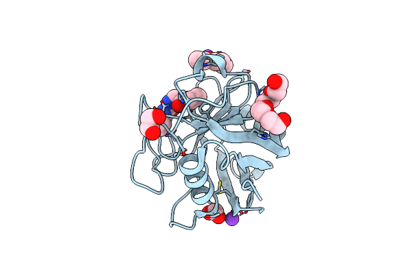 |
Human Cyclophilin D Complexed With Inhibitor
Organism: Homo sapiens
Method: X-RAY DIFFRACTION Resolution:1.45 Å Release Date: 2016-07-20 Classification: ISOMERASE Ligands: 4ZL, K, FMT, PG4, EDO, PGE |
Organism: Homo sapiens
Method: X-RAY DIFFRACTION
Release Date: 2016-07-20
Ligands: 4ZL, K, FMT, PG4, EDO, PGE
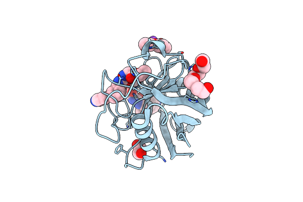 |
Human Cyclophilin D Complexed With Inhibitor.
Organism: Homo sapiens
Method: X-RAY DIFFRACTION Resolution:1.40 Å Release Date: 2016-07-20 Classification: ISOMERASE Ligands: K, PG4, EDO, PGE, 4ZM |
Organism: Homo sapiens
Method: X-RAY DIFFRACTION
Release Date: 2016-07-20
Ligands: K, PG4, EDO, PGE, 4ZM
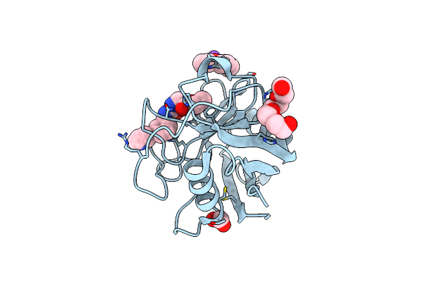 |
Human Cyclophilin D Complexed With Inhibitor
Organism: Homo sapiens
Method: X-RAY DIFFRACTION Resolution:1.80 Å Release Date: 2016-07-20 Classification: ISOMERASE Ligands: K, FMT, PG4, PGE, 4ZP |
Organism: Homo sapiens
Method: X-RAY DIFFRACTION
Release Date: 2016-07-20
Ligands: K, FMT, PG4, PGE, 4ZP
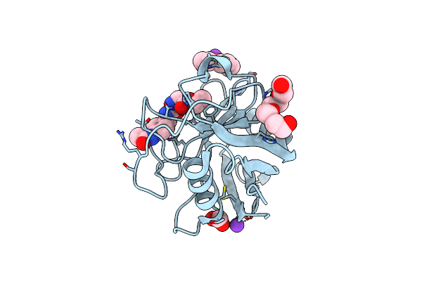 |
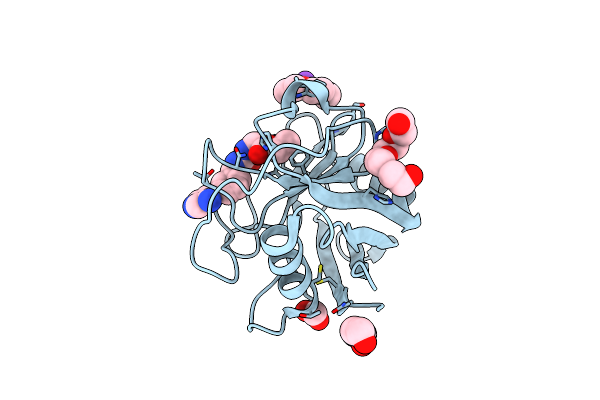
Human Cyclophilin D Complexed With Inhibitor.
Organism: Homo sapiens
Method: X-RAY DIFFRACTION Resolution:1.80 Å Release Date: 2016-07-20 Classification: ISOMERASE Ligands: K, FMT, PG4, PGE, 4ZO |
Organism: Homo sapiens
Method: X-RAY DIFFRACTION
Release Date: 2016-07-20
Ligands: K, FMT, PG4, PGE, 4ZO
 |
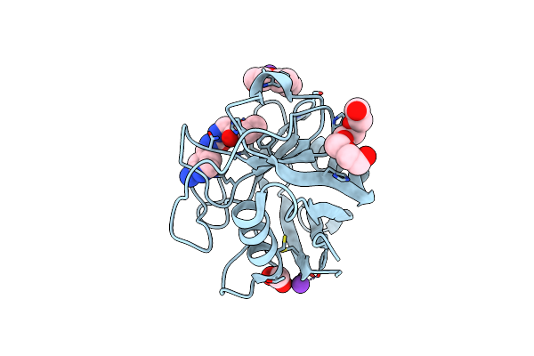
Human Cyclophilin D Complexed With Inhibitor
Organism: Homo sapiens
Method: X-RAY DIFFRACTION Resolution:1.80 Å Release Date: 2016-07-20 Classification: ISOMERASE Ligands: K, FMT, PG4, PGE, PEG, 4ZZ |
Organism: Homo sapiens
Method: X-RAY DIFFRACTION
Release Date: 2016-07-20
Ligands: K, FMT, PG4, PGE, PEG, 4ZZ
 |
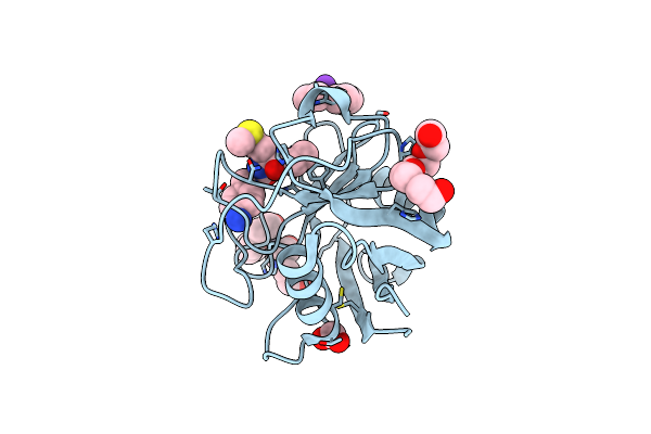
Human Cyclophilin D Complexed With Inhibitor
Organism: Homo sapiens
Method: X-RAY DIFFRACTION Resolution:1.80 Å Release Date: 2016-07-20 Classification: ISOMERASE Ligands: K, FMT, PG4, PGE, 502 |
Organism: Homo sapiens
Method: X-RAY DIFFRACTION
Release Date: 2016-07-20
Ligands: K, FMT, PG4, PGE, 502
 |
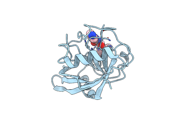
Human Cyclophilin D Complexed With Inhibitor
Organism: Homo sapiens
Method: X-RAY DIFFRACTION Resolution:1.90 Å Release Date: 2016-07-20 Classification: ISOMERASE Ligands: K, FMT, PG4, PGE, 4ZT, PEG |
Organism: Homo sapiens
Method: X-RAY DIFFRACTION
Release Date: 2016-07-20
Ligands: K, FMT, PG4, PGE, 4ZT, PEG
 |
Human Cyclophilin D Complexed With Inhibitor
Organism: Homo sapiens
Method: X-RAY DIFFRACTION Resolution:2.10 Å Release Date: 2016-07-20 Classification: ISOMERASE Ligands: C4Y |
Organism: Homo sapiens
Method: X-RAY DIFFRACTION
Release Date: 2016-07-20
Ligands: C4Y
 |
Type Ii Baeyer-Villiger Monooxygenase.The Oxygenating Constituent Of 3,6-Diketocamphane Monooxygenase From Cam Plasmid Of Pseudomonas Putida In Complex With Fmn.
Organism: Pseudomonas putida
Method: X-RAY DIFFRACTION Resolution:1.93 Å Release Date: 2015-09-09 Classification: OXIDOREDUCTASE Ligands: PIN, GOL, SO4, CL |
Organism: Pseudomonas putida
Method: X-RAY DIFFRACTION
Release Date: 2015-09-09
Ligands: PIN, GOL, SO4, CL
 |
Type Ii Baeyer-Villiger Monooxygenase.The Oxygenating Constituent Of 3,6-Diketocamphane Monooxygenase From Cam Plasmid Of Pseudomonas Putida In Complex With Fmn.
Organism: Pseudomonas putida
Method: X-RAY DIFFRACTION Resolution:1.90 Å Release Date: 2015-08-26 Classification: OXIDOREDUCTASE Ligands: FMN, PG4, PEG, PGE |
Organism: Pseudomonas putida
Method: X-RAY DIFFRACTION
Release Date: 2015-08-26
Ligands: FMN, PG4, PEG, PGE
 |
Crystal Structure Of The Fmn-Reductase Msue From Pseudomonas Putida Kt2440.
Organism: Pseudomonas putida
Method: X-RAY DIFFRACTION Resolution:1.96 Å Release Date: 2014-10-01 Classification: OXIDOREDUCTASE |
Organism: Pseudomonas putida
Method: X-RAY DIFFRACTION
Release Date: 2014-10-01
 |
Insight Into The Mechanism Of Enzymatic Glycosyltransfer With Retention Through The Synthesis And Analysis Of Bisubstrate Glycomimetics Of Trehalose-6-Phosphate Synthase
Organism: Escherichia coli
Method: X-RAY DIFFRACTION Resolution:2.20 Å Release Date: 2010-02-23 Classification: TRANSFERASE Ligands: EDO, UDP, VDO |
Organism: Escherichia coli
Method: X-RAY DIFFRACTION
Release Date: 2010-02-23
Ligands: EDO, UDP, VDO
 |
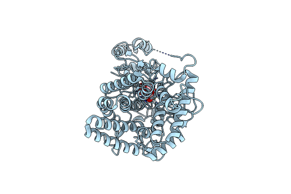
Family 37 Trehalase From Escherichia Coli In Complex With Validoxylamine
Organism: Escherichia coli
Method: X-RAY DIFFRACTION Resolution:2.20 Å Release Date: 2007-02-13 Classification: HYDROLASE Ligands: VDM |
Organism: Escherichia coli
Method: X-RAY DIFFRACTION
Release Date: 2007-02-13
Ligands: VDM
 |
Family 37 Trehalase From Escherichia Coli In Complex With 1- Thiatrehazolin
Organism: Escherichia coli
Method: X-RAY DIFFRACTION Resolution:1.50 Å Release Date: 2007-02-13 Classification: HYDROLASE Ligands: TTZ |
Organism: Escherichia coli
Method: X-RAY DIFFRACTION
Release Date: 2007-02-13
Ligands: TTZ
 |
Trehalose-6-Phosphate From E. Coli Bound With Udp-2-Fluoro Glucose.
Organism: Escherichia coli
Method: X-RAY DIFFRACTION Resolution:2.00 Å Release Date: 2003-12-12 Classification: SYNTHASE Ligands: U2F |
Organism: Escherichia coli
Method: X-RAY DIFFRACTION
Release Date: 2003-12-12
Ligands: U2F
 |
Trehalose-6-Phosphate From E. Coli Bound With Udp-Glucose.
Organism: Escherichia coli
Method: X-RAY DIFFRACTION Resolution:2.00 Å Release Date: 2003-11-13 Classification: SYNTHASE Ligands: UPG |
Organism: Escherichia coli
Method: X-RAY DIFFRACTION
Release Date: 2003-11-13
Ligands: UPG
 |
Trehalose-6-Phosphate Synthase. Otsa
Organism: Escherichia coli
Method: X-RAY DIFFRACTION Resolution:2.43 Å Release Date: 2003-02-07 Classification: SYNTHASE Ligands: UDP, G6P, IMD |
Organism: Escherichia coli
Method: X-RAY DIFFRACTION
Release Date: 2003-02-07
Ligands: UDP, G6P, IMD