

Search Count: 24
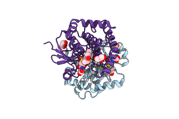 |
Crystal Structure Of Prostaglandin D2 Synthase In Complex With Fragment 1A At 1.84A Resolution.
Organism: Homo sapiens
Method: X-RAY DIFFRACTION Resolution:1.84 Å Release Date: 2021-07-28 Classification: ISOMERASE Ligands: GSH, MG, QPN, GOL |
Organism: Homo sapiens
Method: X-RAY DIFFRACTION
Release Date: 2021-07-28
Ligands: GSH, MG, QPN, GOL
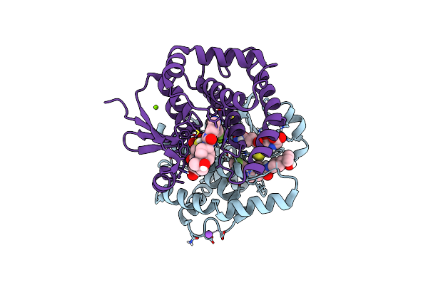 |
H-Pdgs Complexed With A 2-Phenylimidazo[1,2-A]Pyridine-6-Carboxamide Inhibitors
Organism: Homo sapiens
Method: X-RAY DIFFRACTION Resolution:1.88 Å Release Date: 2021-05-26 Classification: Isomerase, Transferase/Inhibitor Ligands: GSH, VH4, MG, NA |
Organism: Homo sapiens
Method: X-RAY DIFFRACTION
Release Date: 2021-05-26
Ligands: GSH, VH4, MG, NA
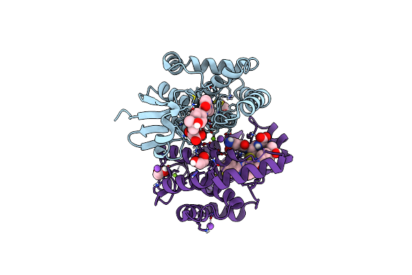 |
H-Pdgs Complexed With A 2-Phenylimidazo[1,2-A]Pyridine-6-Carboxamide Inhibitors
Organism: Homo sapiens
Method: X-RAY DIFFRACTION Resolution:1.13 Å Release Date: 2021-05-26 Classification: Isomerase, Transferase/Inhibitor Ligands: CL, EDO, GSH, VH7, MG, NA |
Organism: Homo sapiens
Method: X-RAY DIFFRACTION
Release Date: 2021-05-26
Ligands: CL, EDO, GSH, VH7, MG, NA
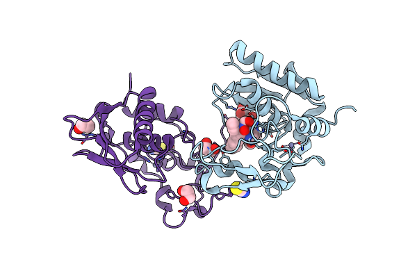 |
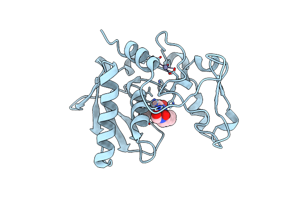
Bmp1 Complexed With A Reverse Hydroxymate - Compound 22
Organism: Homo sapiens
Method: X-RAY DIFFRACTION Resolution:1.45 Å Release Date: 2018-08-08 Classification: HYDROLASE Ligands: SCN, ZN, EVV, EDO |
Organism: Homo sapiens
Method: X-RAY DIFFRACTION
Release Date: 2018-08-08
Ligands: SCN, ZN, EVV, EDO
 |
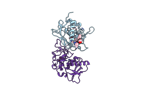
Bmp1 Complexed With A Reverse Hydroxamate - Compound 4
Organism: Homo sapiens
Method: X-RAY DIFFRACTION Resolution:2.33 Å Release Date: 2018-08-08 Classification: HYDROLASE/HYDROLASE INHIBITOR Ligands: ZN, E7D |
Organism: Homo sapiens
Method: X-RAY DIFFRACTION
Release Date: 2018-08-08
Ligands: ZN, E7D
 |
Bmp1 Complexed With A Reverse Hydroxymate - Compound 1
Organism: Homo sapiens
Method: X-RAY DIFFRACTION Resolution:2.05 Å Release Date: 2018-08-08 Classification: HYDROLASE Ligands: EDO, ZN, E8M |
Organism: Homo sapiens
Method: X-RAY DIFFRACTION
Release Date: 2018-08-08
Ligands: EDO, ZN, E8M
 |
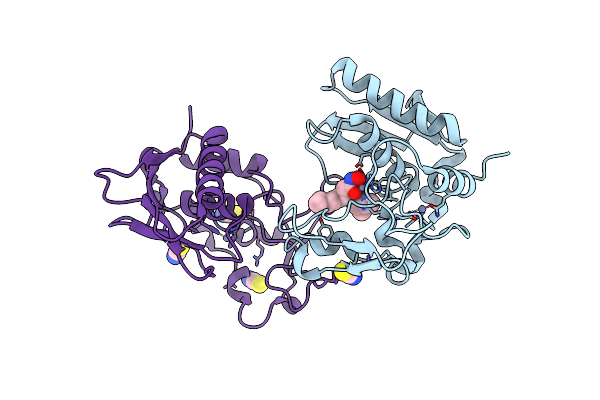
Bmp1 Complexed With (2~{S})-2-[[(1~{R},3~{S},4~{S})-2-[(2~{R})-2-[2-(Oxidanylamino)-2-Oxidanylidene-Ethyl]Heptanoyl]-2-Azabicyclo[2.2.1]Heptan-3-Yl]Carbonylamino]-2-Phenyl-Ethanoic Acid
Organism: Homo sapiens
Method: X-RAY DIFFRACTION Resolution:2.05 Å Release Date: 2018-08-08 Classification: HYDROLASE Ligands: SCN, ZN, E8P, EDO |
Organism: Homo sapiens
Method: X-RAY DIFFRACTION
Release Date: 2018-08-08
Ligands: SCN, ZN, E8P, EDO
 |
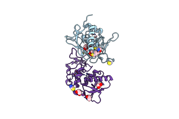
Bmp1 Complexed With A Hydroxamate
Organism: Homo sapiens
Method: X-RAY DIFFRACTION Resolution:1.93 Å Release Date: 2018-08-08 Classification: HYDROLASE Ligands: SCN, ZN, E8J |
Organism: Homo sapiens
Method: X-RAY DIFFRACTION
Release Date: 2018-08-08
Ligands: SCN, ZN, E8J
 |
Bmp1 Complexed With A Hydroxamate - Compound 2
Organism: Homo sapiens
Method: X-RAY DIFFRACTION Resolution:1.75 Å Release Date: 2018-08-08 Classification: HYDROLASE Ligands: SCN, ZN, E8S, EDO |
Organism: Homo sapiens
Method: X-RAY DIFFRACTION
Release Date: 2018-08-08
Ligands: SCN, ZN, E8S, EDO
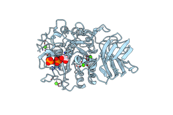 |
Crystal Structure Of Unglycosylated Apo Human Furin @1.89A
Organism: Homo sapiens
Method: X-RAY DIFFRACTION Resolution:1.89 Å Release Date: 2016-05-04 Classification: HYDROLASE Ligands: CA, PO4, EDO |
Organism: Homo sapiens
Method: X-RAY DIFFRACTION
Release Date: 2016-05-04
Ligands: CA, PO4, EDO
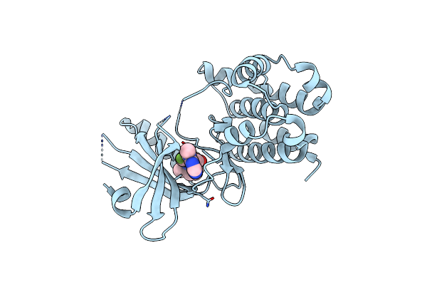 |
Crystal Structure Of Gsk6157 Bound To Perk (R587-R1092, Delete A660-T867) At 2.34A Resolution
Organism: Homo sapiens
Method: X-RAY DIFFRACTION Resolution:2.34 Å Release Date: 2014-09-03 Classification: TRANSFERASE/TRANSFERASE INHIBITOR Ligands: 27D |
Organism: Homo sapiens
Method: X-RAY DIFFRACTION
Release Date: 2014-09-03
Ligands: 27D
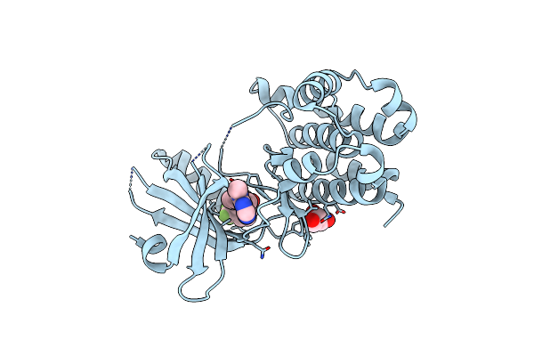 |
Crystal Structure Of Gsk6414 Bound To Perk (R587-R1092, Delete A660-T867) At 2.28 A Resolution
Organism: Homo sapiens
Method: X-RAY DIFFRACTION Resolution:2.28 Å Release Date: 2012-08-08 Classification: TRANSFERASE/TRANSFERASE INHIBITOR Ligands: GOL, 0WH |
Organism: Homo sapiens
Method: X-RAY DIFFRACTION
Release Date: 2012-08-08
Ligands: GOL, 0WH
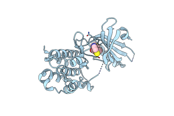 |
Crystal Structure Of Gsk6924 Bound To Perk (R587-R1092, Delete A660-T867) At 2.70 A Resolution
Organism: Homo sapiens
Method: X-RAY DIFFRACTION Resolution:2.70 Å Release Date: 2012-08-08 Classification: TRANSFERASE/TRANSFERASE INHIBITOR Ligands: 924 |
Organism: Homo sapiens
Method: X-RAY DIFFRACTION
Release Date: 2012-08-08
Ligands: 924
 |
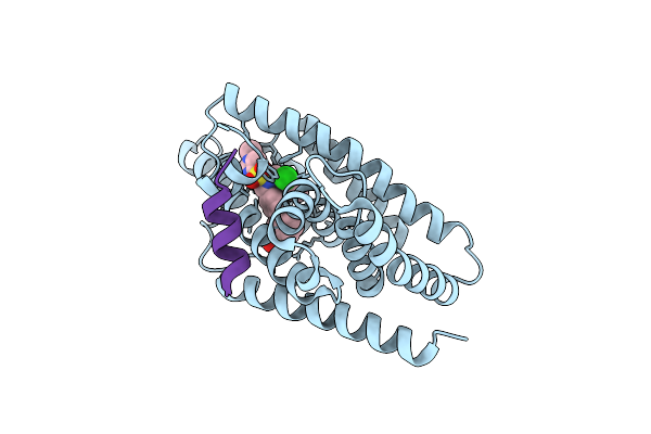
Crystal Structure Of A Deletion Mutant Of Human Reverba Ligand Binding Domain Bound With An Ncor Id1 Peptide Determined To 2.60A
Organism: Homo sapiens
Method: X-RAY DIFFRACTION Resolution:2.60 Å Release Date: 2010-06-30 Classification: Transcription regulator |
Organism: Homo sapiens
Method: X-RAY DIFFRACTION
Release Date: 2010-06-30
 |
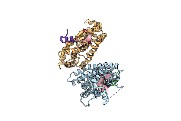
X-Ray Crystal Structure Of A Potent Liver X Receptor Modulator
Organism: Homo sapiens
Method: X-RAY DIFFRACTION Resolution:2.30 Å Release Date: 2010-04-07 Classification: TRANSCRIPTION Ligands: G58 |
Organism: Homo sapiens
Method: X-RAY DIFFRACTION
Release Date: 2010-04-07
Ligands: G58
 |
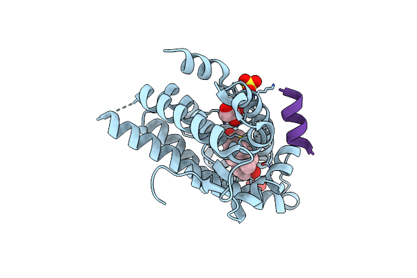
The X-Ray Crystal Structure Of Ppar-Gamma In Complex With An Indole Derivative Modulator, Gsk538, And An Src-1 Peptide
Organism: Homo sapiens
Method: X-RAY DIFFRACTION Resolution:2.10 Å Release Date: 2010-01-12 Classification: TRANSCRIPTION Ligands: 538 |
Organism: Homo sapiens
Method: X-RAY DIFFRACTION
Release Date: 2010-01-12
Ligands: 538
 |
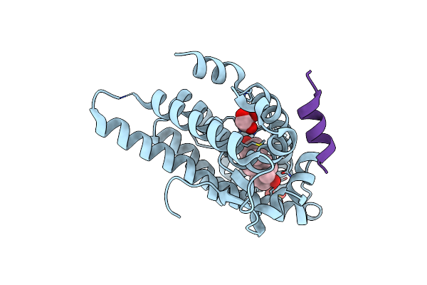
The Wild Type Androgen Receptor Ligand Binding Domain Bound With Testosterone And An Ar 20-30 Peptide
Organism: Homo sapiens
Method: X-RAY DIFFRACTION Resolution:1.87 Å Release Date: 2007-07-24 Classification: HORMONE Ligands: SO4, TES, GOL |
Organism: Homo sapiens
Method: X-RAY DIFFRACTION
Release Date: 2007-07-24
Ligands: SO4, TES, GOL
 |
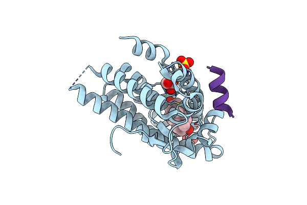
The Wild Type Androgen Receptor Ligand Binding Domain Bound With Testosterone And A Tif2 Box 3 Coactivator Peptide 740-753
Organism: Homo sapiens
Method: X-RAY DIFFRACTION Resolution:1.90 Å Release Date: 2007-07-24 Classification: HORMONE Ligands: SO4, TES, GOL |
Organism: Homo sapiens
Method: X-RAY DIFFRACTION
Release Date: 2007-07-24
Ligands: SO4, TES, GOL
 |
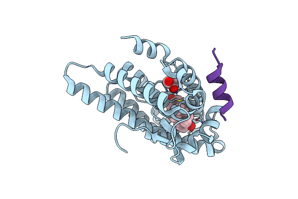
The Androgen Receptor Prostate Cancer Mutant H874Y Ligand Binding Domain Bound With Testosterone And An Ar 20-30 Peptide
Organism: Homo sapiens
Method: X-RAY DIFFRACTION Resolution:1.80 Å Release Date: 2007-07-24 Classification: HORMONE Ligands: SO4, TES, GOL |
Organism: Homo sapiens
Method: X-RAY DIFFRACTION
Release Date: 2007-07-24
Ligands: SO4, TES, GOL
 |
The Androgen Receptor Prostate Cancer Mutant H874Y Ligand Binding Domain Bound With Testosterone And A Tif2 Box3 Coactivator Peptide 740-753
Organism: Homo sapiens
Method: X-RAY DIFFRACTION Resolution:1.92 Å Release Date: 2007-07-24 Classification: HORMONE Ligands: TES, GOL |
Organism: Homo sapiens
Method: X-RAY DIFFRACTION
Release Date: 2007-07-24
Ligands: TES, GOL