
Search Count: 5,051
 |
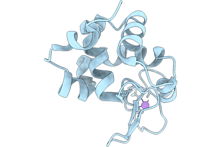
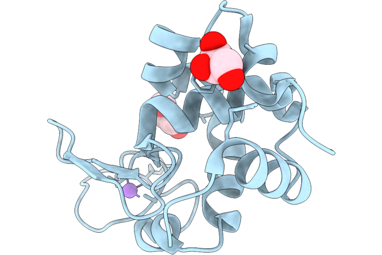
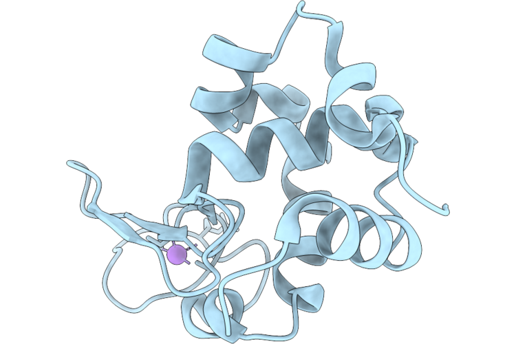
Hen Egg-White Lysozyme (Hewl) Collected At The European Xfel, Spb/Sfx At The Interaction Region Downstream With 9.6 Kev Photon Energy
Organism: Gallus gallus
Method: X-RAY DIFFRACTION Resolution:1.85 Å Release Date: 2026-07-08 Classification: HYDROLASE Ligands: CL, PEG, ACT |
 |
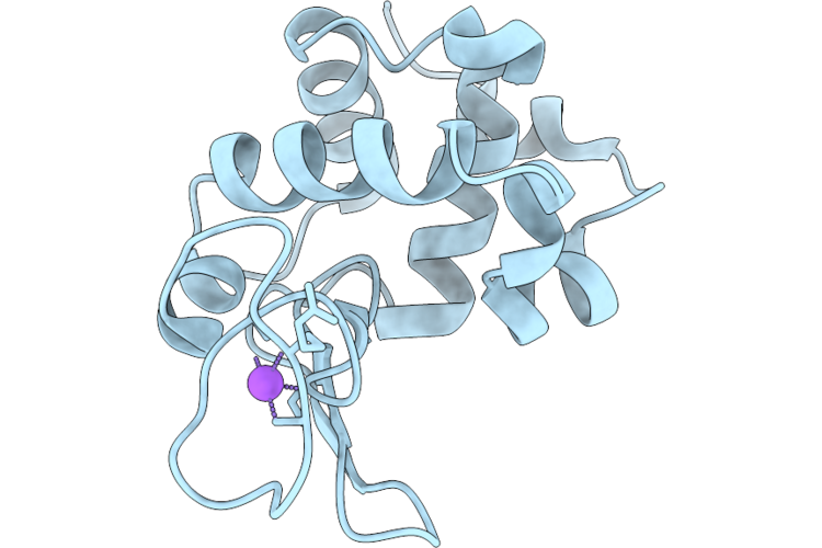
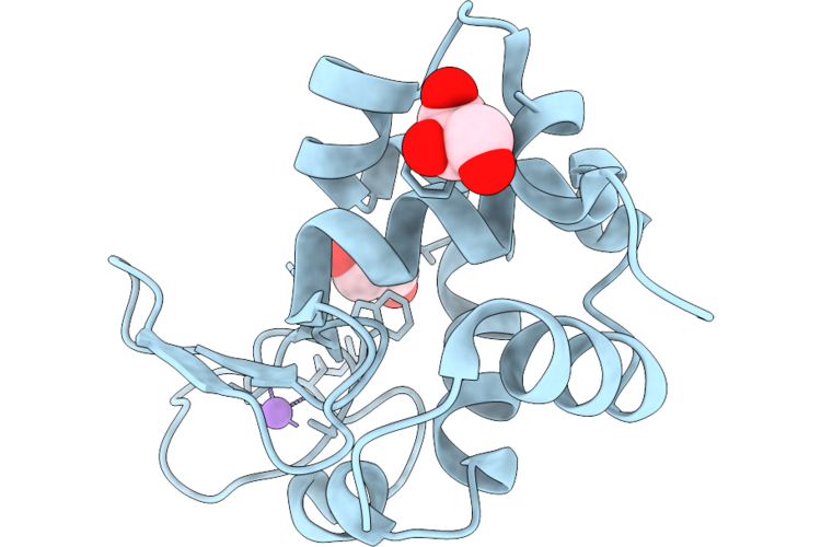
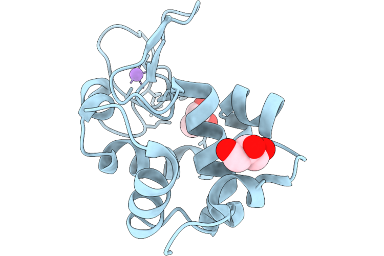
Hen Egg-White Lysozyme (Hewl) Collected At The European Xfel, Spb/Sfx At The Interaction Region Downstream With 12.25 Kev Photon Energy
Organism: Gallus gallus
Method: X-RAY DIFFRACTION Resolution:1.70 Å Release Date: 2026-07-08 Classification: HYDROLASE |
 |
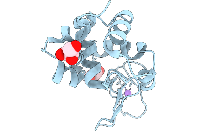
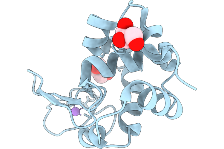
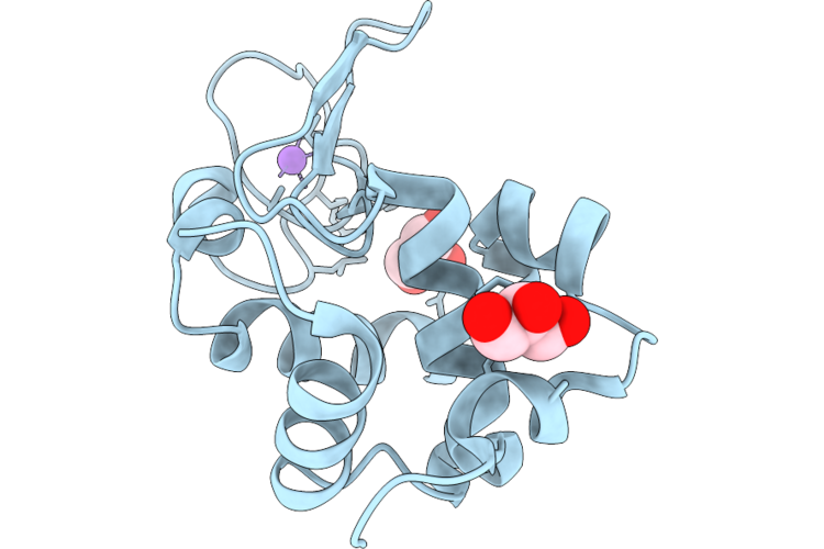
Crystal Structure Of Hen Egg White Lysozyme At 100 Kelvin With Peg 400 (Duplicate)
Organism: Gallus gallus
Method: X-RAY DIFFRACTION Resolution:1.50 Å Release Date: 2026-07-01 Classification: HYDROLASE |
 |
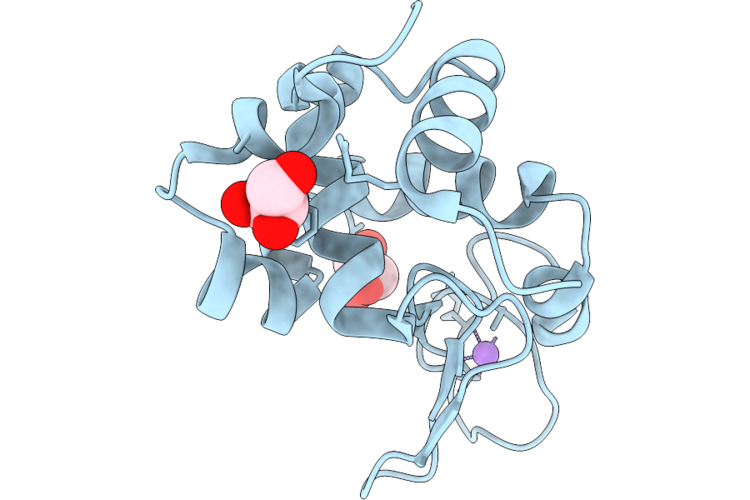
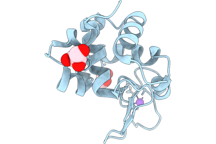
Crystal Structure Of Hen Egg White Lysozyme At 100 Kelvin With Peg 400 (Triplicate)
Organism: Gallus gallus
Method: X-RAY DIFFRACTION Resolution:1.50 Å Release Date: 2026-07-01 Classification: HYDROLASE |
 |
Crystal Structure Of Hen Egg White Lysozyme At 100 Kelvin With Peg 6000
Organism: Gallus gallus
Method: X-RAY DIFFRACTION Resolution:1.50 Å Release Date: 2026-07-01 Classification: HYDROLASE |
 |
Crystal Structure Of Hen Egg White Lysozyme At 100 Kelvin With Peg 6000 (Duplicate)
Organism: Gallus gallus
Method: X-RAY DIFFRACTION Resolution:1.50 Å Release Date: 2026-07-01 Classification: HYDROLASE |
 |
Crystal Structure Of Hen Egg White Lysozyme At 100 Kelvin With Peg 6000 (Triplicate)
Organism: Gallus gallus
Method: X-RAY DIFFRACTION Resolution:1.50 Å Release Date: 2026-06-24 Classification: HYDROLASE |
 |
Crystal Structure Of Hen Egg White Lysozyme At 100 Kelvin With Silicone Oil
Organism: Gallus gallus
Method: X-RAY DIFFRACTION Resolution:1.50 Å Release Date: 2026-06-24 Classification: HYDROLASE |
 |
Crystal Structure Of Hen Egg White Lysozyme At 100 Kelvin With Silicone Oil (Duplicate)
Organism: Gallus gallus
Method: X-RAY DIFFRACTION Resolution:1.50 Å Release Date: 2026-06-24 Classification: HYDROLASE |
 |
Crystal Structure Of Hen Egg White Lysozyme At 100 Kelvin With Silicone Oil (Triplicate)
Organism: Gallus gallus
Method: X-RAY DIFFRACTION Resolution:1.50 Å Release Date: 2026-06-24 Classification: HYDROLASE |
 |
Crystal Structure Of Hen Egg White Lysozyme At 100 Kelvin With Vaseline
Organism: Gallus gallus
Method: X-RAY DIFFRACTION Resolution:1.50 Å Release Date: 2026-06-24 Classification: HYDROLASE |
 |
Crystal Structure Of Hen Egg White Lysozyme At 100 Kelvin With Vaseline (Duplicate)
Organism: Gallus gallus
Method: X-RAY DIFFRACTION Resolution:1.50 Å Release Date: 2026-06-24 Classification: HYDROLASE |
 |
Crystal Structure Of Hen Egg White Lysozyme At 100 Kelvin With Vaseline (Triplicate)
Organism: Gallus gallus
Method: X-RAY DIFFRACTION Resolution:1.50 Å Release Date: 2026-06-24 Classification: HYDROLASE |
 |
Crystal Structure Of Hen Egg White Lysozyme At 300 Kelvin With Apiezon N
Organism: Gallus gallus
Method: X-RAY DIFFRACTION Resolution:1.50 Å Release Date: 2026-06-24 Classification: HYDROLASE |
 |
Crystal Structure Of Hen Egg White Lysozyme At 300 Kelvin With Apiezon N (Duplicate)
Organism: Gallus gallus
Method: X-RAY DIFFRACTION Resolution:1.50 Å Release Date: 2026-06-24 Classification: HYDROLASE |
 |
Crystal Structure Of Hen Egg White Lysozyme At 300 Kelvin With Apiezon N (Triplicate)
Organism: Gallus gallus
Method: X-RAY DIFFRACTION Resolution:1.48 Å Release Date: 2026-06-24 Classification: HYDROLASE |
 |
Crystal Structure Of Hen Egg White Lysozyme At 300 Kelvin With Apiezon T
Organism: Gallus gallus
Method: X-RAY DIFFRACTION Resolution:1.47 Å Release Date: 2026-06-24 Classification: HYDROLASE |
 |
Crystal Structure Of Hen Egg White Lysozyme At 300 Kelvin With Apiezon T (Duplicate)
Organism: Gallus gallus
Method: X-RAY DIFFRACTION Resolution:1.47 Å Release Date: 2026-06-24 Classification: HYDROLASE |
 |
Crystal Structure Of Hen Egg White Lysozyme At 300 Kelvin With Apiezon T (Triplicate)
Organism: Gallus gallus
Method: X-RAY DIFFRACTION Resolution:1.50 Å Release Date: 2026-06-24 Classification: HYDROLASE |
 |
Crystal Structure Of Hen Egg White Lysozyme At 300 Kelvin With Vaseline
Organism: Gallus gallus
Method: X-RAY DIFFRACTION Resolution:1.50 Å Release Date: 2026-06-24 Classification: HYDROLASE |