

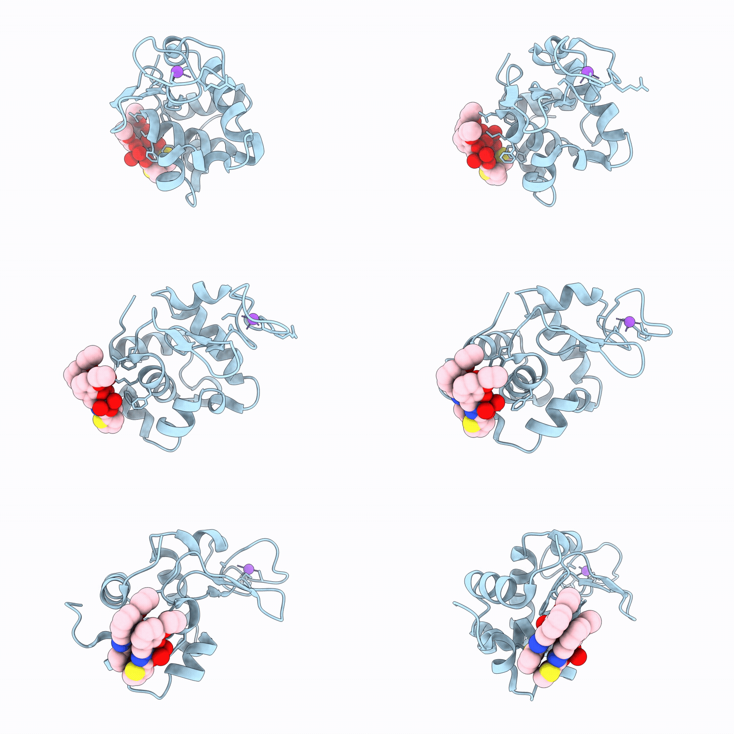
9S2A image
Deposition Date 2025-07-21
Release Date 2026-04-01
Last Version Date 2026-04-08
Entry Detail
PDB ID:
9S2A
Keywords:
Title:
X-ray structure of lysozyme obtained upon reaction with the dioxidovanadium(V) complex with thiophene-2-carboxylic acid (3-ethoxy-2-hydroxybenzylidene)hydrazide
Biological Source:
Source Organism(s):
Gallus gallus (Taxon ID: 9031)
Method Details:
Experimental Method:
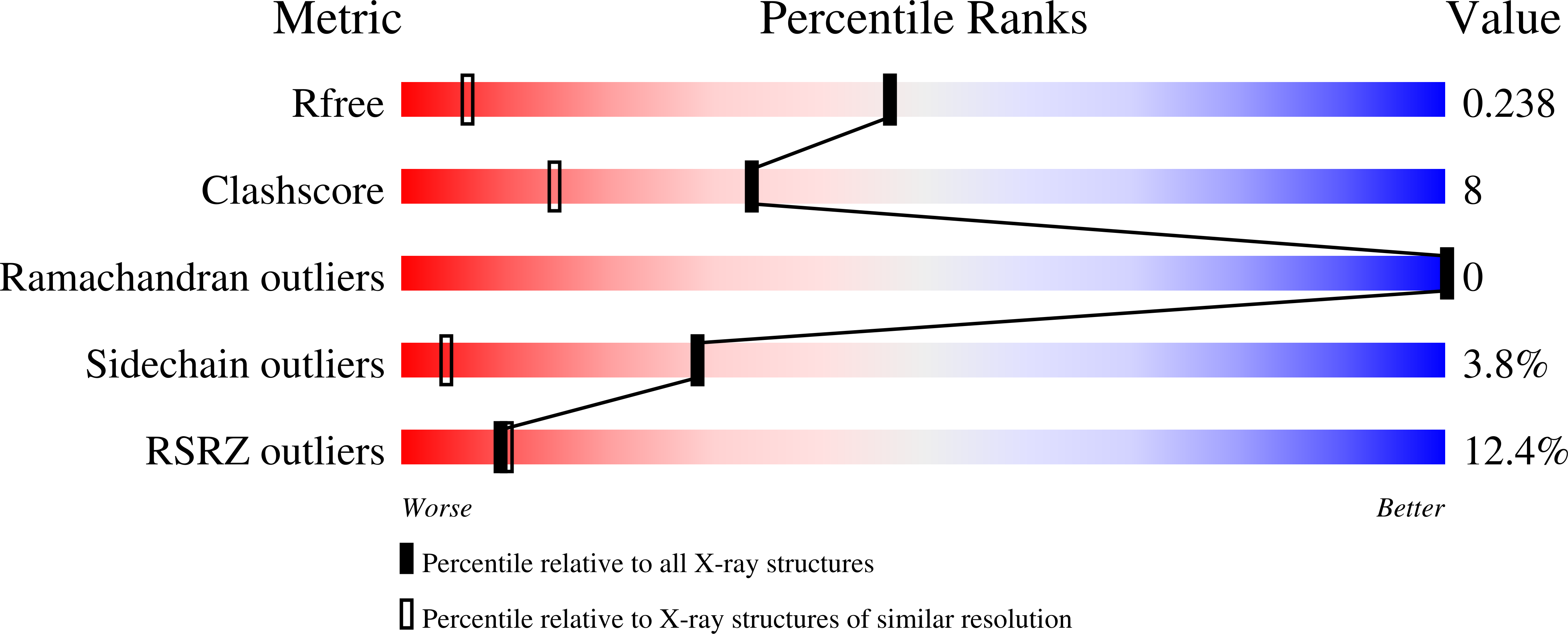
Resolution:
1.37 Å
R-Value Free:
0.23
R-Value Work:
0.19
Space Group:
P 43 21 2
Macromolecular Entities
Structures with similar UniProt ID
Protein Blast
Polymer Type:polypeptide(L)
Molecule:Lysozyme C
Gene (Uniprot):LYZ
Chain IDs:A (auth: AAA)
Chain Length:129
Number of Molecules:1
Biological Source:Gallus gallus
Primary Citation
Binding of Aqueous-Stable, Lipophilic, Hemocompatible Anticancer V V O 2 Metallodrugs with Biological Molecules: X-ray Structures of the Adduct of the V V -hydrazonato Complex with Hen Egg White Lysozyme.
Inorg Chem 65 6412 6433 (2026)
PMID: 41830615 DOI: 10.1021/acs.inorgchem.5c05201

Abstact

Three new water-stable aqueous dioxidovanadium(V) complexes, [(V(V)O(2)L(1-3))M(H(2)O)(n)] (1-3), incorporating hydrazone ligands with different alkali metals (Na(+)/K(+)) as counterions were synthesized and characterized by various physicochemical approaches, including single-crystal X-ray diffraction (SCXRD). Time-dependent spectroscopic/spectrometric techniques were used to determine their aqueous-phase stabilities. Blood compatibility studies were employed to investigate their efficacy and stability with human red blood cells. Lipophilicity and calf thymus (CT)-DNA interaction of 1-3 were investigated using conventional techniques. High-resolution molecular structures of the adduct formed between 1 and hen egg white lysozyme (HEWL) were determined by SCXRD. The structural analysis reveals that the compound self-assembles within protein crystals, forming a dimeric structure that non-covalently interacts with the protein surface. The binding of 1 to HEWL was also evaluated through different spectroscopic methods. Fluorescence data indicate that 1 can also bind the physiologically relevant protein human serum albumin at pH 7.4. Furthermore, the cytotoxicity of 1-3 was evaluated against the lung (A549) and human breast adenocarcinoma (MCF-7) cancer cell lines, as well as an human embryonic kidney cell line (HEK-293) noncancerous cell line. 1 (IC(50) value of 9.2 +/- 0.1 muM) is more effective than the other two complexes. It induces cell death via apoptosis.

Legend

Protein

Chemical

Disease

Primary Citation of related structures
Feedback Form
Name
Email
Institute
Feedback