

Search Count: 16
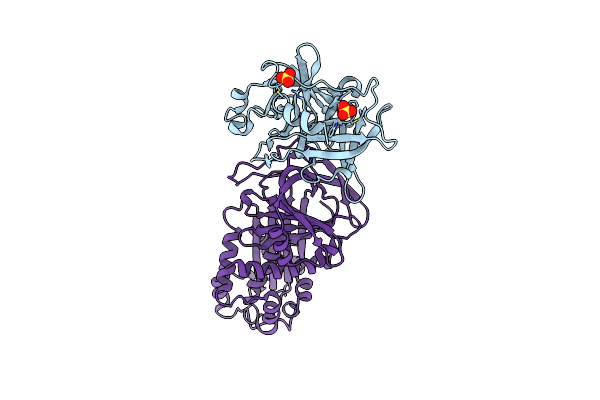 |
Crystal Structure Of A Michaelis Complex Between Plasminogen Activator Inhibitor-1 And Urokinase-Type Plasminogen Activator
Organism: Homo sapiens
Method: X-RAY DIFFRACTION Resolution:2.30 Å Release Date: 2010-12-29 Classification: hydrolase Inhibitor/Hydrolase Ligands: SO4 |
Organism: Homo sapiens
Method: X-RAY DIFFRACTION
Release Date: 2010-12-29
Ligands: SO4
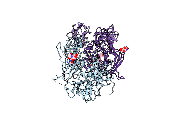 |
Crystal Structure Of Human Factor Viii
Organism: Homo sapiens
Method: X-RAY DIFFRACTION Resolution:3.98 Å Release Date: 2008-04-01 Classification: BLOOD CLOTTING Ligands: CU, CA, NAG |
Organism: Homo sapiens
Method: X-RAY DIFFRACTION
Release Date: 2008-04-01
Ligands: CU, CA, NAG
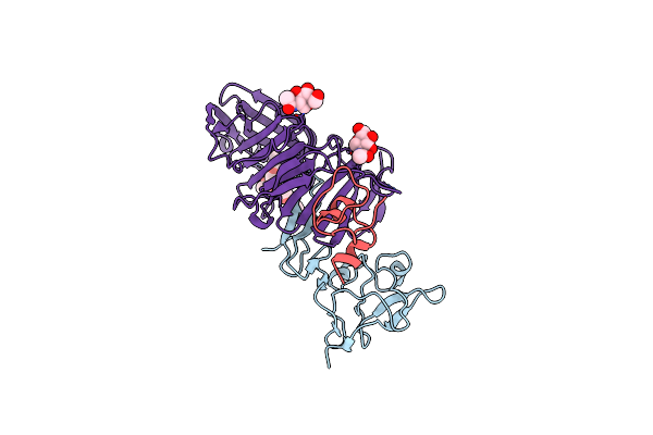 |
Structure Of Urokinase Receptor, Urokinase And Vitronectin Complex
Organism: Homo sapiens
Method: X-RAY DIFFRACTION Resolution:2.80 Å Release Date: 2008-03-25 Classification: IMMUNE SYSTEM Ligands: NAG |
Organism: Homo sapiens
Method: X-RAY DIFFRACTION
Release Date: 2008-03-25
Ligands: NAG
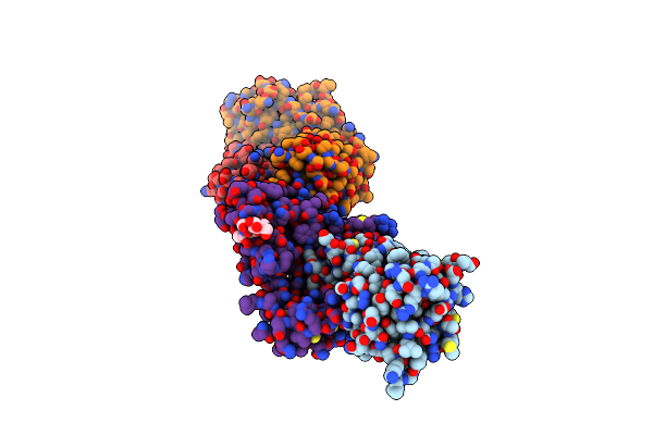 |
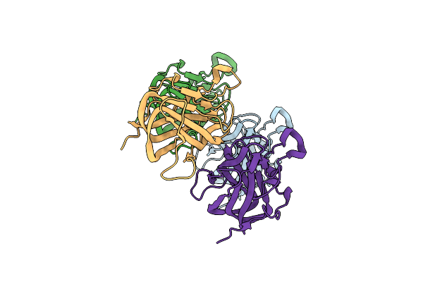
Structure Of Urokinase Receptor, Urokinase And Vitronectin Complex
Organism: Homo sapiens, Mus musculus
Method: X-RAY DIFFRACTION Resolution:2.50 Å Release Date: 2008-03-25 Classification: IMMUNE SYSTEM Ligands: NAG |
Organism: Homo sapiens, Mus musculus
Method: X-RAY DIFFRACTION
Release Date: 2008-03-25
Ligands: NAG
 |
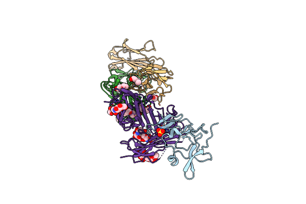
Crystal Structure Of The Bovine Lactadherin C2 Domain
Organism: Bos taurus
Method: X-RAY DIFFRACTION Resolution:2.40 Å Release Date: 2007-08-07 Classification: CELL ADHESION |
Organism: Bos taurus
Method: X-RAY DIFFRACTION
Release Date: 2007-08-07
 |
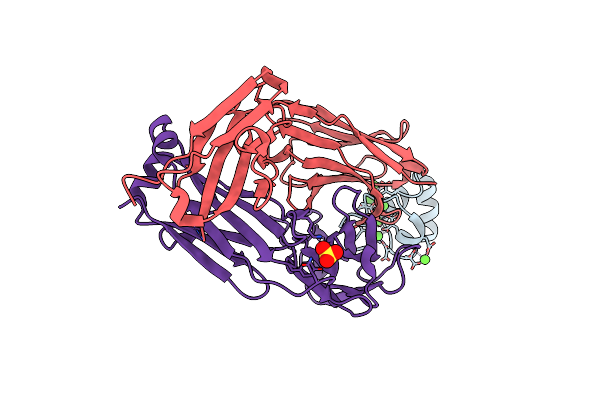
Structure Of Human Urokinase Plasminogen Activator In Complex With Urokinase Receptor And An Anti-Upar Antibody At 1.9 A
Organism: Homo sapiens, Mus musculus
Method: X-RAY DIFFRACTION Resolution:1.90 Å Release Date: 2006-02-21 Classification: IMMUNE SYSTEM, HYDROLASE Ligands: SO4, ETX, EDO, PGE, NDG, PG4 |
Organism: Homo sapiens, Mus musculus
Method: X-RAY DIFFRACTION
Release Date: 2006-02-21
Ligands: SO4, ETX, EDO, PGE, NDG, PG4
 |
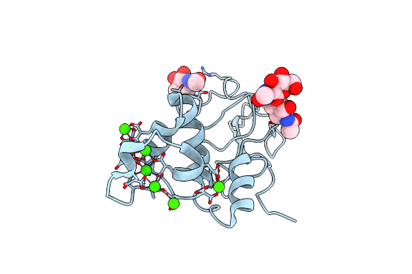
Crystal Structure Of Human Factor Ix Gla Domain In Complex Of An Inhibitory Antibody, 10C12
Organism: Homo sapiens
Method: X-RAY DIFFRACTION Resolution:2.20 Å Release Date: 2004-01-20 Classification: IMMUNE SYSTEM Ligands: SO4, CA |
Organism: Homo sapiens
Method: X-RAY DIFFRACTION
Release Date: 2004-01-20
Ligands: SO4, CA
 |
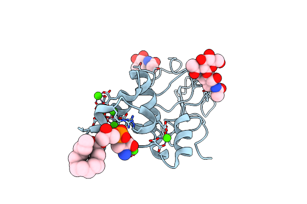
Bovine Prothrombin Fragment 1 In Complex With Calcium Ion
Organism: Bos taurus
Method: X-RAY DIFFRACTION Resolution:1.90 Å Release Date: 2003-09-16 Classification: HYDROLASE Ligands: NAG, CA |
Organism: Bos taurus
Method: X-RAY DIFFRACTION
Release Date: 2003-09-16
Ligands: NAG, CA
 |
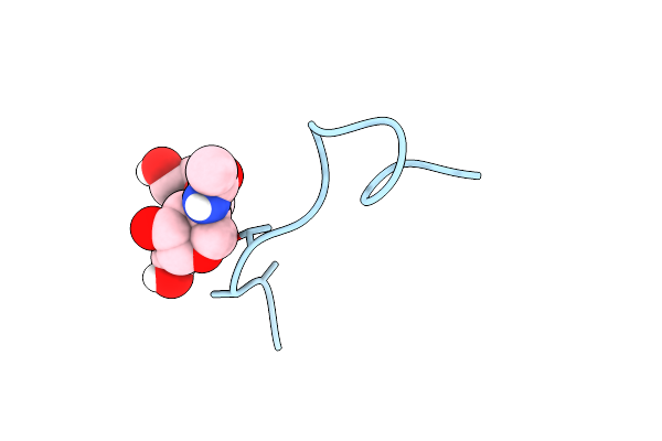
Bovine Prothrombin Fragment 1 In Complex With Calcium And Lysophosphotidylserine
Organism: Bos taurus
Method: X-RAY DIFFRACTION Resolution:2.30 Å Release Date: 2003-09-16 Classification: HYDROLASE Ligands: NAG, CA, CL, LPS |
Organism: Bos taurus
Method: X-RAY DIFFRACTION
Release Date: 2003-09-16
Ligands: NAG, CA, CL, LPS
 |
A Novel Conotoxin From Conus Textile With Unusual Post-Translational Modifications Reduces Presynaptic Calcium Influx, Nmr, 1 Structure, Glycosylated Protein
Organism: Conus textile
Method: SOLUTION NMR Release Date: 1999-06-08 Classification: GAMMA-CARBOXY GLUTAMIC ACID |
Organism: Conus textile
Method: SOLUTION NMR
Release Date: 1999-06-08
 |
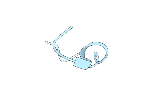
Nmr Structure Of Calcium Bound Conformer Of Conantokin G, Minimized Average Structure
|
Organism: Conus geographus
Method: SOLUTION NMR
Release Date: 1998-04-08
 |
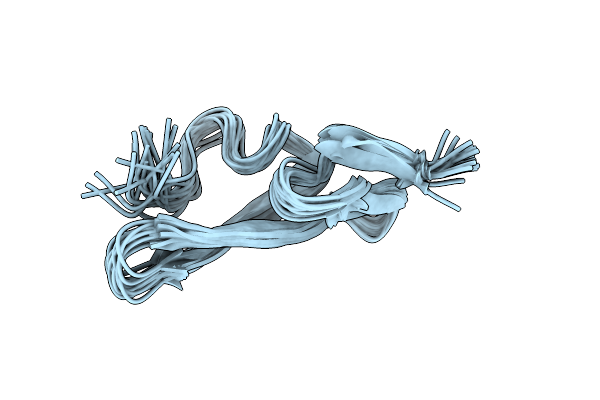
Nmr Structure Of Metal-Free Conantokin G, 1 Structure
Organism: Conus geographus
Method: SOLUTION NMR Release Date: 1997-08-20 Classification: GAMMA-CARBOXYGLUTAMIC ACID |
Organism: Conus geographus
Method: SOLUTION NMR
Release Date: 1997-08-20
 |
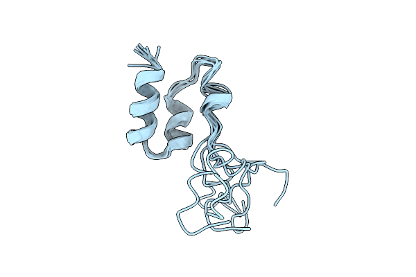
Structure Of The Egf Domain Of P-Selectin, Nmr, 19 Structures
Organism: Homo sapiens
Method: SOLUTION NMR Release Date: 1997-04-01 Classification: CELL ADHESION PROTEIN |
Organism: Homo sapiens
Method: SOLUTION NMR
Release Date: 1997-04-01
 |
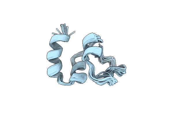
Coagulation Factor, Mg(Ii), Nmr, 7 Structures (Backbone Atoms Only)
Organism: Homo sapiens
Method: SOLUTION NMR Release Date: 1996-11-08 Classification: COAGULATION FACTOR |
Organism: Homo sapiens
Method: SOLUTION NMR
Release Date: 1996-11-08
 |
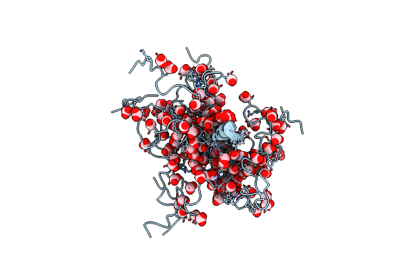
Nmr Structure Of Calcium Ion-Bound Gamma-Carboxy-Glutamic Acid-Rich Domain Of Factor Ix
Organism: Homo sapiens
Method: SOLUTION NMR Release Date: 1996-06-20 Classification: COAGULATION FACTOR |
Organism: Homo sapiens
Method: SOLUTION NMR
Release Date: 1996-06-20
 |
Structure Of The Metal-Free Gamma-Carboxyglutamic Acid-Rich Membrane Binding Region Of Factor Ix By Two-Dimensional Nmr Spectroscopy
Organism: Homo sapiens
Method: SOLUTION NMR Release Date: 1995-07-10 Classification: COAGULATION FACTOR Ligands: FMT |
Organism: Homo sapiens
Method: SOLUTION NMR
Release Date: 1995-07-10
Ligands: FMT