

Search Count: 14
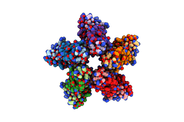 |
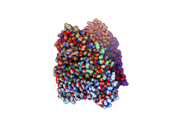
Fitting Of The H2A-H2B Histones In The Electron Microscopy Map Of The Complex Nucleoplasmin:H2A-H2B Histones (1:5).
Organism: Gallus gallus
Method: ELECTRON MICROSCOPY Resolution:19.50 Å Release Date: 2010-11-03 Classification: NUCLEAR PROTEIN |
Organism: Gallus gallus
Method: ELECTRON MICROSCOPY
Release Date: 2010-11-03
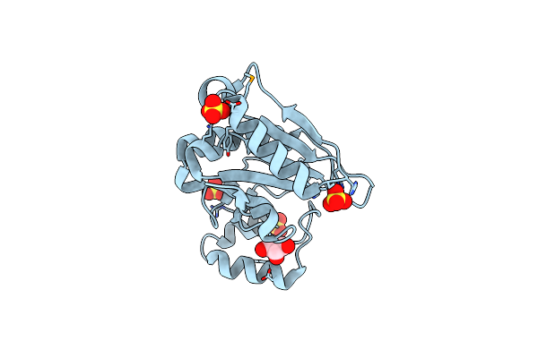 |
Crystal Structure Of Putative Sufu (Suppressor Of Fused Protein) Homolog (Yp_208451.1) From Neisseria Gonorrhoeae Fa 1090 At 1.40 A Resolution
Organism: Neisseria gonorrhoeae
Method: X-RAY DIFFRACTION Resolution:1.40 Å Release Date: 2010-01-26 Classification: PROTEIN BINDING Ligands: SO4, GOL |
Organism: Neisseria gonorrhoeae
Method: X-RAY DIFFRACTION
Release Date: 2010-01-26
Ligands: SO4, GOL
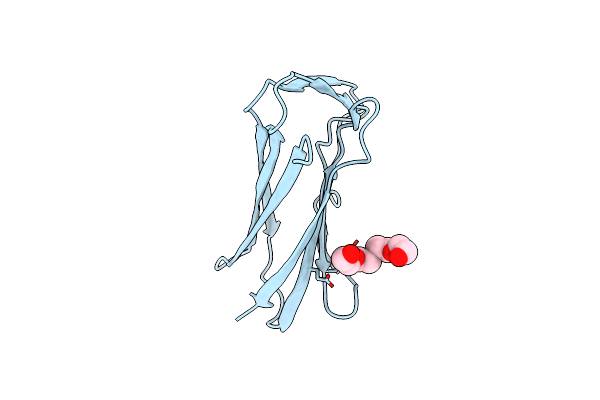 |
Crystal Structure Of An Intracellular Proteinase Inhibitor (Ipi, Bsu11130) From Bacillus Subtilis At 2.61 A Resolution
Organism: Bacillus subtilis
Method: X-RAY DIFFRACTION Resolution:2.61 Å Release Date: 2009-09-15 Classification: PROTEIN BINDING Ligands: PG4 |
Organism: Bacillus subtilis
Method: X-RAY DIFFRACTION
Release Date: 2009-09-15
Ligands: PG4
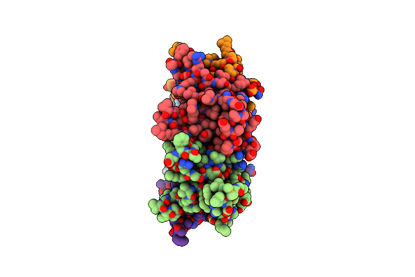 |
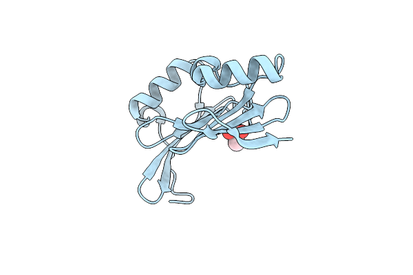
Crystal Structure Of Pleckstrin Homology Domain (Yp_926556.1) From Shewanella Amazonensis Sb2B At 1.99 A Resolution
Organism: Shewanella amazonensis sb2b
Method: X-RAY DIFFRACTION Resolution:1.99 Å Release Date: 2009-06-23 Classification: structural genomics, unknown function Ligands: GOL, PEG |
Organism: Shewanella amazonensis sb2b
Method: X-RAY DIFFRACTION
Release Date: 2009-06-23
Ligands: GOL, PEG
 |
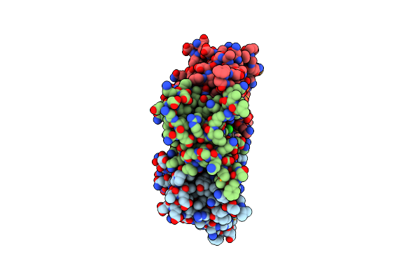
Crystal Structure Of A Putative Periplasmic Protein From Duf2874 Family (Bvu_2987) From Bacteroides Vulgatus Atcc 8482 At 1.85 A Resolution
Organism: Bacteroides vulgatus atcc 8482
Method: X-RAY DIFFRACTION Resolution:1.85 Å Release Date: 2008-08-05 Classification: UNKNOWN FUNCTION Ligands: CAC |
Organism: Bacteroides vulgatus atcc 8482
Method: X-RAY DIFFRACTION
Release Date: 2008-08-05
Ligands: CAC
 |
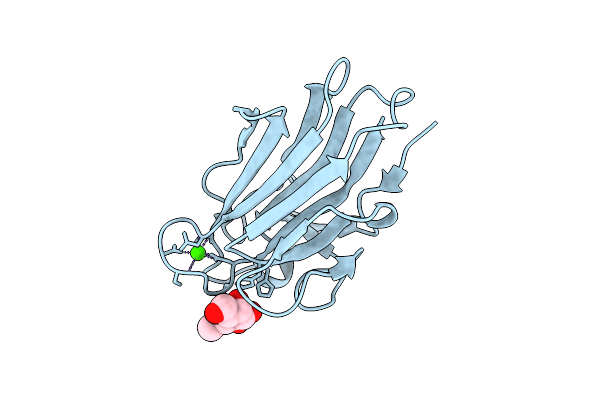
Crystal Structure Of A Duf1696 Family Protein With A Pleckstrin-Homology Domain (Shew_0819) From Shewanella Loihica Pv-4 At 2.00 A Resolution
Organism: Shewanella loihica pv-4
Method: X-RAY DIFFRACTION Resolution:2.00 Å Release Date: 2008-07-29 Classification: UNKNOWN FUNCTION Ligands: MPD, CL |
Organism: Shewanella loihica pv-4
Method: X-RAY DIFFRACTION
Release Date: 2008-07-29
Ligands: MPD, CL
 |
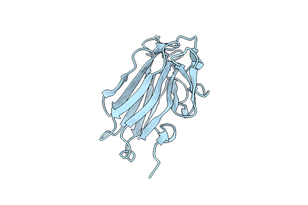
The Structure Of Cbm51 From Clostridium Perfringens Gh95 In Complex With Methyl-Galactose
Organism: Clostridium perfringens
Method: X-RAY DIFFRACTION Resolution:1.90 Å Release Date: 2008-02-19 Classification: SUGAR BINDING PROTEIN Ligands: CA, MBG |
Organism: Clostridium perfringens
Method: X-RAY DIFFRACTION
Release Date: 2008-02-19
Ligands: CA, MBG
 |
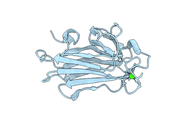
The Structure Of Cbm51 From Clostridium Perfringens Gh95
Organism: Clostridium perfringens
Method: X-RAY DIFFRACTION Resolution:1.50 Å Release Date: 2008-02-19 Classification: SUGAR BINDING PROTEIN Ligands: CA |
Organism: Clostridium perfringens
Method: X-RAY DIFFRACTION
Release Date: 2008-02-19
Ligands: CA
 |
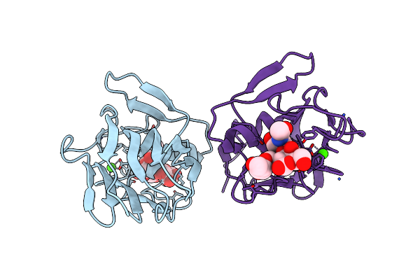
The Structure Of Seleno-Methionine Labelled Cbm51 From Clostridium Perfringens Gh95
Organism: Clostridium perfringens
Method: X-RAY DIFFRACTION Resolution:1.70 Å Release Date: 2008-02-19 Classification: SUGAR BINDING PROTEIN Ligands: CA |
Organism: Clostridium perfringens
Method: X-RAY DIFFRACTION
Release Date: 2008-02-19
Ligands: CA
 |
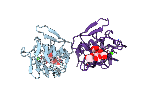
Family 51 Carbohydrate Binding Module From A Family 98 Glycoside Hydrolase Produced By Clostridium Perfringens In Complex With Blood Group A-Trisaccharide Ligand.
Organism: Clostridium perfringens
Method: X-RAY DIFFRACTION Resolution:1.40 Å Release Date: 2008-02-19 Classification: HYDROLASE Ligands: CA |
Organism: Clostridium perfringens
Method: X-RAY DIFFRACTION
Release Date: 2008-02-19
Ligands: CA
 |
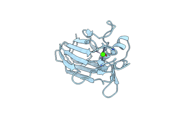
Family 51 Carbohydrate Binding Module From A Family 98 Glycoside Hydrolase Produced By Clostridium Perfringens In Complex With Blood Group B-Trisaccharide Ligand.
Organism: Clostridium perfringens
Method: X-RAY DIFFRACTION Resolution:1.45 Å Release Date: 2008-02-19 Classification: HYDROLASE Ligands: CA |
Organism: Clostridium perfringens
Method: X-RAY DIFFRACTION
Release Date: 2008-02-19
Ligands: CA
 |
Family 51 Carbohydrate Binding Module From A Family 98 Glycoside Hydrolase Produced By Clostridium Perfringens.
Organism: Clostridium perfringens
Method: X-RAY DIFFRACTION Resolution:1.55 Å Release Date: 2008-02-19 Classification: HYDROLASE Ligands: CA |
Organism: Clostridium perfringens
Method: X-RAY DIFFRACTION
Release Date: 2008-02-19
Ligands: CA
 |
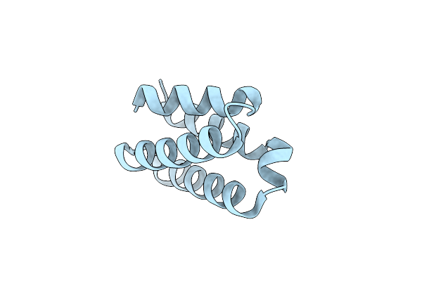
Crystal Structure Of A Ph Domain Containing Bacterial Protein (Exig_2160) From Exiguobacterium Sibiricum 255-15 At 2.42 A Resolution
Organism: Exiguobacterium sibiricum
Method: X-RAY DIFFRACTION Resolution:2.42 Å Release Date: 2007-11-13 Classification: UNKNOWN FUNCTION |
Organism: Exiguobacterium sibiricum
Method: X-RAY DIFFRACTION
Release Date: 2007-11-13
 |
Crystal Structure Of Protein Spy2152 From Streptococcus Pyogenes
Organism: Streptococcus pyogenes
Method: X-RAY DIFFRACTION Resolution:2.15 Å Release Date: 2006-03-07 Classification: STRUCTURAL GENOMICS, UNKNOWN FUNCTION |
Organism: Streptococcus pyogenes
Method: X-RAY DIFFRACTION
Release Date: 2006-03-07