

Search Count: 10
 |
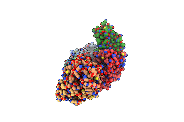
Structure Of Phosphopantetheine Adenylyltransferase (Ppat) From Enterobacter Spp. With The 17-Mer Expression Tag Bound In The Substrate Binding Site Of A Neighbouring Molecule At 2.10 A Resolution.
Organism: Enterobacter sp. 638
Method: X-RAY DIFFRACTION Resolution:2.10 Å Release Date: 2025-04-16 Classification: TRANSFERASE Ligands: CIT |
Organism: Enterobacter sp. 638
Method: X-RAY DIFFRACTION
Release Date: 2025-04-16
Ligands: CIT
 |
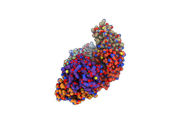
Structure Of Phosphopantetheine Adenylyltransferase (Ppat) From Enterobacter Spp. With The Expression Tag Bound In The Substrate Binding Site Of A Neighbouring Molecule At 2.20 A Resolution.
Organism: Enterobacter sp. 638
Method: X-RAY DIFFRACTION Resolution:2.20 Å Release Date: 2024-12-11 Classification: TRANSFERASE Ligands: PAE, GOL |
Organism: Enterobacter sp. 638
Method: X-RAY DIFFRACTION
Release Date: 2024-12-11
Ligands: PAE, GOL
 |
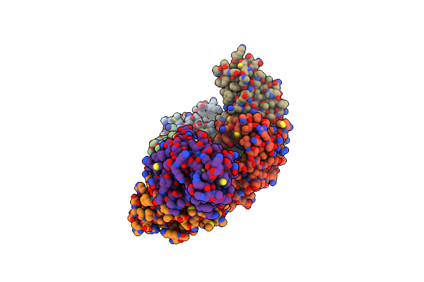
Structure Of Phosphopantetheine Adenylyltransferase (Ppat) From Enterobacter Spp. With The Expression Tag Bound In The Substrate Binding Site Of A Neighbouring Molecule At 2.39 A Resolution.
Organism: Enterobacter sp. 638
Method: X-RAY DIFFRACTION Resolution:2.39 Å Release Date: 2024-08-21 Classification: TRANSFERASE Ligands: PAE, GOL, EDO |
Organism: Enterobacter sp. 638
Method: X-RAY DIFFRACTION
Release Date: 2024-08-21
Ligands: PAE, GOL, EDO
 |
Structure Of Phosphopantetheine Adenylyltransferase (Ppat) From Enterobacter Spp. With The Expression Tag Bound In The Substrate Binding Site Of A Neighbouring Molecule At 2.37 A Resolution.
Organism: Enterobacter sp. 638
Method: X-RAY DIFFRACTION Resolution:2.37 Å Release Date: 2024-08-21 Classification: TRANSFERASE Ligands: PAE, GOL, EDO |
Organism: Enterobacter sp. 638
Method: X-RAY DIFFRACTION
Release Date: 2024-08-21
Ligands: PAE, GOL, EDO
 |
Structure Of Phosphopantetheine Adenylyltransferase (Ppat) From Enterobacter Spp. With The 17-Mer Expression Tag Bound In The Substrate Binding Site Of A Neighbouring Molecule At 2.60 A Resolution.
Organism: Enterobacter sp. 638
Method: X-RAY DIFFRACTION Resolution:2.60 Å Release Date: 2024-08-21 Classification: TRANSFERASE Ligands: CIT, GOL |
Organism: Enterobacter sp. 638
Method: X-RAY DIFFRACTION
Release Date: 2024-08-21
Ligands: CIT, GOL
 |
Structure Of Phosphopantetheine Adenylyltransferase (Ppat) From Enterobacter Spp. With The Expression Tag Bound In The Substrate Binding Site Of A Neighbouring Molecule At 2.25 A Resolution.
Organism: Enterobacter sp. 638
Method: X-RAY DIFFRACTION Resolution:2.25 Å Release Date: 2024-08-21 Classification: TRANSFERASE Ligands: PAE, GOL, EDO |
Organism: Enterobacter sp. 638
Method: X-RAY DIFFRACTION
Release Date: 2024-08-21
Ligands: PAE, GOL, EDO
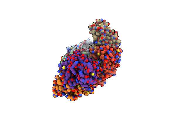 |
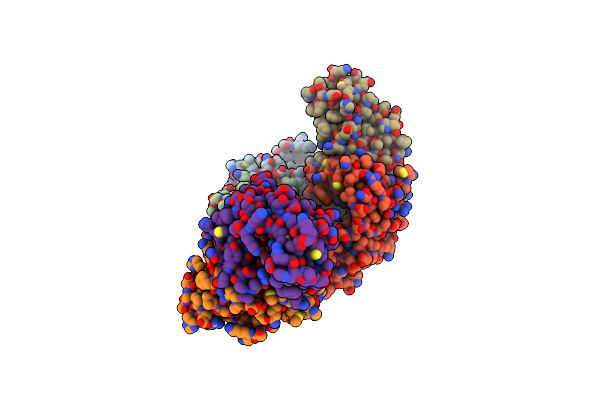
Structure Of Phosphopantetheine Adenylyltransferase (Ppat) From Enterobacter Sp. With The Expression Tag Bound In The Substrate Binding Site Of A Neighbouring Molecule At 2.65 A Resolution.
Organism: Enterobacter sp. 638
Method: X-RAY DIFFRACTION Resolution:2.65 Å Release Date: 2024-06-05 Classification: TRANSFERASE Ligands: PAE, GOL, EDO |
Organism: Enterobacter sp. 638
Method: X-RAY DIFFRACTION
Release Date: 2024-06-05
Ligands: PAE, GOL, EDO
 |
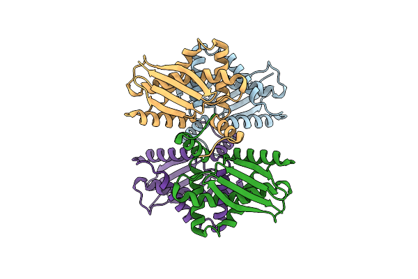
Structure Of Phosphopantetheine Adenylyltransferase (Ppat) From Enterobacter Sp. With The Expression Tag Bound In The Substrate Binding Site Of A Neighbouring Molecule At 2.41 A Resolution.
Organism: Enterobacter sp. 638
Method: X-RAY DIFFRACTION Resolution:2.41 Å Release Date: 2024-06-05 Classification: TRANSFERASE Ligands: PAE, GOL, EDO |
Organism: Enterobacter sp. 638
Method: X-RAY DIFFRACTION
Release Date: 2024-06-05
Ligands: PAE, GOL, EDO
 |
Cryo-Em Structure Of Enterobacter Sp. 638 Bcsd
Organism: Enterobacter sp. 638
Method: ELECTRON MICROSCOPY Release Date: 2023-12-20 Classification: CYTOSOLIC PROTEIN |
Organism: Enterobacter sp. 638
Method: ELECTRON MICROSCOPY
Release Date: 2023-12-20
 |
Crystal Structure Of An Enolase From Enterobacter Sp. 638 (Efi Target Efi-501662) With Bound Mg
Organism: Enterobacter sp. 638
Method: X-RAY DIFFRACTION Resolution:1.80 Å Release Date: 2011-09-14 Classification: LYASE Ligands: MG, GOL, CL |
Organism: Enterobacter sp. 638
Method: X-RAY DIFFRACTION
Release Date: 2011-09-14
Ligands: MG, GOL, CL