

Search Count: 20
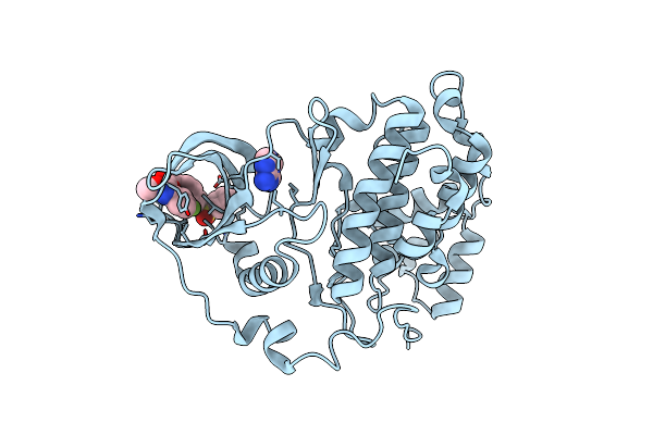 |
The Structure Of Ck2Alpha With Cch503 Bound
Organism: Homo sapiens
Method: X-RAY DIFFRACTION Resolution:1.47 Å Release Date: 2019-06-19 Classification: TRANSFERASE Ligands: 503, ATP |
Organism: Homo sapiens
Method: X-RAY DIFFRACTION
Release Date: 2019-06-19
Ligands: 503, ATP
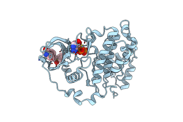 |
The Structure Of Ck2Alpha With Cch507 Bound
Organism: Homo sapiens
Method: X-RAY DIFFRACTION Resolution:1.60 Å Release Date: 2019-06-19 Classification: TRANSFERASE Ligands: E8K, ATP |
Organism: Homo sapiens
Method: X-RAY DIFFRACTION
Release Date: 2019-06-19
Ligands: E8K, ATP
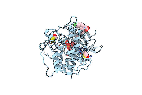 |
Human Pdk1-Pkczeta Kinase Chimera In Complex With Allosteric Compound Ps315 Bound To The Pif-Pocket
Organism: Homo sapiens
Method: X-RAY DIFFRACTION Resolution:1.85 Å Release Date: 2014-05-28 Classification: TRANSFERASE Ligands: ATP, 31S, DMS, DTD |
Organism: Homo sapiens
Method: X-RAY DIFFRACTION
Release Date: 2014-05-28
Ligands: ATP, 31S, DMS, DTD
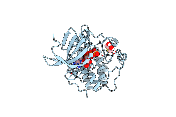 |
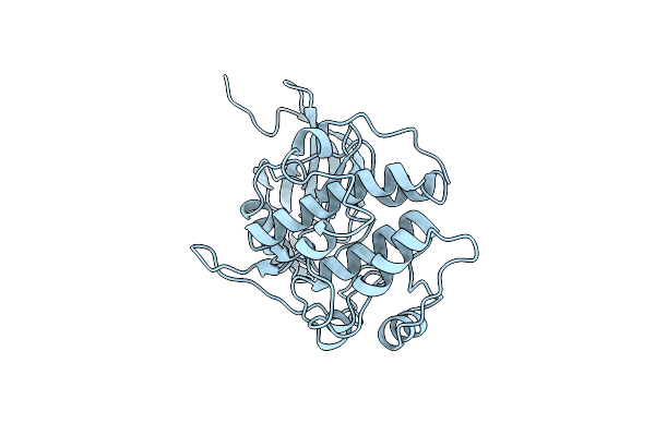
Human Pdk1-Pkczeta Kinase Chimera
Organism: Homo sapiens
Method: X-RAY DIFFRACTION Resolution:1.25 Å Release Date: 2014-05-28 Classification: TRANSFERASE Ligands: ATP, DTD, NA |
Organism: Homo sapiens
Method: X-RAY DIFFRACTION
Release Date: 2014-05-28
Ligands: ATP, DTD, NA
 |
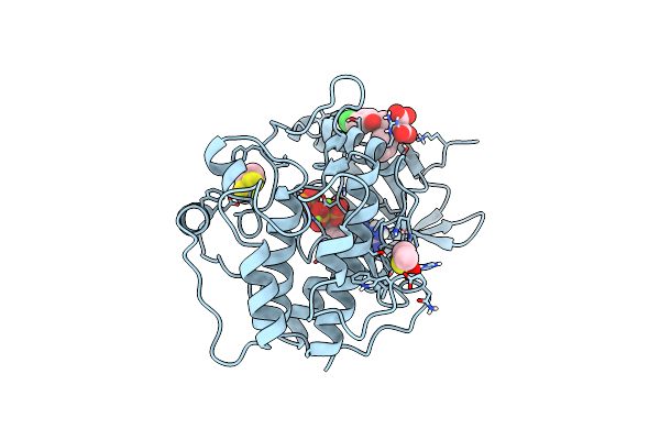
Candida Albicans Pkh Kinase Domain
Organism: Candida albicans
Method: X-RAY DIFFRACTION Resolution:3.16 Å Release Date: 2013-10-23 Classification: TRANSFERASE |
Organism: Candida albicans
Method: X-RAY DIFFRACTION
Release Date: 2013-10-23
 |
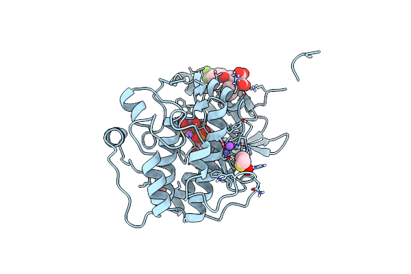
Human Pdk1 Kinase Domain In Complex With Allosteric Compound Ps182 Bound To The Pif-Pocket
Organism: Homo sapiens
Method: X-RAY DIFFRACTION Resolution:1.43 Å Release Date: 2012-10-03 Classification: TRANSFERASE Ligands: ATP, MJF, DMS, MG, DTD |
Organism: Homo sapiens
Method: X-RAY DIFFRACTION
Release Date: 2012-10-03
Ligands: ATP, MJF, DMS, MG, DTD
 |
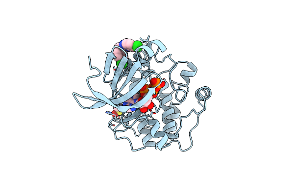
Human Pdk1 Kinase Domain In Complex With Allosteric Compound Ps210 Bound To The Pif-Pocket
Organism: Homo sapiens
Method: X-RAY DIFFRACTION Resolution:1.68 Å Release Date: 2012-10-03 Classification: TRANSFERASE Ligands: ATP, 21O, DMS, MN, NA, CL |
Organism: Homo sapiens
Method: X-RAY DIFFRACTION
Release Date: 2012-10-03
Ligands: ATP, 21O, DMS, MN, NA, CL
 |
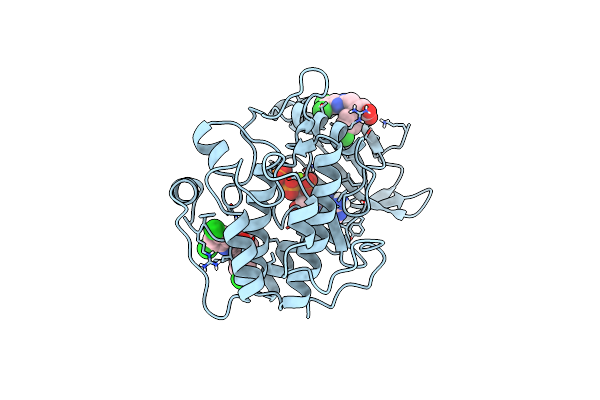
Human Pdk1 Kinase Domain In Complex With Allosteric Activator Ps114 Bound To The Pif-Pocket
Organism: Homo sapiens
Method: X-RAY DIFFRACTION Resolution:2.00 Å Release Date: 2011-12-07 Classification: TRANSFERASE Ligands: ATP, A06, DMS, CL |
Organism: Homo sapiens
Method: X-RAY DIFFRACTION
Release Date: 2011-12-07
Ligands: ATP, A06, DMS, CL
 |
Human Pdk1 Kinase Domain In Complex With Allosteric Activator Ps171 Bound To The Pif-Pocket
Organism: Homo sapiens
Method: X-RAY DIFFRACTION Resolution:1.85 Å Release Date: 2011-12-07 Classification: TRANSFERASE Ligands: ATP, AZ7, MG, CL |
Organism: Homo sapiens
Method: X-RAY DIFFRACTION
Release Date: 2011-12-07
Ligands: ATP, AZ7, MG, CL
 |
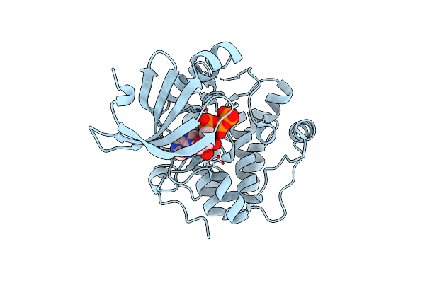
Acoustically Mounted Porcine Insulin Microcrystals Yield An X-Ray Sad Structure
Organism: Sus scrofa
Method: X-RAY DIFFRACTION Resolution:1.80 Å Release Date: 2011-05-18 Classification: HORMONE Ligands: ZN |
Organism: Sus scrofa
Method: X-RAY DIFFRACTION
Release Date: 2011-05-18
Ligands: ZN
 |
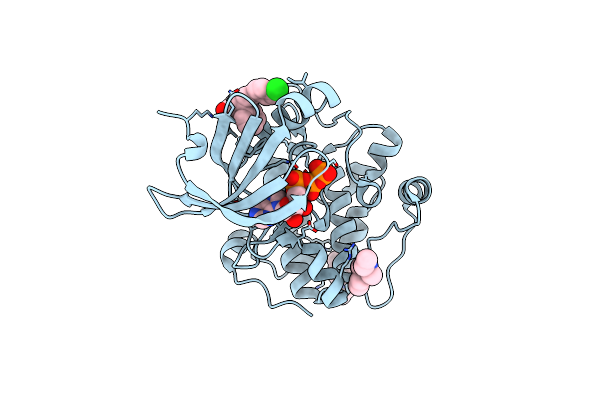
Crystal Structure Of A Mutant Of Human Pdk1 Kinase Domain In Complex With Atp
Organism: Homo sapiens
Method: X-RAY DIFFRACTION Resolution:1.91 Å Release Date: 2009-09-15 Classification: TRANSFERASE Ligands: ATP |
Organism: Homo sapiens
Method: X-RAY DIFFRACTION
Release Date: 2009-09-15
Ligands: ATP
 |
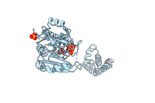
Crystal Structure Of Human Pdk1 Kinase Domain In Complex With An Allosteric Activator Bound To The Pif-Pocket
Organism: Homo sapiens
Method: X-RAY DIFFRACTION Resolution:1.90 Å Release Date: 2009-09-15 Classification: TRANSFERASE Ligands: ATP, P47, EPE |
Organism: Homo sapiens
Method: X-RAY DIFFRACTION
Release Date: 2009-09-15
Ligands: ATP, P47, EPE
 |
Crystal Structure Of Primase Repb'
Organism: Plasmid rsf1010
Method: X-RAY DIFFRACTION Resolution:1.99 Å Release Date: 2009-04-28 Classification: REPLICATION Ligands: SO4, DPO |
Organism: Plasmid rsf1010
Method: X-RAY DIFFRACTION
Release Date: 2009-04-28
Ligands: SO4, DPO
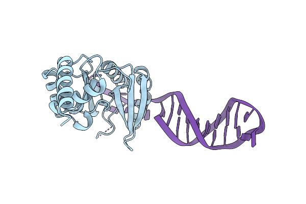 |
Crystal Structure Of The Catalytic Domain Of Primase Repb' In Complex With Initiator Dna
Organism: Plasmid rsf1010
Method: X-RAY DIFFRACTION Resolution:2.70 Å Release Date: 2009-04-28 Classification: REPLICATION/DNA |
Organism: Plasmid rsf1010
Method: X-RAY DIFFRACTION
Release Date: 2009-04-28
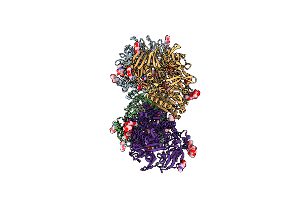 |
Porcine Dipeptidyl Peptidase Iv (Cd26) In Complex With 7-Benzyl-1,3-Dimethyl-8-Piperazin-1-Yl-3,7-Dihydro-Purine-2,6-Dione (Bdpx)
Organism: Sus scrofa
Method: X-RAY DIFFRACTION Resolution:2.11 Å Release Date: 2006-02-28 Classification: HYDROLASE Ligands: NAG, SO4, SC3 |
Organism: Sus scrofa
Method: X-RAY DIFFRACTION
Release Date: 2006-02-28
Ligands: NAG, SO4, SC3
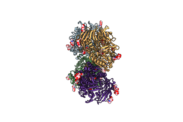 |
Porcine Dipeptidyl Peptidase Iv (Cd26) In Complex With The Tripeptide Tert-Butyl-Gly-L-Pro-L-Ile (Tbu-Gpi)
Organism: Sus scrofa
Method: X-RAY DIFFRACTION Resolution:2.75 Å Release Date: 2006-02-28 Classification: HYDROLASE/HYDROLASE INHIBITOR Ligands: 0QG, NAG, SO4 |
Organism: Sus scrofa
Method: X-RAY DIFFRACTION
Release Date: 2006-02-28
Ligands: 0QG, NAG, SO4
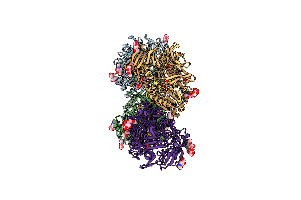 |
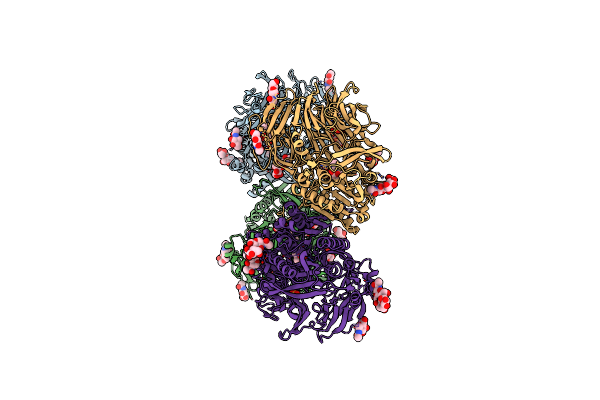
Porcine Dipeptidyl Peptidase Iv (Cd26) In Complex With 4-(2-Aminoethyl)-Benzene Sulphonyl Fluoride (Aebsf)
Organism: Sus scrofa
Method: X-RAY DIFFRACTION Resolution:1.95 Å Release Date: 2006-02-28 Classification: hydrolase/hydrolase inhibitor Ligands: NAG, SO4, AES |
Organism: Sus scrofa
Method: X-RAY DIFFRACTION
Release Date: 2006-02-28
Ligands: NAG, SO4, AES
 |
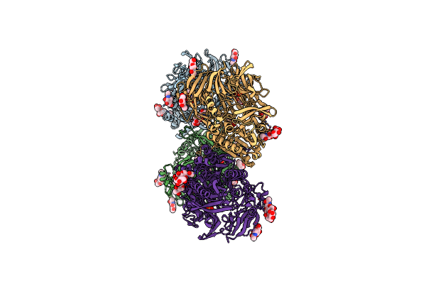
Porcine Dipeptidyl Peptidase Iv (Cd26) In Complex With L-Pro-Boro-L-Pro (Boropro)
Organism: Sus scrofa
Method: X-RAY DIFFRACTION Resolution:2.56 Å Release Date: 2006-02-28 Classification: HYDROLASE Ligands: NAG, SO4, BPR |
Organism: Sus scrofa
Method: X-RAY DIFFRACTION
Release Date: 2006-02-28
Ligands: NAG, SO4, BPR
 |
Crystal Structure Of Porcine Dipeptidyl Peptidase Iv (Cd26)
Organism: Sus scrofa
Method: X-RAY DIFFRACTION Resolution:1.80 Å Release Date: 2003-05-06 Classification: HYDROLASE Ligands: NAG, SO4 |
Organism: Sus scrofa
Method: X-RAY DIFFRACTION
Release Date: 2003-05-06
Ligands: NAG, SO4
 |
Crystal Structure Of Porcine Dipeptidyl Peptidase Iv (Cd26) In Complex With A Peptidomimetic Inhibitor
Organism: Sus scrofa
Method: X-RAY DIFFRACTION Resolution:2.84 Å Release Date: 2003-05-06 Classification: HYDROLASE Ligands: NAG, SO4, PHI, P2Y |
Organism: Sus scrofa
Method: X-RAY DIFFRACTION
Release Date: 2003-05-06
Ligands: NAG, SO4, PHI, P2Y