

Search Count: 20
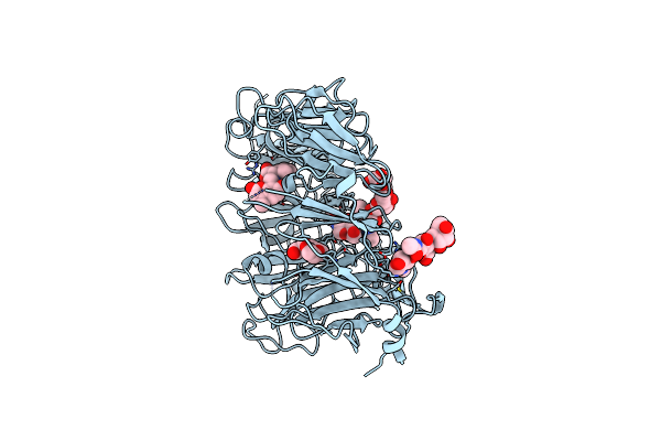 |
Crystal Structure Of The Vps10P Domain Of Human Sortilin/Nts3 In Complex With Af40431
Organism: Homo sapiens
Method: X-RAY DIFFRACTION Resolution:2.70 Å Release Date: 2014-02-12 Classification: SIGNALING PROTEIN Ligands: PG4, 2ET |
Organism: Homo sapiens
Method: X-RAY DIFFRACTION
Release Date: 2014-02-12
Ligands: PG4, 2ET
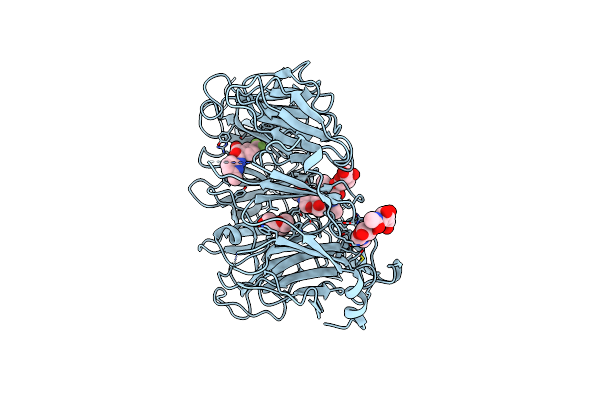 |
Crystal Structure Of The Vps10P Domain Of Human Sortilin/Nts3 In Complex With Af38469
Organism: Homo sapiens
Method: X-RAY DIFFRACTION Resolution:2.70 Å Release Date: 2014-01-22 Classification: SIGNALING PROTEIN Ligands: PGE, 2JQ |
Organism: Homo sapiens
Method: X-RAY DIFFRACTION
Release Date: 2014-01-22
Ligands: PGE, 2JQ
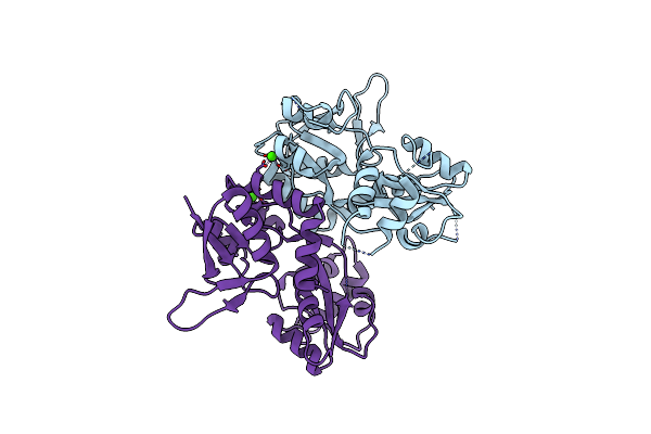 |
Structure Of The Ligand-Binding Core Of The Ionotropic Glutamate Receptor-Like Glurdelta2 In The Apo Form
Organism: Rattus norvegicus
Method: X-RAY DIFFRACTION Resolution:2.75 Å Release Date: 2007-08-07 Classification: RECEPTOR Ligands: CA |
Organism: Rattus norvegicus
Method: X-RAY DIFFRACTION
Release Date: 2007-08-07
Ligands: CA
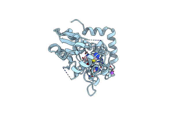 |
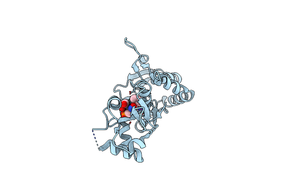
Structure Of The Ligand-Binding Core Of The Ionotropic Glutamate Receptor-Like Glurdelta2 In Complex With D-Serine
Organism: Rattus norvegicus
Method: X-RAY DIFFRACTION Resolution:1.74 Å Release Date: 2007-08-07 Classification: RECEPTOR Ligands: DSN, NA, CL, SCN |
Organism: Rattus norvegicus
Method: X-RAY DIFFRACTION
Release Date: 2007-08-07
Ligands: DSN, NA, CL, SCN
 |
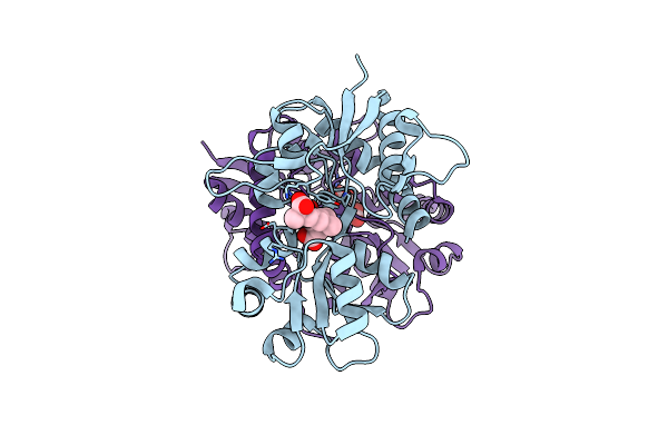
Crystal Structure Of The Ligand-Binding Core Of Iglur5 In Complex With The Antagonist (S)-Atpo At 1.85 A Resolution
Organism: Rattus norvegicus
Method: X-RAY DIFFRACTION Resolution:1.85 Å Release Date: 2007-07-03 Classification: MEMBRANE PROTEIN Ligands: AT1, GOL |
Organism: Rattus norvegicus
Method: X-RAY DIFFRACTION
Release Date: 2007-07-03
Ligands: AT1, GOL
 |
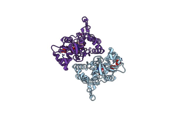
Crystal Structure Of The Ligand-Binding Core Of Iglur5 In Complex With The Partial Agonist Domoic Acid At 2.5 A Resolution
Organism: Rattus norvegicus
Method: X-RAY DIFFRACTION Resolution:2.50 Å Release Date: 2007-07-03 Classification: MEMBRANE PROTEIN Ligands: DOQ |
Organism: Rattus norvegicus
Method: X-RAY DIFFRACTION
Release Date: 2007-07-03
Ligands: DOQ
 |
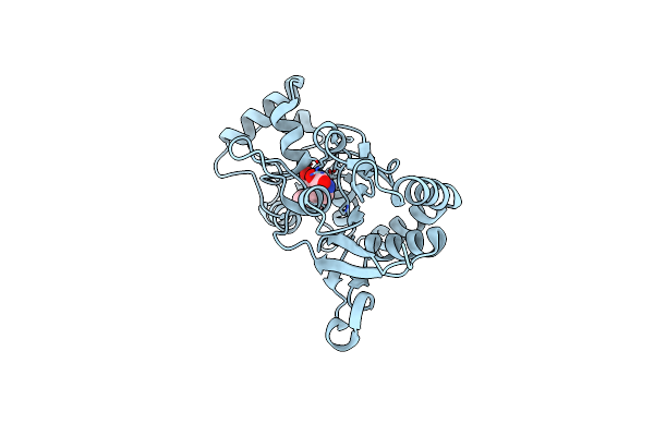
X-Ray Structure Of The Glur2 Ligand Binding Core (S1S2J) In Complex With 2-Bn-Tet-Ampa At 2.26A Resolution
Organism: Rattus norvegicus
Method: X-RAY DIFFRACTION Resolution:2.26 Å Release Date: 2007-06-19 Classification: MEMBRANE PROTEIN Ligands: SO4, GLU, MP9 |
Organism: Rattus norvegicus
Method: X-RAY DIFFRACTION
Release Date: 2007-06-19
Ligands: SO4, GLU, MP9
 |
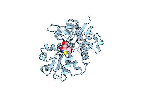
Crystal Structure Of The Glur2 Ligand Binding Core (S1S2J-Y450W) Mutant In Complex With The Partial Agonist Kainic Acid At 2.1 A Resolution
Organism: Rattus norvegicus
Method: X-RAY DIFFRACTION Resolution:2.10 Å Release Date: 2005-08-30 Classification: MEMBRANE PROTEIN Ligands: KAI |
Organism: Rattus norvegicus
Method: X-RAY DIFFRACTION
Release Date: 2005-08-30
Ligands: KAI
 |
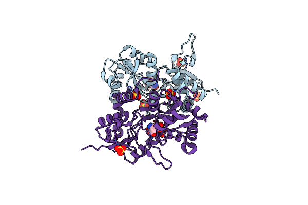
X-Ray Structure Of The Glur2 Ligand-Binding Core (S1S2J) In Complex With (S)-Thio-Atpa At 2.2 A Resolution.
Organism: Rattus norvegicus
Method: X-RAY DIFFRACTION Resolution:2.17 Å Release Date: 2005-08-09 Classification: MEMBRANE PROTEIN Ligands: U1K |
Organism: Rattus norvegicus
Method: X-RAY DIFFRACTION
Release Date: 2005-08-09
Ligands: U1K
 |
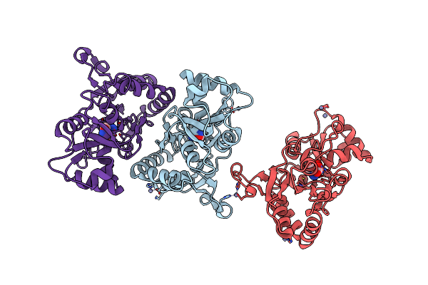
Crystal Structure Of The Kainate Receptor Glur5 Ligand-Binding Core In Complex With (S)-Glutamate
Organism: Rattus norvegicus
Method: X-RAY DIFFRACTION Resolution:1.95 Å Release Date: 2005-02-01 Classification: MEMBRANE PROTEIN Ligands: SO4, GLU |
Organism: Rattus norvegicus
Method: X-RAY DIFFRACTION
Release Date: 2005-02-01
Ligands: SO4, GLU
 |
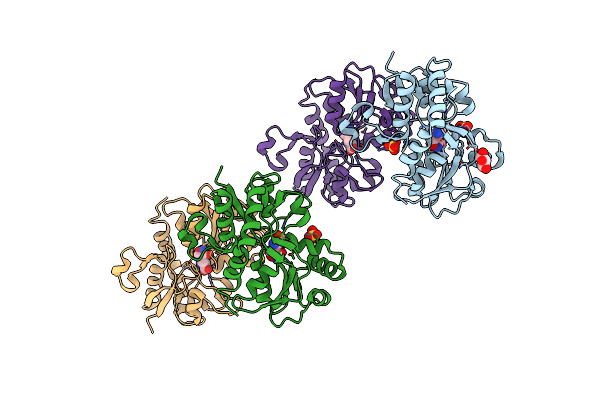
X-Ray Structure Of The Glur2 Ligand-Binding Core (S1S2J) In Complex With (S)-Des-Me-Ampa At 1.97 A Resolution, Crystallization In The Presence Of Zinc Acetate
Organism: Rattus norvegicus
Method: X-RAY DIFFRACTION Resolution:1.97 Å Release Date: 2003-07-08 Classification: MEMBRANE PROTEIN Ligands: ZN, SHI |
Organism: Rattus norvegicus
Method: X-RAY DIFFRACTION
Release Date: 2003-07-08
Ligands: ZN, SHI
 |
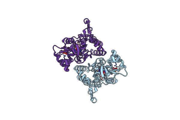
X-Ray Structure Of The Glur2 Ligand-Binding Core (S1S2J) In Complex With (S)-Des-Me-Ampa At 1.46 A Resolution. Crystallization In The Presence Of Lithium Sulfate.
Organism: Rattus norvegicus
Method: X-RAY DIFFRACTION Resolution:1.46 Å Release Date: 2003-07-01 Classification: MEMBRANE PROTEIN Ligands: SO4, SHI, GOL |
Organism: Rattus norvegicus
Method: X-RAY DIFFRACTION
Release Date: 2003-07-01
Ligands: SO4, SHI, GOL
 |
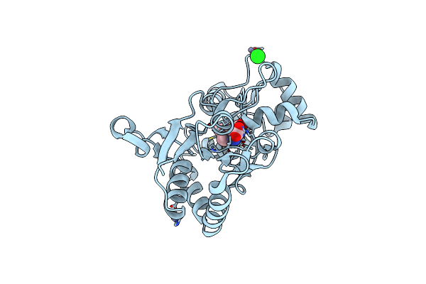
X-Ray Structure Of The Glur2 Ligand-Binding Core (S1S2J) In Complex With (S)-Atpa At 1.9 A Resolution. Crystallization Without Zinc Ions.
Organism: Rattus norvegicus
Method: X-RAY DIFFRACTION Resolution:1.90 Å Release Date: 2003-03-11 Classification: MEMBRANE PROTEIN Ligands: SO4, CE2 |
Organism: Rattus norvegicus
Method: X-RAY DIFFRACTION
Release Date: 2003-03-11
Ligands: SO4, CE2
 |
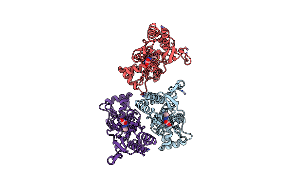
X-Ray Structure Of The Glur2 Ligand-Binding Core (S1S2J) In Complex With The Antagonist (S)-Atpo At 2.1 A Resolution.
Organism: Rattus norvegicus
Method: X-RAY DIFFRACTION Resolution:2.10 Å Release Date: 2003-03-04 Classification: MEMBRANE PROTEIN Ligands: AT1, SO4, ACT |
Organism: Rattus norvegicus
Method: X-RAY DIFFRACTION
Release Date: 2003-03-04
Ligands: AT1, SO4, ACT
 |
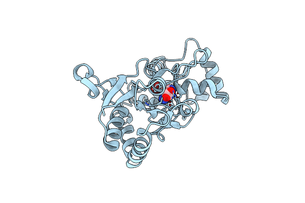
X-Ray Structure Of The Glur2 Ligand-Binding Core (S1S2J) In Complex With (S)-Atpa At 1.85 A Resolution. Crystallization With Zinc Ions.
Organism: Rattus norvegicus
Method: X-RAY DIFFRACTION Resolution:1.85 Å Release Date: 2003-03-04 Classification: MEMBRANE PROTEIN Ligands: ZN, CL, CE2 |
Organism: Rattus norvegicus
Method: X-RAY DIFFRACTION
Release Date: 2003-03-04
Ligands: ZN, CL, CE2
 |
X-Ray Structure Of The Glur2 Ligand Binding Core (S1S2J) In Complex With 2-Me-Tet-Ampa At 1.85 A Resolution.
Organism: Rattus norvegicus
Method: X-RAY DIFFRACTION Resolution:1.85 Å Release Date: 2002-09-18 Classification: MEMBRANE PROTEIN Ligands: ZN, BN1 |
Organism: Rattus norvegicus
Method: X-RAY DIFFRACTION
Release Date: 2002-09-18
Ligands: ZN, BN1
 |
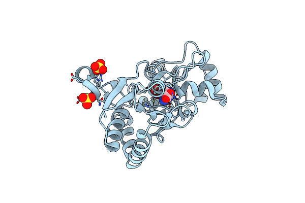
X-Ray Structure Of The Glur2 Ligand Binding Core (S1S2J) In Complex With Br-Hibo At 1.65 A Resolution
Organism: Rattus norvegicus
Method: X-RAY DIFFRACTION Resolution:1.65 Å Release Date: 2002-09-18 Classification: MEMBRANE PROTEIN Ligands: BRH |
Organism: Rattus norvegicus
Method: X-RAY DIFFRACTION
Release Date: 2002-09-18
Ligands: BRH
 |
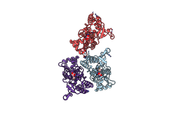
X-Ray Structure Of The Glur2 Ligand Binding Core (S1S2J-Y702F) In Complex With Br-Hibo At 1.73 A Resolution
Organism: Rattus norvegicus
Method: X-RAY DIFFRACTION Resolution:1.73 Å Release Date: 2002-09-18 Classification: MEMBRANE PROTEIN Ligands: SO4, BRH |
Organism: Rattus norvegicus
Method: X-RAY DIFFRACTION
Release Date: 2002-09-18
Ligands: SO4, BRH
 |
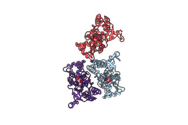
X-Ray Structure Of The Glur2 Ligand Binding Core (S1S2J) In Complex With Acpa At 1.46 A Resolution
Organism: Rattus norvegicus
Method: X-RAY DIFFRACTION Resolution:1.46 Å Release Date: 2002-09-18 Classification: MEMBRANE PROTEIN Ligands: ZN, AM1, ACT |
Organism: Rattus norvegicus
Method: X-RAY DIFFRACTION
Release Date: 2002-09-18
Ligands: ZN, AM1, ACT
 |
X-Ray Structure Of The Glur2 Ligand Binding Core (S1S2J-Y702F) In Complex With Acpa At 1.95 A Resolution
Organism: Rattus norvegicus
Method: X-RAY DIFFRACTION Resolution:1.95 Å Release Date: 2002-09-18 Classification: MEMBRANE PROTEIN Ligands: ZN, AM1, ACT |
Organism: Rattus norvegicus
Method: X-RAY DIFFRACTION
Release Date: 2002-09-18
Ligands: ZN, AM1, ACT