
Search Count: 290
 |
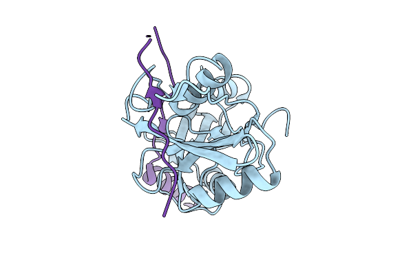
Microed Structure Of Proteinase K From Microcrystals Frozen By Traditional Cryoem Methods
Organism: Parengyodontium album
Method: ELECTRON CRYSTALLOGRAPHY Resolution:2.20 Å Release Date: 2026-07-01 Classification: HYDROLASE Ligands: CA |
 |
Microed Structure Of Proteinase K From Spray-Frozen Microcrystals
Organism: Parengyodontium album
Method: ELECTRON CRYSTALLOGRAPHY Resolution:2.50 Å Release Date: 2026-07-01 Classification: HYDROLASE |
 |
Urate Oxidase From Aspergillus Flavus With Its Inhibitor 9-Methyl Uric Acid By Continuous Serial Electron Diffraction (Serialed)
Organism: Aspergillus flavus
Method: ELECTRON CRYSTALLOGRAPHY Resolution:1.25 Å Release Date: 2026-04-29 Classification: OXIDOREDUCTASE Ligands: MUA |
 |
Urate Oxidase From Aspergillus Flavus With Its Substrate Uric Acid By Continuous Serial Electron Diffraction (Serialed)
Organism: Aspergillus flavus
Method: ELECTRON CRYSTALLOGRAPHY Resolution:1.75 Å Release Date: 2026-04-29 Classification: OXIDOREDUCTASE Ligands: OXY, URC |
 |
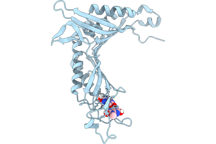
Structure Of Human Mth1 In Complex With 8Dg By Continuous Serial Electron Diffraction (Serialed)
Organism: Homo sapiens
Method: ELECTRON CRYSTALLOGRAPHY Resolution:1.66 Å Release Date: 2026-04-22 Classification: HYDROLASE Ligands: SO4, 8DG |
 |
Structure Of Human Mth1 In Complex With 8Dg By Microed Using High Electron Fluence
Organism: Homo sapiens
Method: ELECTRON CRYSTALLOGRAPHY Resolution:2.32 Å Release Date: 2026-04-22 Classification: HYDROLASE Ligands: 8DG, SO4 |
 |
Structure Of Human Mth1 In Complex With 8Dg By Microed Using Low Electron Fluence
Organism: Homo sapiens
Method: ELECTRON CRYSTALLOGRAPHY Resolution:2.86 Å Release Date: 2026-04-22 Classification: HYDROLASE Ligands: 8DG, SO4 |
 |
Structure Of Lysozyme By Continuous Serial Electron Diffraction (Serialed)
Organism: Gallus gallus
Method: ELECTRON CRYSTALLOGRAPHY Resolution:0.83 Å Release Date: 2026-04-22 Classification: HYDROLASE Ligands: ACT, CL |
 |
Serial Electron Diffraction (Serialed) Structure Of Y122F Mutant Ribonucleotide Reductase R2 From E. Coli In Its Oxidised (Met) Form
Organism: Escherichia coli
Method: ELECTRON CRYSTALLOGRAPHY Resolution:1.80 Å Release Date: 2026-04-22 Classification: OXIDOREDUCTASE Ligands: FE |
 |
Serial Electron Diffraction (Serialed) Structure Of Y122F Mutant Ribonucleotide Reductase R2 From E. Coli In Its Oxidised (Met) Form (Re-Oxidised)
Organism: Escherichia coli
Method: ELECTRON CRYSTALLOGRAPHY Resolution:2.00 Å Release Date: 2026-04-22 Classification: OXIDOREDUCTASE Ligands: FE |
 |
Serial Electron Diffraction (Serialed) Structure Of Ribonucleotide Reductase R2 From E. Coli In Its Oxidised (Met) Form
Organism: Escherichia coli
Method: ELECTRON CRYSTALLOGRAPHY Resolution:1.80 Å Release Date: 2026-04-22 Classification: OXIDOREDUCTASE Ligands: FE |
 |
Serial Electron Diffraction (Serialed) Structure Of Ribonucleotide Reductase R2 From E. Coli In Its Oxidised (Met) Form (Re-Oxidised)
Organism: Escherichia coli
Method: ELECTRON CRYSTALLOGRAPHY Resolution:1.70 Å Release Date: 2026-04-22 Classification: OXIDOREDUCTASE Ligands: FE |
 |
Serial Electron Diffraction (Serialed) Structure Of Y122F Mutant Ribonucleotide Reductase R2 From E. Coli In Its Reduced (Red) Form
Organism: Escherichia coli
Method: ELECTRON CRYSTALLOGRAPHY Resolution:1.80 Å Release Date: 2026-04-22 Classification: OXIDOREDUCTASE Ligands: FE2 |
 |
Serial Electron Diffraction (Serialed) Structure Of Ribonucleotide Reductase R2 From E. Coli In Its Reduced (Red) Form
Organism: Escherichia coli
Method: ELECTRON CRYSTALLOGRAPHY Resolution:1.80 Å Release Date: 2026-04-22 Classification: OXIDOREDUCTASE Ligands: FE2 |
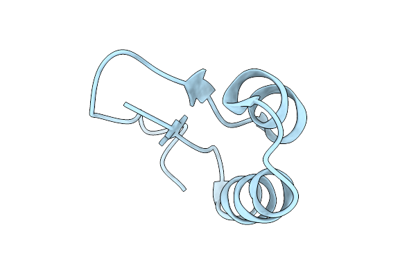 |
Ab Initio Structure Of Crambin By Microed At 0.85A
Organism: Crambe hispanica subsp. abyssinica
Method: ELECTRON CRYSTALLOGRAPHY Resolution:0.85 Å Release Date: 2026-02-25 Classification: PLANT PROTEIN |
 |
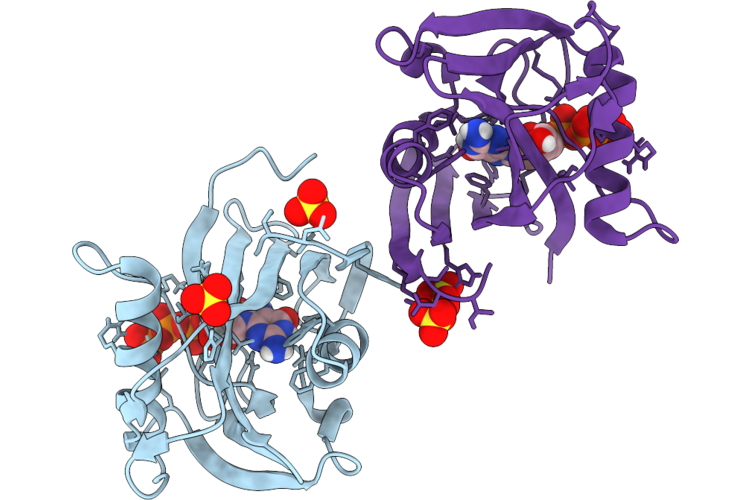
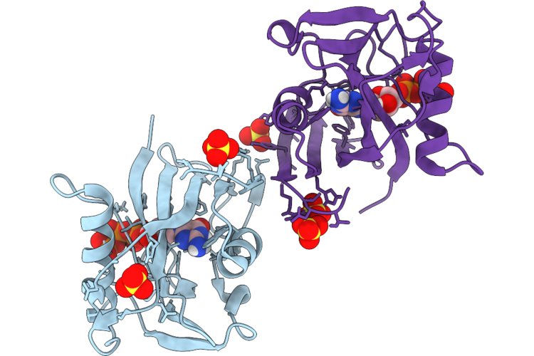
Micro-Ed Structure Of The Nsh2-Csh2 Tandem Domain Of Shp2 In Complex With The Bis-Phosphorylated Py627-Py659-Gab1 (613-694) Peptide
Organism: Homo sapiens
Method: ELECTRON CRYSTALLOGRAPHY Resolution:3.20 Å Release Date: 2025-12-31 Classification: SIGNALING PROTEIN |
 |
Structure Of Lysozyme-N,N',N"-Triacetylchitotriose Complex Determined Using Reyes For Automated Microed
Organism: Gallus gallus
Method: ELECTRON CRYSTALLOGRAPHY Resolution:2.20 Å Release Date: 2025-12-10 Classification: HYDROLASE Ligands: CL, NA |
 |
In Situ Microed Structure Of Human Eosinophil Major Basic Protein-1
Organism: Homo sapiens
Method: ELECTRON CRYSTALLOGRAPHY Resolution:3.00 Å Release Date: 2025-09-03 Classification: IMMUNE SYSTEM Ligands: CL |
 |
In Situ Microed Structure Of Il-5 Activated Human Eosinophil Major Basic Protein-1
Organism: Homo sapiens
Method: ELECTRON CRYSTALLOGRAPHY Resolution:3.18 Å Release Date: 2025-09-03 Classification: IMMUNE SYSTEM Ligands: CL |
 |
In Situ Microed Structure Of Il-33 Activated Human Eosinophil Major Basic Protein-1
Organism: Homo sapiens
Method: ELECTRON CRYSTALLOGRAPHY Resolution:3.18 Å Release Date: 2025-09-03 Classification: IMMUNE SYSTEM Ligands: CL |