

Search Count: 14
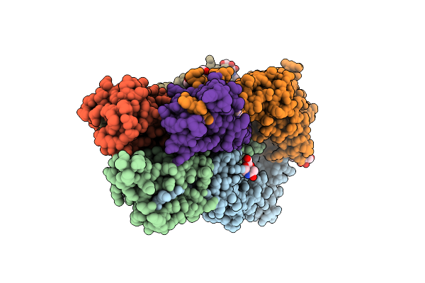 |
Crystal Structure Of A H5N1 Influenza Virus Hemagglutinin With Cbs1117
Organism: Influenza a virus (a/chicken/vietnam/4/2003(h5n1)), Influenza a virus (a/chicken/vietnam/30/2003(h5n1))
Method: X-RAY DIFFRACTION Resolution:2.20 Å Release Date: 2020-07-01 Classification: VIRAL PROTEIN Ligands: NAG, R3P |
Organism: Influenza a virus (a/chicken/vietnam/4/2003(h5n1)), Influenza a virus (a/chicken/vietnam/30/2003(h5n1))
Method: X-RAY DIFFRACTION
Release Date: 2020-07-01
Ligands: NAG, R3P
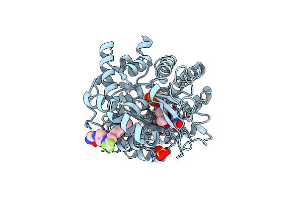 |
Crystal Structure Of Human Dhodh With Osu-03012
Organism: Homo sapiens
Method: X-RAY DIFFRACTION Resolution:1.40 Å Release Date: 2019-11-13 Classification: OXIDOREDUCTASE/OXIDOREDUCTASE INHIBITOR Ligands: ORO, FMN, SO4, M4J |
Organism: Homo sapiens
Method: X-RAY DIFFRACTION
Release Date: 2019-11-13
Ligands: ORO, FMN, SO4, M4J
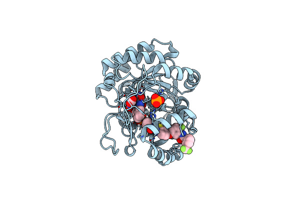 |
Crystal Structure Of Human Dhodh With Tak-632
Organism: Homo sapiens
Method: X-RAY DIFFRACTION Resolution:2.70 Å Release Date: 2019-11-13 Classification: OXIDOREDUCTASE/OXIDOREDUCTASE INHIBITOR Ligands: ORO, FMN, PO4, 1SU |
Organism: Homo sapiens
Method: X-RAY DIFFRACTION
Release Date: 2019-11-13
Ligands: ORO, FMN, PO4, 1SU
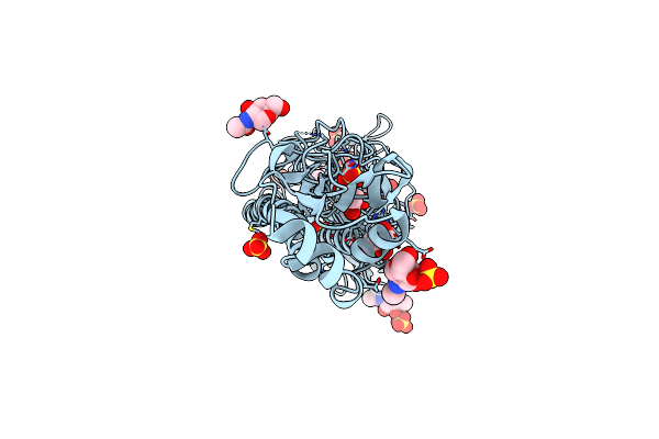 |
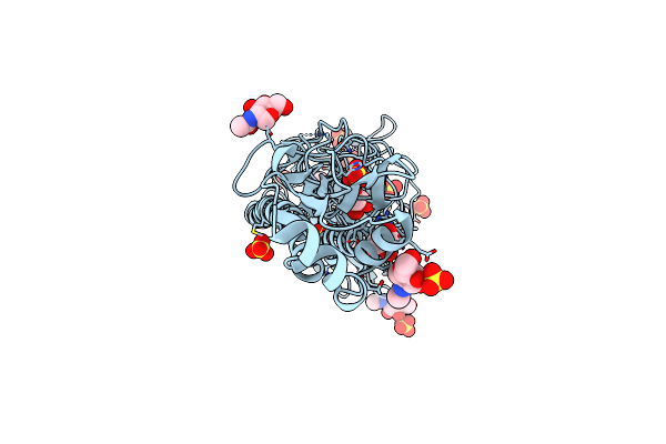
Crystal Structure Of A H5N1 Influenza Virus Hemagglutinin At Ph 6.0
Organism: Influenza a virus (a/mallard/vietnam/3/2003(h5n1))
Method: X-RAY DIFFRACTION Resolution:2.11 Å Release Date: 2019-11-13 Classification: VIRAL PROTEIN Ligands: SO4, GOL, NAG |
Organism: Influenza a virus (a/mallard/vietnam/3/2003(h5n1))
Method: X-RAY DIFFRACTION
Release Date: 2019-11-13
Ligands: SO4, GOL, NAG
 |
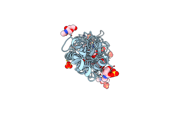
Crystal Structure Of A H5N1 Influenza Virus Hemagglutinin At Ph 5.5
Organism: Influenza a virus (a/chicken/vietnam/4/2003(h5n1))
Method: X-RAY DIFFRACTION Resolution:2.30 Å Release Date: 2019-11-13 Classification: VIRAL PROTEIN Ligands: SO4, GOL, NAG |
Organism: Influenza a virus (a/chicken/vietnam/4/2003(h5n1))
Method: X-RAY DIFFRACTION
Release Date: 2019-11-13
Ligands: SO4, GOL, NAG
 |
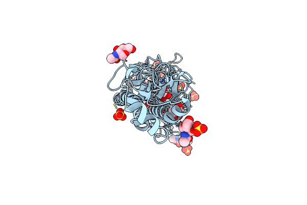
Crystal Structure Of A H5N1 Influenza Virus Hemagglutinin At Ph 6.5
Organism: Influenza a virus (a/chicken/vietnam/4/2003(h5n1))
Method: X-RAY DIFFRACTION Resolution:2.39 Å Release Date: 2019-11-13 Classification: VIRAL PROTEIN Ligands: SO4, GOL, NAG |
Organism: Influenza a virus (a/chicken/vietnam/4/2003(h5n1))
Method: X-RAY DIFFRACTION
Release Date: 2019-11-13
Ligands: SO4, GOL, NAG
 |
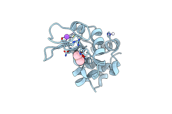
Crystal Structure Of A H5N1 Influenza Virus Hemagglutinin At Ph 7.0
Organism: Influenza a virus (a/chicken/vietnam/4/2003(h5n1))
Method: X-RAY DIFFRACTION Resolution:2.87 Å Release Date: 2019-11-13 Classification: VIRAL PROTEIN Ligands: SO4, GOL, NAG |
Organism: Influenza a virus (a/chicken/vietnam/4/2003(h5n1))
Method: X-RAY DIFFRACTION
Release Date: 2019-11-13
Ligands: SO4, GOL, NAG
 |
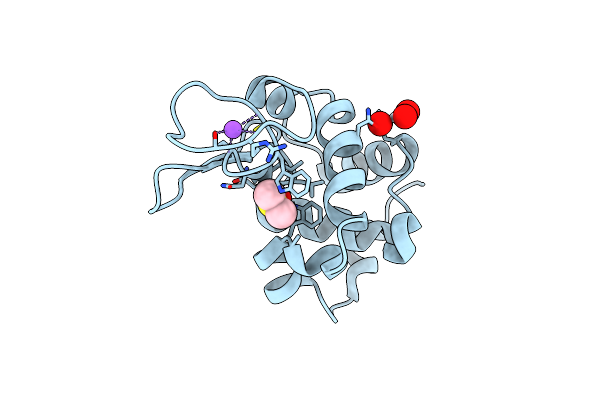
The X-Ray Structure Of The Adduct Formed In The Reaction Between Hen Egg White Lysozyme A Compound I, A Platin(Ii) Compound Containing A O, S Bidentate Ligand
Organism: Gallus gallus
Method: X-RAY DIFFRACTION Resolution:1.75 Å Release Date: 2016-12-07 Classification: HYDROLASE Ligands: DMS, PT, CL, NA |
Organism: Gallus gallus
Method: X-RAY DIFFRACTION
Release Date: 2016-12-07
Ligands: DMS, PT, CL, NA
 |
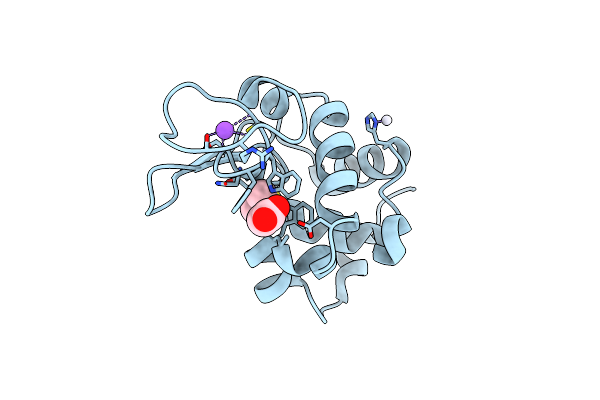
The X-Ray Structure Of The Adduct Formed In The Reaction Between Hen Egg White Lysozyme And Compound 3, A Platin(Ii) Compound Containing A O, S Bidentate Ligand
Organism: Gallus gallus
Method: X-RAY DIFFRACTION Resolution:1.78 Å Release Date: 2016-12-07 Classification: HYDROLASE Ligands: DMS, 6B6, NA, CL |
Organism: Gallus gallus
Method: X-RAY DIFFRACTION
Release Date: 2016-12-07
Ligands: DMS, 6B6, NA, CL
 |
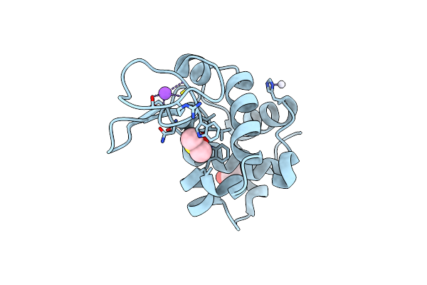
The X-Ray Structure Of The Adduct Formed In The Reaction Between Hen Egg White Lysozyme A Compound 2, A Platin(Ii) Compound Containing A O, S Bidentate Ligand
Organism: Gallus gallus
Method: X-RAY DIFFRACTION Resolution:1.75 Å Release Date: 2016-12-07 Classification: HYDROLASE Ligands: PT, DMS, CL, NA, ACT |
Organism: Gallus gallus
Method: X-RAY DIFFRACTION
Release Date: 2016-12-07
Ligands: PT, DMS, CL, NA, ACT
 |
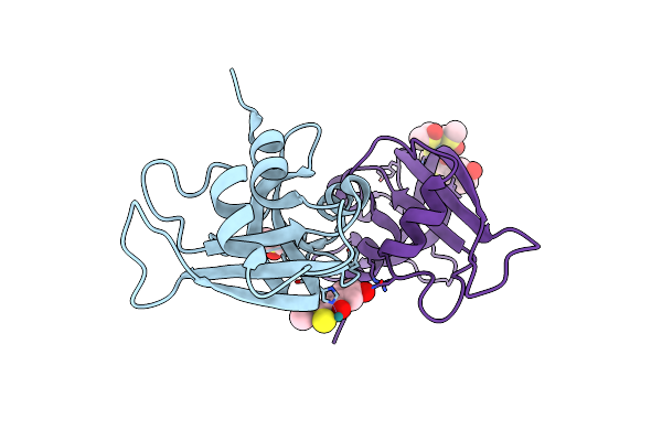
The X-Ray Structure Of The Adduct Formed In The Reaction Between Hen Egg White Lysozyme And Compound 4, A Platin(Ii) Compound Containing A O, S Bidentate Ligand
Organism: Gallus gallus
Method: X-RAY DIFFRACTION Resolution:1.85 Å Release Date: 2016-12-07 Classification: HYDROLASE Ligands: DMS, PT, CL, ACT, NA |
Organism: Gallus gallus
Method: X-RAY DIFFRACTION
Release Date: 2016-12-07
Ligands: DMS, PT, CL, ACT, NA
 |
The X-Ray Structure Of The Adduct Formed In The Reaction Between Bovine Pancreatic Ribonuclease And Compound I, A Piano-Stool Organometallic Ru(Ii) Arene Compound Containing An O,S-Chelating Ligand
Organism: Bos taurus
Method: X-RAY DIFFRACTION Resolution:1.79 Å Release Date: 2016-08-03 Classification: HYDROLASE Ligands: RU, 6L6, DMS |
Organism: Bos taurus
Method: X-RAY DIFFRACTION
Release Date: 2016-08-03
Ligands: RU, 6L6, DMS
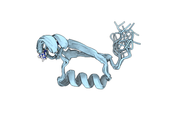 |
Solution Structure Of The C-Terminal Domain (Monomer) Of The Hpv45 Oncoprotein E7
Organism: Human papillomavirus
Method: SOLUTION NMR Release Date: 2006-10-17 Classification: ONCOPROTEIN, VIRUS/VIRAL PROTEIN Ligands: ZN |
Organism: Human papillomavirus
Method: SOLUTION NMR
Release Date: 2006-10-17
Ligands: ZN
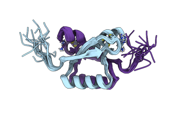 |
Nmr Structure Of The C-Terminal Domain (Dimer) Of Hpv45 Oncoprotein E7
Organism: Human papillomavirus type 45
Method: SOLUTION NMR Release Date: 2006-08-08 Classification: ONCOPROTEIN, VIRUS/VIRAL PROTEIN Ligands: ZN |
Organism: Human papillomavirus type 45
Method: SOLUTION NMR
Release Date: 2006-08-08
Ligands: ZN