

Search Count: 49
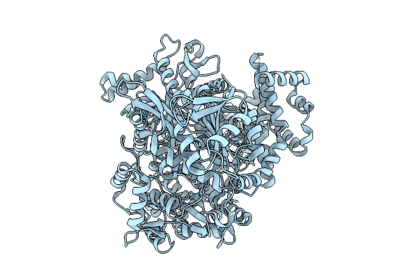 |
Hmus Heme Dechelatase: Disordered Domain 1, Heme Free.
Organism: Bacteroides thetaiotaomicron
Method: ELECTRON MICROSCOPY Release Date: 2025-09-24 Classification: METAL TRANSPORT |
Organism: Bacteroides thetaiotaomicron
Method: ELECTRON MICROSCOPY
Release Date: 2025-09-24
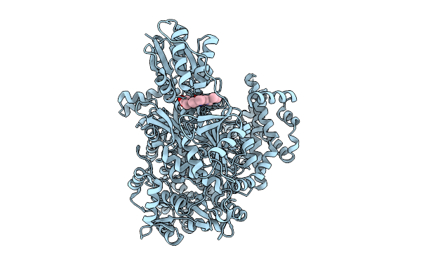 |
A Widespread Heme Dechelatase In Healthy And Pathogenic Human Microbiomes.
Organism: Bacteroides
Method: ELECTRON MICROSCOPY Release Date: 2025-08-20 Classification: METAL TRANSPORT Ligands: HEM, NA |
Organism: Bacteroides
Method: ELECTRON MICROSCOPY
Release Date: 2025-08-20
Ligands: HEM, NA
 |
Crystal Structure Of An Alpha/Beta-Hydrolase Enzyme From Chloroflexus Sp. Ms-G (202)
Organism: Chloroflexus sp. ms-g
Method: X-RAY DIFFRACTION Resolution:2.19 Å Release Date: 2022-12-28 Classification: HYDROLASE |
Organism: Chloroflexus sp. ms-g
Method: X-RAY DIFFRACTION
Release Date: 2022-12-28
 |
Crystal Structure Of An Alpha/Beta-Hydrolase Enzyme From Candidatus Kryptobacter Tengchongensis (306)
Organism: Candidatus kryptobacter tengchongensis
Method: X-RAY DIFFRACTION Resolution:1.89 Å Release Date: 2022-12-28 Classification: HYDROLASE Ligands: PO4 |
Organism: Candidatus kryptobacter tengchongensis
Method: X-RAY DIFFRACTION
Release Date: 2022-12-28
Ligands: PO4
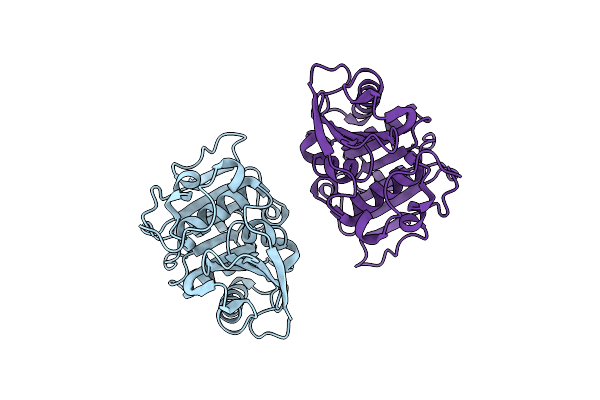 |
Crystal Structure Of A Cutinase Enzyme From Marinactinospora Thermotolerans Dsm45154 (606)
Organism: Marinactinospora thermotolerans
Method: X-RAY DIFFRACTION Resolution:1.93 Å Release Date: 2022-12-28 Classification: HYDROLASE |
Organism: Marinactinospora thermotolerans
Method: X-RAY DIFFRACTION
Release Date: 2022-12-28
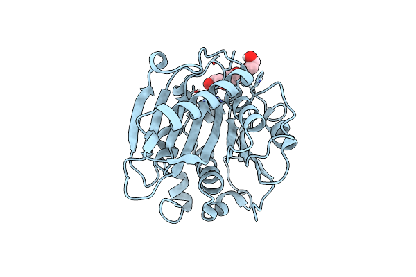 |
Crystal Structure Of A Cutinase Enzyme From Saccharopolyspora Flava (611)
Organism: Saccharopolyspora flava
Method: X-RAY DIFFRACTION Resolution:1.56 Å Release Date: 2022-12-28 Classification: HYDROLASE Ligands: PG4 |
Organism: Saccharopolyspora flava
Method: X-RAY DIFFRACTION
Release Date: 2022-12-28
Ligands: PG4
 |
Crystal Structure Of A Cutinase Enzyme From Thermobifida Fusca Ntu22 (702)
Organism: Thermobifida fusca
Method: X-RAY DIFFRACTION Resolution:1.64 Å Release Date: 2022-12-28 Classification: HYDROLASE Ligands: PEG |
Organism: Thermobifida fusca
Method: X-RAY DIFFRACTION
Release Date: 2022-12-28
Ligands: PEG
 |
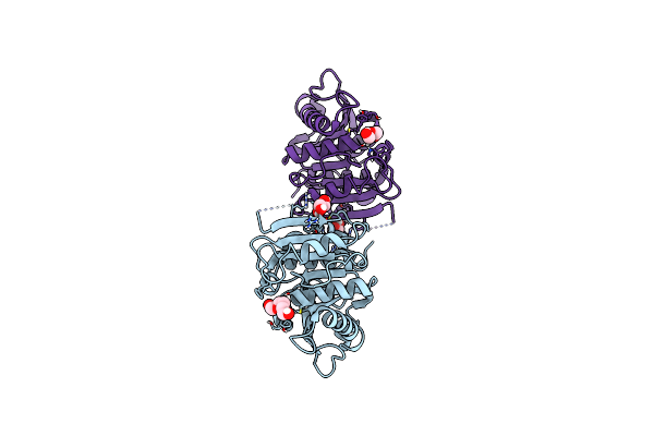
Crystal Structure Of Cutinase 1 From Thermobifida Fusca Dsm44342 (703)
Organism: Thermobifida fusca
Method: X-RAY DIFFRACTION Resolution:1.51 Å Release Date: 2022-12-28 Classification: HYDROLASE Ligands: PG4 |
Organism: Thermobifida fusca
Method: X-RAY DIFFRACTION
Release Date: 2022-12-28
Ligands: PG4
 |
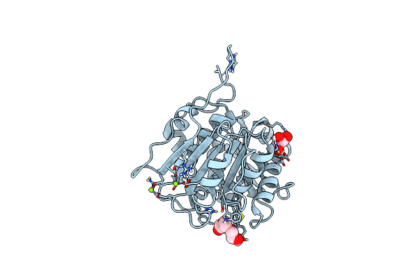
Crystal Structure Of A Cutinase Enzyme From Thermobifida Fusca Yx (705)
Organism: Thermobifida fusca yx
Method: X-RAY DIFFRACTION Resolution:1.43 Å Release Date: 2022-12-28 Classification: HYDROLASE Ligands: GOL, PG4, PEG |
Organism: Thermobifida fusca yx
Method: X-RAY DIFFRACTION
Release Date: 2022-12-28
Ligands: GOL, PG4, PEG
 |
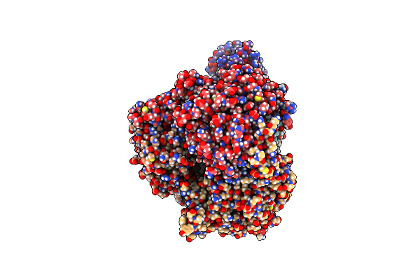
Crystal Structure Of A Cutinase Enzyme From Thermobifida Cellulosilytica Tb100 (711)
Organism: Thermobifida cellulosilytica tb100
Method: X-RAY DIFFRACTION Resolution:1.78 Å Release Date: 2022-12-28 Classification: HYDROLASE Ligands: GOL, PG4, MG |
Organism: Thermobifida cellulosilytica tb100
Method: X-RAY DIFFRACTION
Release Date: 2022-12-28
Ligands: GOL, PG4, MG
 |
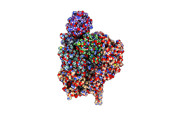
Crystal Structure Of Tpado In A Substrate-Free State
Organism: Comamonas sp., Gallus gallus
Method: X-RAY DIFFRACTION Resolution:2.28 Å Release Date: 2022-04-06 Classification: OXIDOREDUCTASE Ligands: FES, FE |
Organism: Comamonas sp., Gallus gallus
Method: X-RAY DIFFRACTION
Release Date: 2022-04-06
Ligands: FES, FE
 |
Crystal Structure Of Tpado In Complex With Tpa
Organism: Comamonas sp., Gallus gallus
Method: X-RAY DIFFRACTION Resolution:2.08 Å Release Date: 2022-03-30 Classification: OXIDOREDUCTASE Ligands: FES, SO4, UB7, FE |
Organism: Comamonas sp., Gallus gallus
Method: X-RAY DIFFRACTION
Release Date: 2022-03-30
Ligands: FES, SO4, UB7, FE
 |
Crystal Structure Of Tpado In Complex With 2-Oh-Tpa
Organism: Comamonas sp., Gallus gallus
Method: X-RAY DIFFRACTION Resolution:1.95 Å Release Date: 2022-03-30 Classification: OXIDOREDUCTASE Ligands: SO4, FES, FE, 8IB |
Organism: Comamonas sp., Gallus gallus
Method: X-RAY DIFFRACTION
Release Date: 2022-03-30
Ligands: SO4, FES, FE, 8IB
 |
Crystal Structure Of F501H/H506E Variant Of 2-Ketopropyl Coenzyme M Oxidoreductase/Carboxylase (2-Kpcc) From Xanthobacter Autotrophicus
Organism: Xanthobacter autotrophicus py2
Method: X-RAY DIFFRACTION Resolution:1.80 Å Release Date: 2021-07-21 Classification: OXIDOREDUCTASE Ligands: COM, FDA, MG |
Organism: Xanthobacter autotrophicus py2
Method: X-RAY DIFFRACTION
Release Date: 2021-07-21
Ligands: COM, FDA, MG
 |
Crystal Structure Of F501H Variant Of 2-Ketopropyl Coenzyme M Oxidoreductase/Carboxylase (2-Kpcc) From Xanthobacter Autotrophicus
Organism: Xanthobacter autotrophicus py2
Method: X-RAY DIFFRACTION Resolution:1.85 Å Release Date: 2021-07-21 Classification: OXIDOREDUCTASE Ligands: FDA, COM, MG |
Organism: Xanthobacter autotrophicus py2
Method: X-RAY DIFFRACTION
Release Date: 2021-07-21
Ligands: FDA, COM, MG
 |
Crystal Structure Of Gcoa T296A Bound To Guaiacol
Organism: Amycolatopsis sp. (strain atcc 39116 / 75iv2)
Method: X-RAY DIFFRACTION Resolution:1.88 Å Release Date: 2021-02-17 Classification: OXIDOREDUCTASE Ligands: HEM, JZ3, EPE |
Organism: Amycolatopsis sp. (strain atcc 39116 / 75iv2)
Method: X-RAY DIFFRACTION
Release Date: 2021-02-17
Ligands: HEM, JZ3, EPE
 |
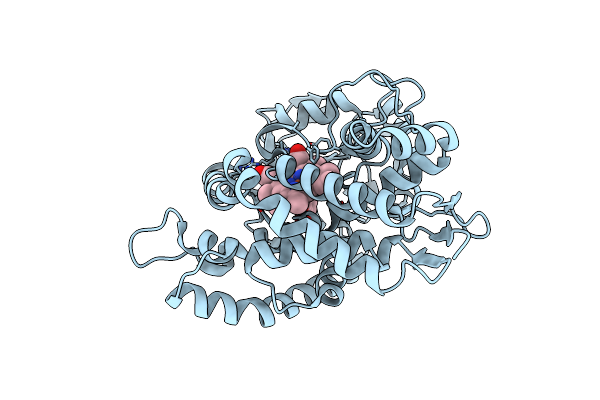
Crystal Structure Of Gcoa T296G Bound To Guaiacol
Organism: Amycolatopsis sp. (strain atcc 39116 / 75iv2)
Method: X-RAY DIFFRACTION Resolution:1.80 Å Release Date: 2021-02-17 Classification: OXIDOREDUCTASE Ligands: HEM, JZ3 |
Organism: Amycolatopsis sp. (strain atcc 39116 / 75iv2)
Method: X-RAY DIFFRACTION
Release Date: 2021-02-17
Ligands: HEM, JZ3
 |
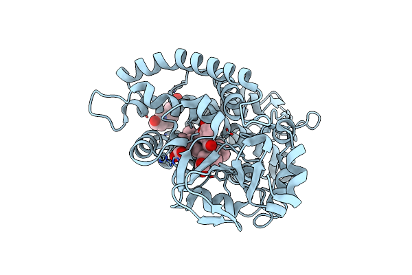
Crystal Structure Of Gcoa T296S Bound To Guaiacol
Organism: Amycolatopsis sp. (strain atcc 39116 / 75iv2)
Method: X-RAY DIFFRACTION Resolution:1.64 Å Release Date: 2021-02-17 Classification: OXIDOREDUCTASE Ligands: HEM, JZ3 |
Organism: Amycolatopsis sp. (strain atcc 39116 / 75iv2)
Method: X-RAY DIFFRACTION
Release Date: 2021-02-17
Ligands: HEM, JZ3
 |
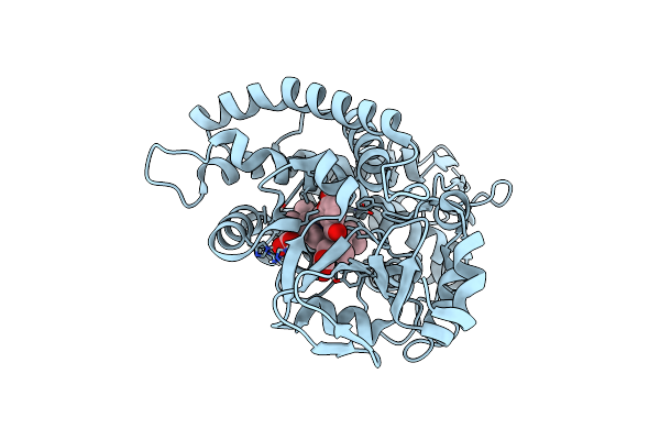
Crystal Structure Of Gcoa T296A Bound To P-Vanillin
Organism: Amycolatopsis sp. (strain atcc 39116 / 75iv2)
Method: X-RAY DIFFRACTION Resolution:1.80 Å Release Date: 2021-02-17 Classification: OXIDOREDUCTASE Ligands: HEM, V55, EPE |
Organism: Amycolatopsis sp. (strain atcc 39116 / 75iv2)
Method: X-RAY DIFFRACTION
Release Date: 2021-02-17
Ligands: HEM, V55, EPE
 |
Crystal Structure Of Gcoa T296G Bound To P-Vanillin
Organism: Amycolatopsis sp. (strain atcc 39116 / 75iv2)
Method: X-RAY DIFFRACTION Resolution:1.70 Å Release Date: 2021-02-17 Classification: OXIDOREDUCTASE Ligands: V55, HEM |
Organism: Amycolatopsis sp. (strain atcc 39116 / 75iv2)
Method: X-RAY DIFFRACTION
Release Date: 2021-02-17
Ligands: V55, HEM