

Search Count: 24
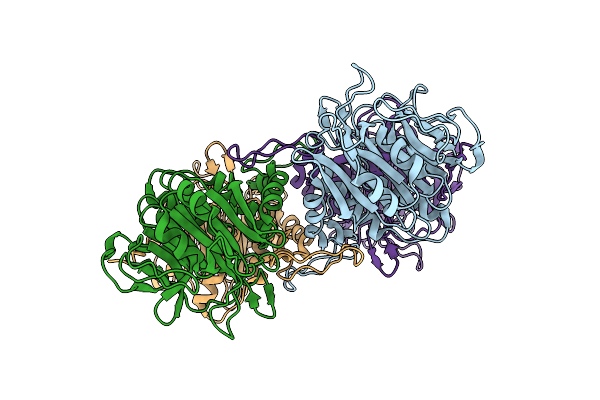 |
Bile Salt Hydrolase A From Lactobacillus Acidophilus
Organism: Lactobacillus acidophilus
Method: X-RAY DIFFRACTION Resolution:2.15 Å Release Date: 2023-01-25 Classification: HYDROLASE |
Organism: Lactobacillus acidophilus
Method: X-RAY DIFFRACTION
Release Date: 2023-01-25
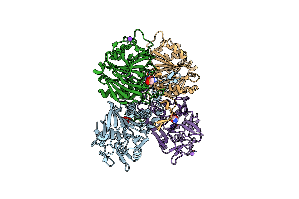 |
Bile Salt Hydrolase A From Lactobacillus Gasseri With Taurine Bound
Organism: Lactobacillus gasseri
Method: X-RAY DIFFRACTION Resolution:2.05 Å Release Date: 2023-01-25 Classification: HYDROLASE Ligands: TAU, K |
Organism: Lactobacillus gasseri
Method: X-RAY DIFFRACTION
Release Date: 2023-01-25
Ligands: TAU, K
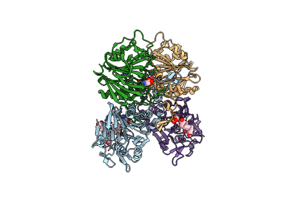 |
Bile Salt Hydrolase A From Lactobacillus Gasseri With Chenodeoxycholate And Taurine Bound
Organism: Lactobacillus gasseri
Method: X-RAY DIFFRACTION Resolution:1.35 Å Release Date: 2023-01-25 Classification: HYDROLASE Ligands: TAU, JN3 |
Organism: Lactobacillus gasseri
Method: X-RAY DIFFRACTION
Release Date: 2023-01-25
Ligands: TAU, JN3
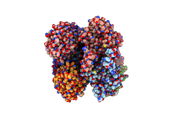 |
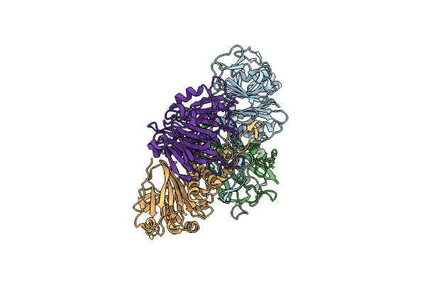
Bile Salt Hydrolase B From Lactobacillus Gasseri
Organism: Lactobacillus gasseri
Method: X-RAY DIFFRACTION Resolution:1.56 Å Release Date: 2023-01-25 Classification: HYDROLASE Ligands: MG |
Organism: Lactobacillus gasseri
Method: X-RAY DIFFRACTION
Release Date: 2023-01-25
Ligands: MG
 |
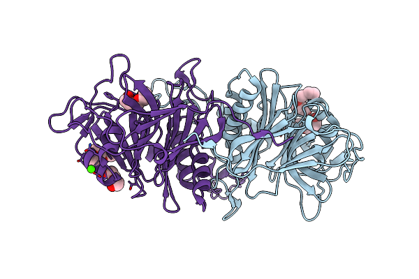
Bile Salt Hydrolase C From Lactobacillus Johnsonii
Organism: Lactobacillus johnsonii
Method: X-RAY DIFFRACTION Resolution:1.45 Å Release Date: 2023-01-25 Classification: HYDROLASE |
Organism: Lactobacillus johnsonii
Method: X-RAY DIFFRACTION
Release Date: 2023-01-25
 |
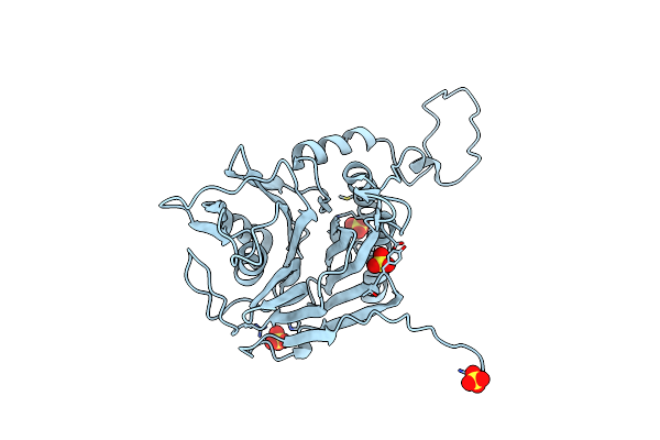
Bile Salt Hydrolase From Lactobacillus Ingluviei
Organism: Lactobacillus ingluviei
Method: X-RAY DIFFRACTION Resolution:1.43 Å Release Date: 2023-01-25 Classification: HYDROLASE Ligands: PG4, PEG, PGE, CA |
Organism: Lactobacillus ingluviei
Method: X-RAY DIFFRACTION
Release Date: 2023-01-25
Ligands: PG4, PEG, PGE, CA
 |
Bile Salt Hydrolase From Lactobacillus Reuteri
Organism: Lactobacillus reuteri
Method: X-RAY DIFFRACTION Resolution:1.66 Å Release Date: 2023-01-25 Classification: HYDROLASE Ligands: SO4 |
Organism: Lactobacillus reuteri
Method: X-RAY DIFFRACTION
Release Date: 2023-01-25
Ligands: SO4
 |
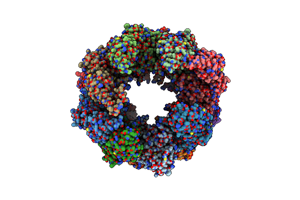
An Asymmetric Unit Map From Electron Cryo-Microscopy Of Haliotis Diversicolor Molluscan Hemocyanin Isoform 1 (Hdh1)
Organism: Haliotis diversicolor
Method: ELECTRON MICROSCOPY Release Date: 2013-04-17 Classification: OXYGEN TRANSPORT |
Organism: Haliotis diversicolor
Method: ELECTRON MICROSCOPY
Release Date: 2013-04-17
 |
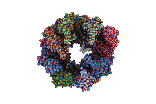
Symmetry-Free Cryo-Em Map Of Tric In The Nucleotide-Free (Apo) State
Organism: Bos taurus
Method: ELECTRON MICROSCOPY Resolution:10.50 Å Release Date: 2012-02-15 Classification: CHAPERONE |
Organism: Bos taurus
Method: ELECTRON MICROSCOPY
Release Date: 2012-02-15
 |
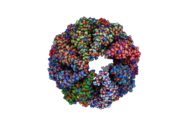
Model Refined Against The Symmetry-Free Cryo-Em Map Of Tric-Amp-Pnp
Organism: Bos taurus
Method: ELECTRON MICROSCOPY Resolution:10.70 Å Release Date: 2012-02-15 Classification: CHAPERONE |
Organism: Bos taurus
Method: ELECTRON MICROSCOPY
Release Date: 2012-02-15
 |
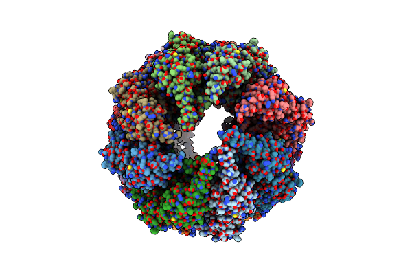
Model Built Against Symmetry-Free Cryo-Em Map Of Tric-Adp-Alfx
Organism: Bos taurus
Method: ELECTRON MICROSCOPY Resolution:13.90 Å Release Date: 2012-02-15 Classification: CHAPERONE |
Organism: Bos taurus
Method: ELECTRON MICROSCOPY
Release Date: 2012-02-15
 |
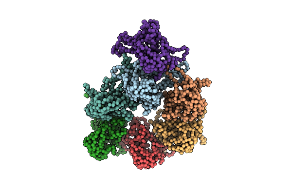
Model Refined Against Symmetry-Free Cryo-Em Map Of Tric-Adp
Organism: Bos taurus
Method: ELECTRON MICROSCOPY Resolution:11.30 Å Release Date: 2012-02-15 Classification: CHAPERONE |
Organism: Bos taurus
Method: ELECTRON MICROSCOPY
Release Date: 2012-02-15
 |
De Novo Model Of Bacteriophage P22 Procapsid Coat Protein
Organism: Enterobacteria phage p22
Method: ELECTRON MICROSCOPY Resolution:3.80 Å Release Date: 2011-02-02 Classification: VIRUS |
Organism: Enterobacteria phage p22
Method: ELECTRON MICROSCOPY
Release Date: 2011-02-02
 |
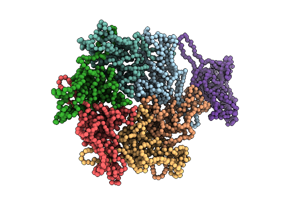
De Novo Model Of Bacteriophage P22 Virion Coat Protein
Organism: Enterobacteria phage p22
Method: ELECTRON MICROSCOPY Resolution:4.00 Å Release Date: 2011-02-02 Classification: VIRUS |
Organism: Enterobacteria phage p22
Method: ELECTRON MICROSCOPY
Release Date: 2011-02-02
 |
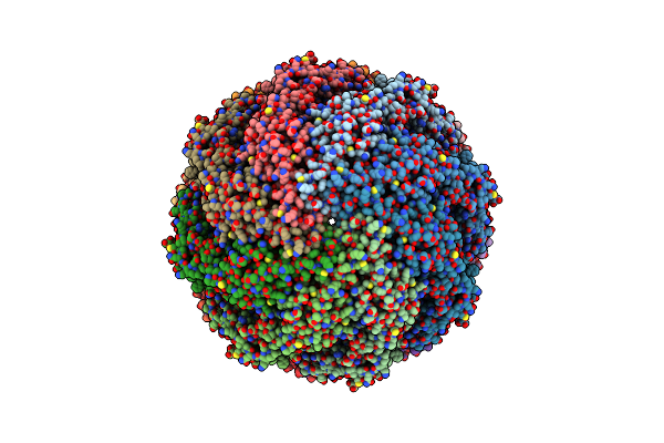
Capsid Structure Of The Infectious Prochlorococcus Cyanophage P-Ssp7
Organism: Prochlorococcus phage p-ssp7
Method: ELECTRON MICROSCOPY Resolution:4.60 Å Release Date: 2010-06-16 Classification: VIRUS |
Organism: Prochlorococcus phage p-ssp7
Method: ELECTRON MICROSCOPY
Release Date: 2010-06-16
 |
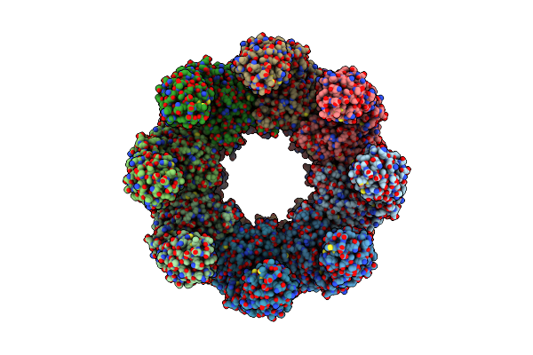
Atomic Model Of Mm-Cpn In The Closed State
Organism: Methanococcus maripaludis
Method: ELECTRON MICROSCOPY Release Date: 2010-03-16 Classification: CHAPERONE |
Organism: Methanococcus maripaludis
Method: ELECTRON MICROSCOPY
Release Date: 2010-03-16
 |
Atomic Model Of The Lidless Mm-Cpn In The Open State
Organism: Methanococcus maripaludis
Method: ELECTRON MICROSCOPY Release Date: 2010-02-02 Classification: CHAPERONE |
Organism: Methanococcus maripaludis
Method: ELECTRON MICROSCOPY
Release Date: 2010-02-02
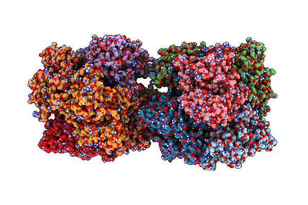 |
Scorpion Hemocyanin Resting State Pseudo Atomic Model Built Based On Cryo-Em Density Map
Organism: Androctonus australis
Method: ELECTRON MICROSCOPY Release Date: 2009-06-02 Classification: OXYGEN BINDING |
Organism: Androctonus australis
Method: ELECTRON MICROSCOPY
Release Date: 2009-06-02
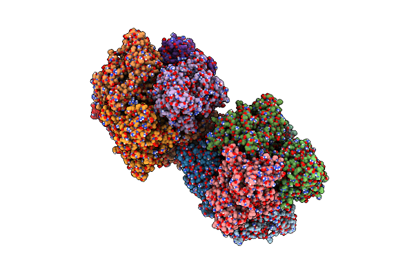 |
Scorpion Hemocyanin Activated State Pseudo Atomic Model Built Based On Cryo-Em Density Map
Organism: Androctonus australis
Method: ELECTRON MICROSCOPY Release Date: 2009-06-02 Classification: OXYGEN BINDING |
Organism: Androctonus australis
Method: ELECTRON MICROSCOPY
Release Date: 2009-06-02
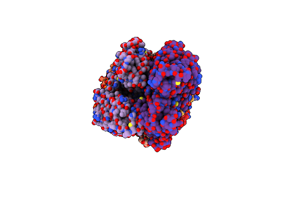 |
Actin Filament Model In The Extended Form Of Acromsomal Bundle In The Limulus Sperm
Organism: Limulus polyphemus
Method: ELECTRON MICROSCOPY Release Date: 2008-11-18 Classification: CONTRACTILE PROTEIN/STRUCTURAL PROTEIN |
Organism: Limulus polyphemus
Method: ELECTRON MICROSCOPY
Release Date: 2008-11-18