

Search Count: 25
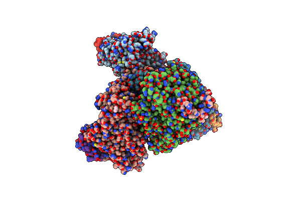 |
Structural Basis For The Recruitment Of Glycogen Synthase By Glycogenin
Organism: Caenorhabditis elegans
Method: X-RAY DIFFRACTION Resolution:2.60 Å Release Date: 2014-07-09 Classification: TRANSFERASE Ligands: SO4, GOL |
Organism: Caenorhabditis elegans
Method: X-RAY DIFFRACTION
Release Date: 2014-07-09
Ligands: SO4, GOL
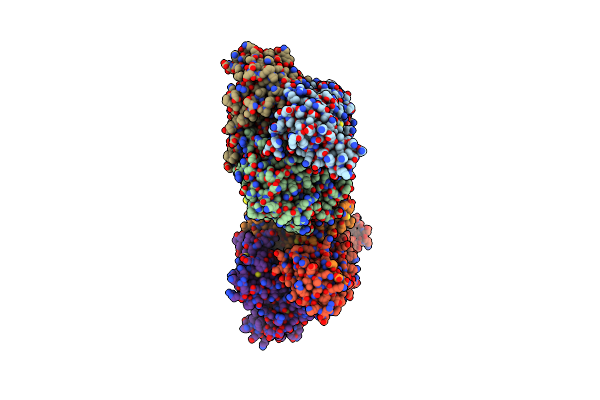 |
Structure Of The Heterotrimeric Lkb1-Stradalpha-Mo25Alpha Complex
Organism: Homo sapiens
Method: X-RAY DIFFRACTION Resolution:2.65 Å Release Date: 2009-12-15 Classification: TRANSFERASE/METAL-BINDING PROTEIN Ligands: ANP, SO4 |
Organism: Homo sapiens
Method: X-RAY DIFFRACTION
Release Date: 2009-12-15
Ligands: ANP, SO4
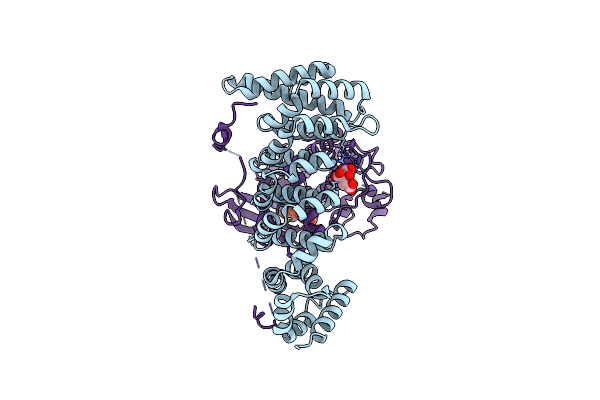 |
Structure Of Strad And Mo25
Organism: Homo sapiens
Method: X-RAY DIFFRACTION Resolution:2.35 Å Release Date: 2009-06-16 Classification: SIGNALING PROTEIN/SIGNALING PROTEIN Ligands: CIT, ATP |
Organism: Homo sapiens
Method: X-RAY DIFFRACTION
Release Date: 2009-06-16
Ligands: CIT, ATP
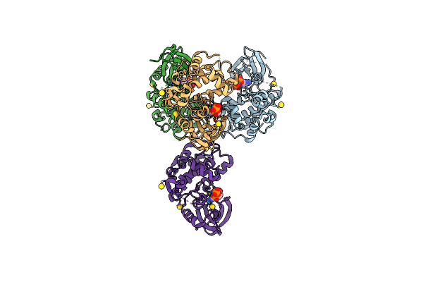 |
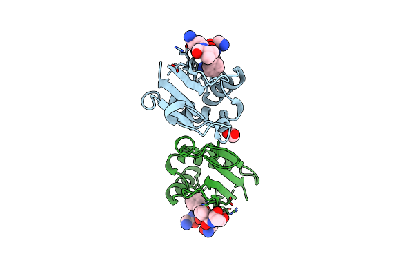
Structure Of The Osr1 Kinase, A Hypertension Drug Target
Organism: Homo sapiens
Method: X-RAY DIFFRACTION Resolution:2.15 Å Release Date: 2008-07-08 Classification: TRANSFERASE Ligands: AU, ANP, MG |
Organism: Homo sapiens
Method: X-RAY DIFFRACTION
Release Date: 2008-07-08
Ligands: AU, ANP, MG
 |
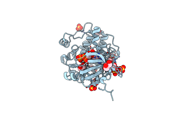
Structural Insights Into The Recognition Of Substrates And Activators By The Osr1 Kinase
Organism: Homo sapiens
Method: X-RAY DIFFRACTION Resolution:1.70 Å Release Date: 2007-07-03 Classification: TRANSFERASE Ligands: ACT |
Organism: Homo sapiens
Method: X-RAY DIFFRACTION
Release Date: 2007-07-03
Ligands: ACT
 |
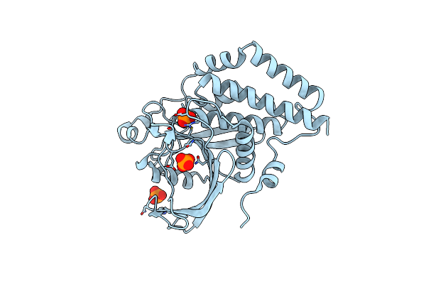
Structure Of Pdk1-S241A Mutant Kinase Domain
Organism: Homo sapiens
Method: X-RAY DIFFRACTION Resolution:1.95 Å Release Date: 2005-02-23 Classification: TRANSFERASE Ligands: GOL, SO4, ATP |
Organism: Homo sapiens
Method: X-RAY DIFFRACTION
Release Date: 2005-02-23
Ligands: GOL, SO4, ATP
 |
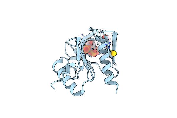
Crystal Structure Of Ptpl1 Human Tyrosine Phosphatase Mutated In Colorectal Cancer - Evidence For A Second Phosphotyrosine Substrate Recognition Pocket
Organism: Homo sapiens
Method: X-RAY DIFFRACTION Resolution:1.85 Å Release Date: 2004-12-14 Classification: HYDROLASE Ligands: PO4 |
Organism: Homo sapiens
Method: X-RAY DIFFRACTION
Release Date: 2004-12-14
Ligands: PO4
 |
Crystal Structure Of The Pdk1 Pleckstrin Homology (Ph) Domain Bound To Inositol (1,3,4,5)-Tetrakisphosphate
Organism: Homo sapiens
Method: X-RAY DIFFRACTION Resolution:1.50 Å Release Date: 2004-11-19 Classification: TRANSFERASE Ligands: GOL, AU, 4IP |
Organism: Homo sapiens
Method: X-RAY DIFFRACTION
Release Date: 2004-11-19
Ligands: GOL, AU, 4IP
 |
Crystal Structure Of The Pdk1 Pleckstrin Homology (Ph) Domain Bound To Dic4-Phosphatidylinositol (3,4,5)-Trisphosphate
Organism: Homo sapiens
Method: X-RAY DIFFRACTION Resolution:1.45 Å Release Date: 2004-11-19 Classification: TRANSFERASE Ligands: 4PT |
Organism: Homo sapiens
Method: X-RAY DIFFRACTION
Release Date: 2004-11-19
Ligands: 4PT
 |
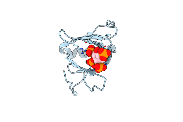
Crystal Structure Of The Pdk1 Pleckstrin Homology (Ph) Domain
Organism: Homo sapiens
Method: X-RAY DIFFRACTION Resolution:1.45 Å Release Date: 2004-11-19 Classification: TRANSFERASE Ligands: GOL, SO4 |
Organism: Homo sapiens
Method: X-RAY DIFFRACTION
Release Date: 2004-11-19
Ligands: GOL, SO4
 |
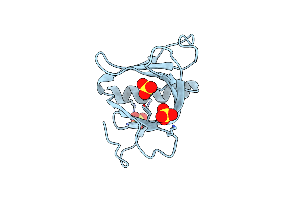
Crystal Structure Of The Pepp1 Pleckstrin Homology Domain In Complex With Inositol 1,3,4,5-Tetrakisphosphate
Organism: Homo sapiens
Method: X-RAY DIFFRACTION Resolution:2.27 Å Release Date: 2004-10-20 Classification: SIGNALING PROTEIN Ligands: 4IP |
Organism: Homo sapiens
Method: X-RAY DIFFRACTION
Release Date: 2004-10-20
Ligands: 4IP
 |
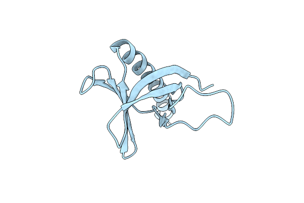
Crystal Structure Of The Pleckstrin Homology (Ph) Domain Of Pepp1
Organism: Homo sapiens
Method: X-RAY DIFFRACTION Resolution:1.48 Å Release Date: 2004-10-07 Classification: SIGNAL TRANSDUCTION Ligands: SO4 |
Organism: Homo sapiens
Method: X-RAY DIFFRACTION
Release Date: 2004-10-07
Ligands: SO4
 |
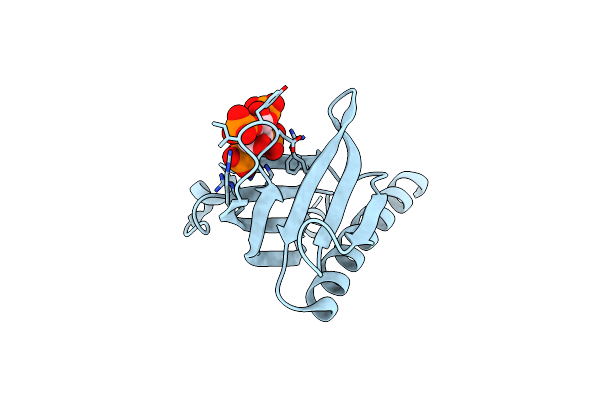
Crystal Structure Of The Pleckstrin Homology Domain Of Pkb Alpha
Organism: Homo sapiens
Method: X-RAY DIFFRACTION Resolution:1.65 Å Release Date: 2004-09-16 Classification: TRANSFERASE |
Organism: Homo sapiens
Method: X-RAY DIFFRACTION
Release Date: 2004-09-16
 |
High Resolution Crystal Structure Of The Pleckstrin Homology Domain Of Protein Kinase B/Akt Bound To Ins(1,3,4,5)-Tetrakisphophate
Organism: Homo sapiens
Method: X-RAY DIFFRACTION Resolution:0.98 Å Release Date: 2004-09-16 Classification: TRANSFERASE Ligands: 4IP |
Organism: Homo sapiens
Method: X-RAY DIFFRACTION
Release Date: 2004-09-16
Ligands: 4IP
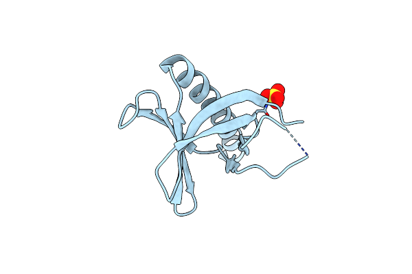 |
Crystal Structure Of The Ph Domain Of Pkb Alpha In Complex With A Sulfate Molecule
Organism: Homo sapiens
Method: X-RAY DIFFRACTION Resolution:1.25 Å Release Date: 2004-09-16 Classification: TRANSFERASE Ligands: SO4 |
Organism: Homo sapiens
Method: X-RAY DIFFRACTION
Release Date: 2004-09-16
Ligands: SO4
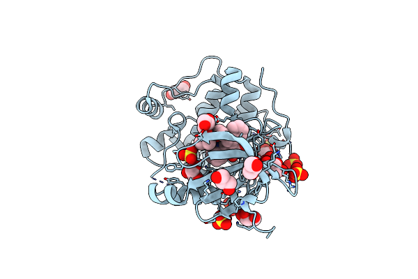 |
Structure Of Human Pdk1 Kinase Domain In Complex With Ly333531
Organism: Homo sapiens
Method: X-RAY DIFFRACTION Resolution:1.70 Å Release Date: 2004-03-04 Classification: TRANSFERASE Ligands: GOL, SO4, LY4 |
Organism: Homo sapiens
Method: X-RAY DIFFRACTION
Release Date: 2004-03-04
Ligands: GOL, SO4, LY4
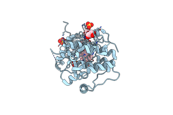 |
Structure Of Human Pdk1 Kinase Domain In Complex With Bim-2
Organism: Homo sapiens
Method: X-RAY DIFFRACTION Resolution:1.90 Å Release Date: 2004-03-04 Classification: TRANSFERASE Ligands: GOL, BI2, SO4 |
Organism: Homo sapiens
Method: X-RAY DIFFRACTION
Release Date: 2004-03-04
Ligands: GOL, BI2, SO4
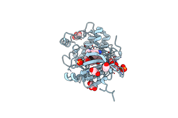 |
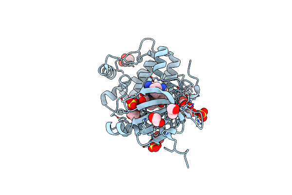
Structure Of Human Pdk1 Kinase Domain In Complex With Bim-1
Organism: Homo sapiens
Method: X-RAY DIFFRACTION Resolution:2.50 Å Release Date: 2004-03-04 Classification: TRANSFERASE Ligands: GOL, SO4, BI1 |
Organism: Homo sapiens
Method: X-RAY DIFFRACTION
Release Date: 2004-03-04
Ligands: GOL, SO4, BI1
 |
Structure Of Human Pdk1 Kinase Domain In Complex With Bim-3
Organism: Spodoptera frugiperda
Method: X-RAY DIFFRACTION Resolution:1.95 Å Release Date: 2004-03-04 Classification: TRANSFERASE Ligands: GOL, SO4, BI3 |
Organism: Spodoptera frugiperda
Method: X-RAY DIFFRACTION
Release Date: 2004-03-04
Ligands: GOL, SO4, BI3
 |
Structure Of Human Pdk1 Kinase Domain In Complex With Bim-8
Organism: Homo sapiens
Method: X-RAY DIFFRACTION Resolution:2.81 Å Release Date: 2004-03-04 Classification: TRANSFERASE Ligands: GOL, SO4, BI8 |
Organism: Homo sapiens
Method: X-RAY DIFFRACTION
Release Date: 2004-03-04
Ligands: GOL, SO4, BI8