

Search Count: 29
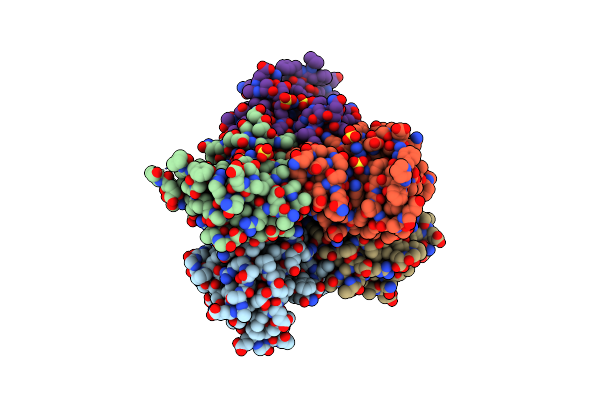 |
R80A Mutant Of Nucleoside Diphosphate Kinase From Mycobacterium Tuberculosis
Organism: Mycobacterium tuberculosis (strain atcc 25177 / h37ra)
Method: X-RAY DIFFRACTION Resolution:2.20 Å Release Date: 2019-02-27 Classification: TRANSFERASE Ligands: SO4, TAM |
Organism: Mycobacterium tuberculosis (strain atcc 25177 / h37ra)
Method: X-RAY DIFFRACTION
Release Date: 2019-02-27
Ligands: SO4, TAM
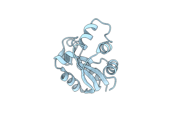 |
Crystal Form I Of The D93N Mutant Of Nucleoside Diphosphate Kinase From Mycobacterium Tuberculosis
Organism: Mycobacterium tuberculosis
Method: X-RAY DIFFRACTION Resolution:2.80 Å Release Date: 2013-03-13 Classification: TRANSFERASE |
Organism: Mycobacterium tuberculosis
Method: X-RAY DIFFRACTION
Release Date: 2013-03-13
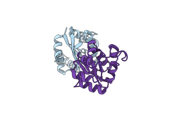 |
Crystal Form Ii Of The D93N Mutant Of Nucleoside Diphosphate Kinase From Mycobacterium Tuberculosis
Organism: Mycobacterium tuberculosis
Method: X-RAY DIFFRACTION Resolution:2.81 Å Release Date: 2013-03-13 Classification: TRANSFERASE |
Organism: Mycobacterium tuberculosis
Method: X-RAY DIFFRACTION
Release Date: 2013-03-13
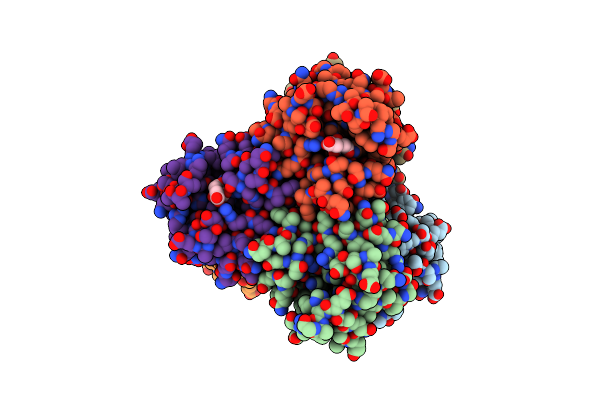 |
R80N Mutant Of Nucleoside Diphosphate Kinase From Mycobacterium Tuberculosis
Organism: Mycobacterium tuberculosis
Method: X-RAY DIFFRACTION Resolution:1.90 Å Release Date: 2013-03-13 Classification: TRANSFERASE Ligands: CIT |
Organism: Mycobacterium tuberculosis
Method: X-RAY DIFFRACTION
Release Date: 2013-03-13
Ligands: CIT
 |
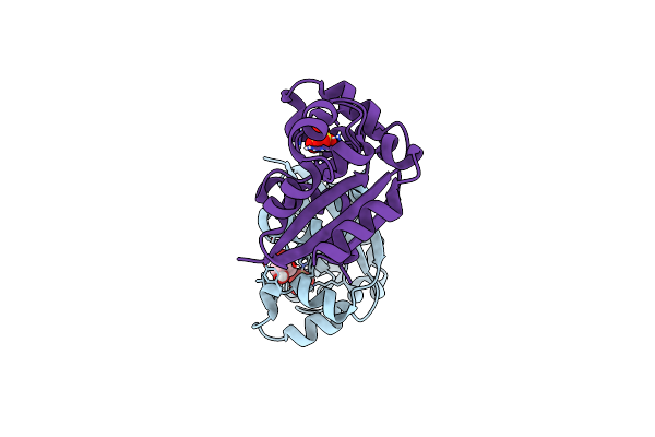
Orthorhombic Crystal Form C222 Of The Aquifex Aeolicus Nucleoside Diphosphate Kinase
Organism: Aquifex aeolicus
Method: X-RAY DIFFRACTION Resolution:1.47 Å Release Date: 2012-03-14 Classification: TRANSFERASE Ligands: SO4 |
Organism: Aquifex aeolicus
Method: X-RAY DIFFRACTION
Release Date: 2012-03-14
Ligands: SO4
 |
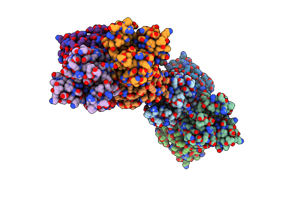
Orthorhombic Crystal Form P21212 Of The Aquifex Aeolicus Nucleoside Diphosphate Kinase
Organism: Aquifex aeolicus
Method: X-RAY DIFFRACTION Resolution:1.37 Å Release Date: 2012-03-14 Classification: TRANSFERASE Ligands: GOL, SO4 |
Organism: Aquifex aeolicus
Method: X-RAY DIFFRACTION
Release Date: 2012-03-14
Ligands: GOL, SO4
 |
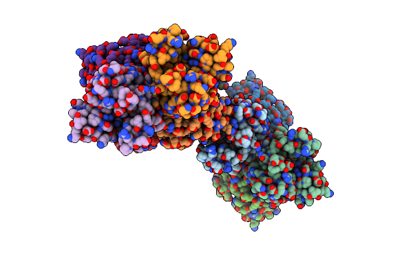
Hexagonal Form P6122 Of The Aquifex Aeolicus Nucleoside Diphosphate Kinase (First Stage Of Radiation Damage)
Organism: Aquifex aeolicus
Method: X-RAY DIFFRACTION Resolution:2.30 Å Release Date: 2012-03-14 Classification: TRANSFERASE |
Organism: Aquifex aeolicus
Method: X-RAY DIFFRACTION
Release Date: 2012-03-14
 |
Hexagonal Form P6122 Of The Aquifex Aeolicus Nucleoside Diphosphate Kinase (Final Stage Of Radiation Damage)
Organism: Aquifex aeolicus
Method: X-RAY DIFFRACTION Resolution:2.30 Å Release Date: 2012-03-14 Classification: TRANSFERASE |
Organism: Aquifex aeolicus
Method: X-RAY DIFFRACTION
Release Date: 2012-03-14
 |
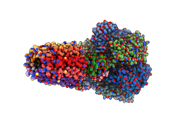
Hexagonal Crystal Form P61 Of The Aquifex Aeolicus Nucleoside Diphosphate Kinase
Organism: Aquifex aeolicus
Method: X-RAY DIFFRACTION Resolution:2.10 Å Release Date: 2012-02-29 Classification: TRANSFERASE |
Organism: Aquifex aeolicus
Method: X-RAY DIFFRACTION
Release Date: 2012-02-29
 |
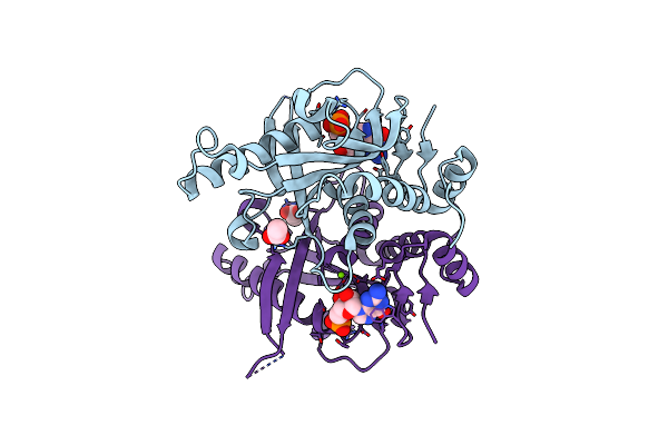
Rotor Architecture In The F(1)-C(10)-Ring Complex Of The Yeast F-Atp Synthase
Organism: Saccharomyces cerevisiae
Method: X-RAY DIFFRACTION Resolution:6.50 Å Release Date: 2012-01-18 Classification: HYDROLASE Ligands: ANP, MG |
Organism: Saccharomyces cerevisiae
Method: X-RAY DIFFRACTION
Release Date: 2012-01-18
Ligands: ANP, MG
 |
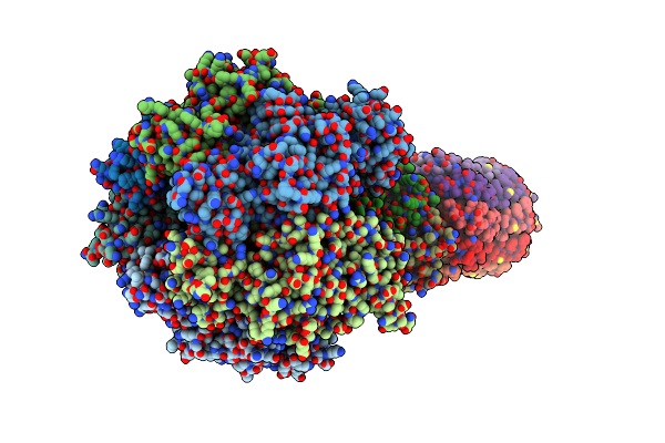
Saccharomyces Cerevisiae Hypoxanthine-Guanine Phosphoribosyltransferase In Complex With Gmp (Monoclinic Crystal Form)
Organism: Saccharomyces cerevisiae
Method: X-RAY DIFFRACTION Resolution:1.80 Å Release Date: 2011-05-04 Classification: TRANSFERASE Ligands: ACT, 5GP, MG |
Organism: Saccharomyces cerevisiae
Method: X-RAY DIFFRACTION
Release Date: 2011-05-04
Ligands: ACT, 5GP, MG
 |
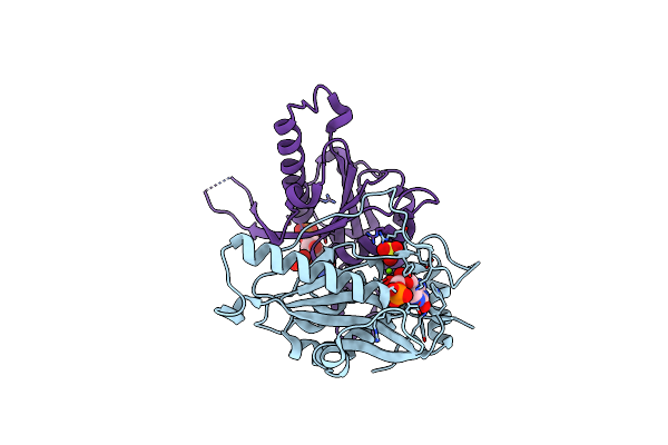
The Mg.Adp Inhibited State Of The Yeast F1C10 Atp Synthase
Organism: Saccharomyces cerevisiae
Method: X-RAY DIFFRACTION Resolution:3.43 Å Release Date: 2010-07-07 Classification: HYDROLASE Ligands: ATP, MG, ADP |
Organism: Saccharomyces cerevisiae
Method: X-RAY DIFFRACTION
Release Date: 2010-07-07
Ligands: ATP, MG, ADP
 |
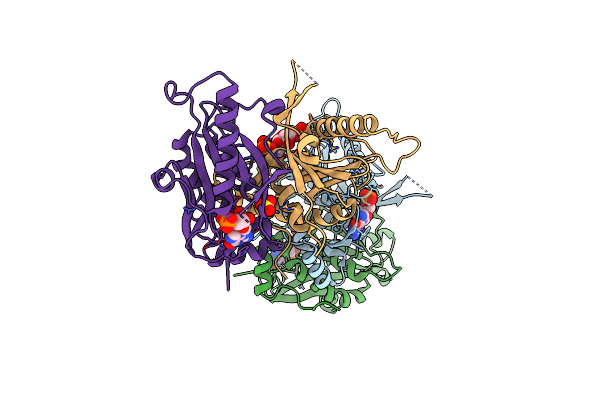
Saccharomyces Cerevisiae Hypoxanthine-Guanine Phosphoribosyltransferase In Complex With Gmp (Guanosine 5'- Monophosphate) (Tetragonal Crystal Form)
Organism: Saccharomyces cerevisiae
Method: X-RAY DIFFRACTION Resolution:2.30 Å Release Date: 2009-11-17 Classification: TRANSFERASE Ligands: 5GP, SO4, MG |
Organism: Saccharomyces cerevisiae
Method: X-RAY DIFFRACTION
Release Date: 2009-11-17
Ligands: 5GP, SO4, MG
 |
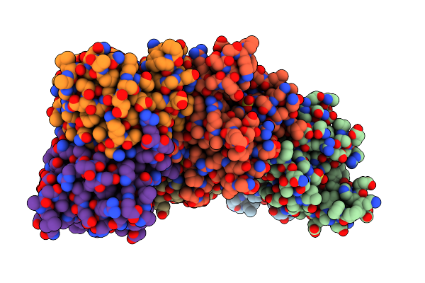
Saccharomyces Cerevisiae Hypoxanthine-Guanine Phosphoribosyltransferase In Complex With Gmp (Guanosine 5'- Monophosphate) (Orthorhombic Crystal Form)
Organism: Saccharomyces cerevisiae
Method: X-RAY DIFFRACTION Resolution:3.45 Å Release Date: 2009-11-17 Classification: TRANSFERASE Ligands: 5GP, SO4 |
Organism: Saccharomyces cerevisiae
Method: X-RAY DIFFRACTION
Release Date: 2009-11-17
Ligands: 5GP, SO4
 |
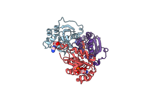
Escherichia Coli Nucleoside Diphosphate Kinase
Organism: Escherichia coli
Method: X-RAY DIFFRACTION Resolution:1.62 Å Release Date: 2007-04-10 Classification: SIGNALING PROTEIN,TRANSFERASE Ligands: SO4 |
Organism: Escherichia coli
Method: X-RAY DIFFRACTION
Release Date: 2007-04-10
Ligands: SO4
 |
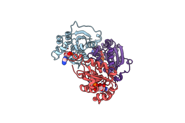
Human Nucleoside Diphosphate Kinase A Complexed With Adp
Organism: Homo sapiens
Method: X-RAY DIFFRACTION Resolution:2.15 Å Release Date: 2006-09-19 Classification: SIGNALING PROTEIN,TRANSFERASE Ligands: ADP |
Organism: Homo sapiens
Method: X-RAY DIFFRACTION
Release Date: 2006-09-19
Ligands: ADP
 |
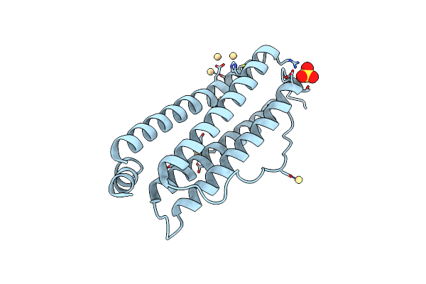
S120G Mutant Of Human Nucleoside Diphosphate Kinase A Complexed With Adp
Organism: Homo sapiens
Method: X-RAY DIFFRACTION Resolution:2.40 Å Release Date: 2006-09-19 Classification: SIGNALING PROTEIN,TRANSFERASE Ligands: ADP |
Organism: Homo sapiens
Method: X-RAY DIFFRACTION
Release Date: 2006-09-19
Ligands: ADP
 |
Recombinant Mouse L-Chain Ferritin
Organism: Mus musculus
Method: X-RAY DIFFRACTION Resolution:1.60 Å Release Date: 2001-03-06 Classification: IRON STORAGE Ligands: CD, SO4 |
Organism: Mus musculus
Method: X-RAY DIFFRACTION
Release Date: 2001-03-06
Ligands: CD, SO4
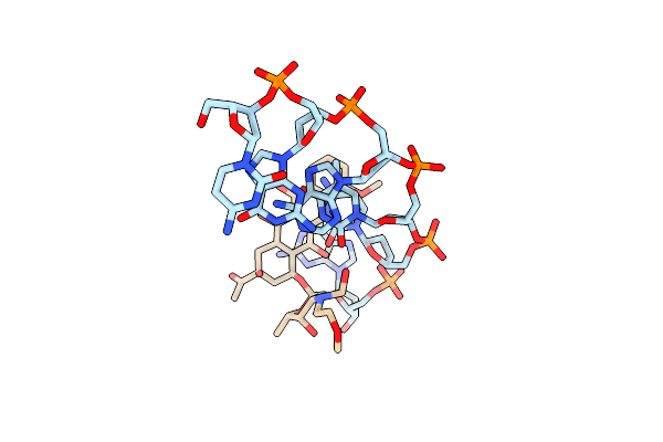 |
Release Of The Cyano Moiety In The Crystal Structure Of N-Cyanomethyl-N-(2-Methoxyethyl)-Daunomycin Complexed With D(Cgatcg)
Method: X-RAY DIFFRACTION
Resolution:1.54 Å Release Date: 1999-09-15 Classification: DNA Ligands: DM9 |
Method: X-RAY DIFFRACTION
Release Date: 1999-09-15
Ligands: DM9
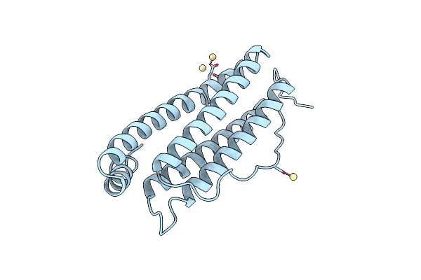 |
Cubic Crystal Structure Recombinant Horse L Apoferritin
Organism: Equus caballus
Method: X-RAY DIFFRACTION Resolution:2.05 Å Release Date: 1997-03-12 Classification: IRON STORAGE Ligands: CD |
Organism: Equus caballus
Method: X-RAY DIFFRACTION
Release Date: 1997-03-12
Ligands: CD