

Search Count: 28
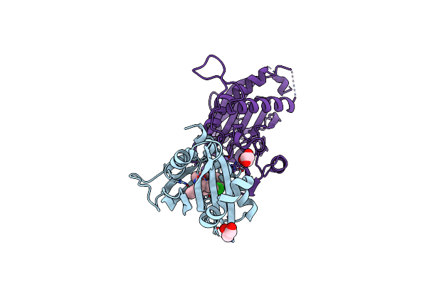 |
Structure Of An Orthogonal Pyr1*:Hab1* Chemical-Induced Dimerization Module In Complex With Mandipropamid
Organism: Arabidopsis thaliana
Method: X-RAY DIFFRACTION Resolution:2.40 Å Release Date: 2023-11-08 Classification: PLANT PROTEIN Ligands: GOL, 3UZ |
Organism: Arabidopsis thaliana
Method: X-RAY DIFFRACTION
Release Date: 2023-11-08
Ligands: GOL, 3UZ
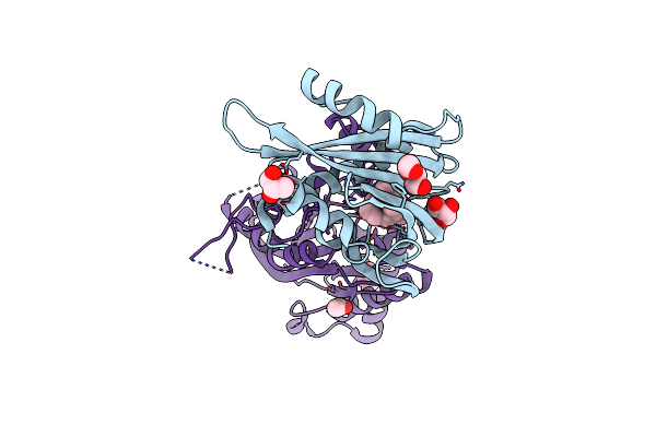 |
An Engineered Pyl2-Based Win 55,212-2 Synthetic Cannabinoid Sensor With A Stabilized Hab1 Variant
Organism: Arabidopsis thaliana
Method: X-RAY DIFFRACTION Resolution:1.90 Å Release Date: 2022-06-01 Classification: PLANT PROTEIN Ligands: WI5, PEG, GOL, MG, ACT, CL |
Organism: Arabidopsis thaliana
Method: X-RAY DIFFRACTION
Release Date: 2022-06-01
Ligands: WI5, PEG, GOL, MG, ACT, CL
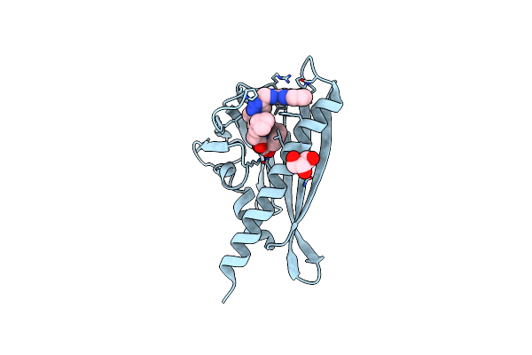 |
Pyl10 Bound To The Aba Pan-Antagonist 4A
Organism: Arabidopsis thaliana
Method: X-RAY DIFFRACTION Resolution:1.77 Å Release Date: 2021-09-29 Classification: PLANT PROTEIN/ANTAGONIST Ligands: GOL, ZLA |
Organism: Arabidopsis thaliana
Method: X-RAY DIFFRACTION
Release Date: 2021-09-29
Ligands: GOL, ZLA
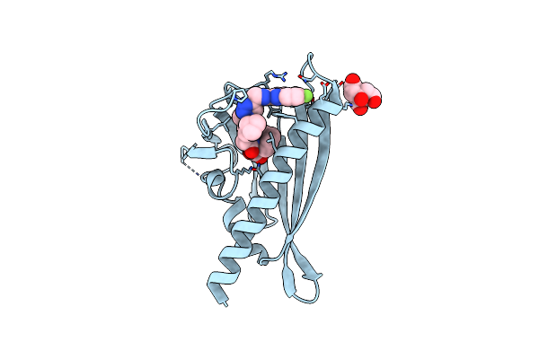 |
Pyl10 Bound To The Aba Pan-Antagonist Antabactin
Organism: Arabidopsis thaliana
Method: X-RAY DIFFRACTION Resolution:1.80 Å Release Date: 2021-09-29 Classification: PLANT PROTEIN/ANTAGONIST Ligands: ZLG, CIT |
Organism: Arabidopsis thaliana
Method: X-RAY DIFFRACTION
Release Date: 2021-09-29
Ligands: ZLG, CIT
 |
Pyl10 Bound To The Selective Agonist Hexabactin
Organism: Arabidopsis thaliana
Method: X-RAY DIFFRACTION Resolution:2.00 Å Release Date: 2020-02-12 Classification: PLANT PROTEIN Ligands: L6V |
Organism: Arabidopsis thaliana
Method: X-RAY DIFFRACTION
Release Date: 2020-02-12
Ligands: L6V
 |
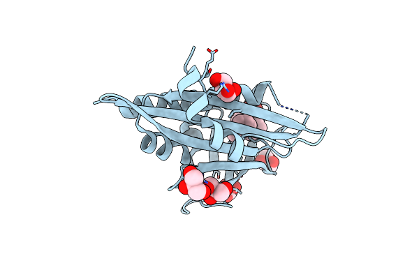
Pyl10 Bound To The Aba Pan-Agonist 3Cb
Organism: Arabidopsis thaliana
Method: X-RAY DIFFRACTION Resolution:2.35 Å Release Date: 2019-11-06 Classification: PLANT PROTEIN Ligands: L6P |
Organism: Arabidopsis thaliana
Method: X-RAY DIFFRACTION
Release Date: 2019-11-06
Ligands: L6P
 |
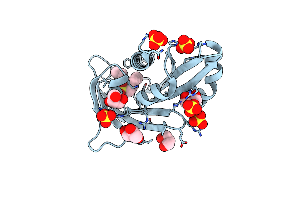
1.5 A Crystal Structure Of Pyr1 Bound To Pyrabactin
Organism: Arabidopsis thaliana
Method: X-RAY DIFFRACTION Resolution:1.52 Å Release Date: 2018-01-24 Classification: HORMONE RECEPTOR Ligands: PYV, GOL |
Organism: Arabidopsis thaliana
Method: X-RAY DIFFRACTION
Release Date: 2018-01-24
Ligands: PYV, GOL
 |
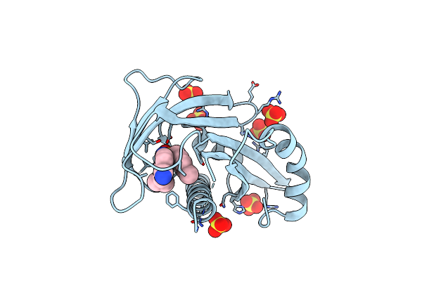
Pyr1 Bound To The Rationally Designed Agonist 4M
Organism: Arabidopsis thaliana
Method: X-RAY DIFFRACTION Resolution:1.93 Å Release Date: 2017-11-29 Classification: HORMONE RECEPTOR Ligands: SO4, 8KP, GOL |
Organism: Arabidopsis thaliana
Method: X-RAY DIFFRACTION
Release Date: 2017-11-29
Ligands: SO4, 8KP, GOL
 |
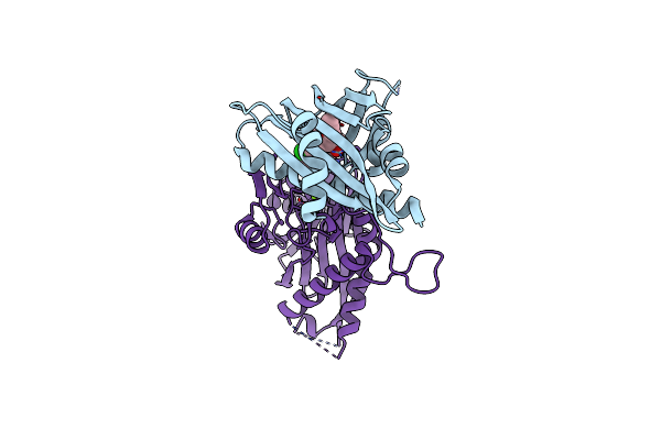
Pyr1 Bound To The Rationally Designed Agonist Cyanabactin
Organism: Arabidopsis thaliana
Method: X-RAY DIFFRACTION Resolution:1.63 Å Release Date: 2017-11-22 Classification: HORMONE RECEPTOR Ligands: SO4, 8KM |
Organism: Arabidopsis thaliana
Method: X-RAY DIFFRACTION
Release Date: 2017-11-22
Ligands: SO4, 8KM
 |
An Engineered Pyr1 Mandipropamid Receptor In Complex With Mandipropamid And Hab1
Organism: Arabidopsis thaliana
Method: X-RAY DIFFRACTION Resolution:2.25 Å Release Date: 2015-02-11 Classification: HYDROLASE/HYDROLASE INTHIBITOR Ligands: 3UZ, MG |
Organism: Arabidopsis thaliana
Method: X-RAY DIFFRACTION
Release Date: 2015-02-11
Ligands: 3UZ, MG
 |
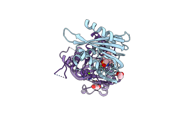
Crystal Structure Of The Abscisic Acid Receptor Pyr1 In Complex With An Antagonist As6
Organism: Arabidopsis thaliana
Method: X-RAY DIFFRACTION Resolution:2.30 Å Release Date: 2014-05-07 Classification: HORMONE RECEPTOR Ligands: 6AS |
Organism: Arabidopsis thaliana
Method: X-RAY DIFFRACTION
Release Date: 2014-05-07
Ligands: 6AS
 |
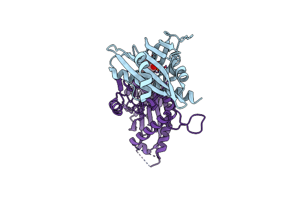
X-Ray Crystal Structure Of The Pyl2-Quinabactin-Hab1 Ternary Complex
Organism: Arabidopsis thaliana
Method: X-RAY DIFFRACTION Resolution:1.98 Å Release Date: 2013-08-07 Classification: Hydrolase/receptor/inhibitor Ligands: A1O, ACT, MG, GOL |
Organism: Arabidopsis thaliana
Method: X-RAY DIFFRACTION
Release Date: 2013-08-07
Ligands: A1O, ACT, MG, GOL
 |
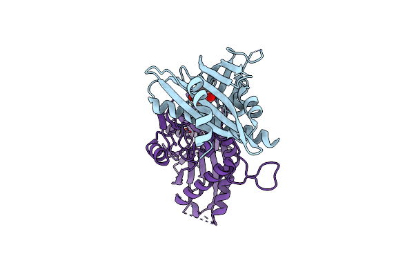
Structure Of The Pyr1 His60Pro Mutant In Complex With The Hab1 Phosphatase And Abscisic Acid
Organism: Arabidopsis thaliana
Method: X-RAY DIFFRACTION Resolution:2.10 Å Release Date: 2012-06-13 Classification: RECEPTOR/HYDROLASE Ligands: A8S, MN, SO4 |
Organism: Arabidopsis thaliana
Method: X-RAY DIFFRACTION
Release Date: 2012-06-13
Ligands: A8S, MN, SO4
 |
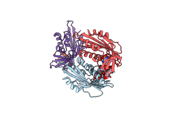
Crystal Structure Of The Pyr1 Abscisic Acid Receptor In Complex With The Hab1 Type 2C Phosphatase Catalytic Domain
Organism: Arabidopsis thaliana
Method: X-RAY DIFFRACTION Resolution:1.80 Å Release Date: 2011-03-16 Classification: PROTEIN BINDING Ligands: A8S, MN |
Organism: Arabidopsis thaliana
Method: X-RAY DIFFRACTION
Release Date: 2011-03-16
Ligands: A8S, MN
 |
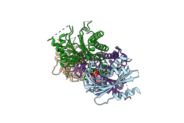
Crystal Structure Of The Abscisic Receptor Pyl2 In Complex With Pyrabactin
Organism: Arabidopsis thaliana
Method: X-RAY DIFFRACTION Resolution:1.85 Å Release Date: 2010-08-25 Classification: hormone binding protein Ligands: PYV, EPE |
Organism: Arabidopsis thaliana
Method: X-RAY DIFFRACTION
Release Date: 2010-08-25
Ligands: PYV, EPE
 |
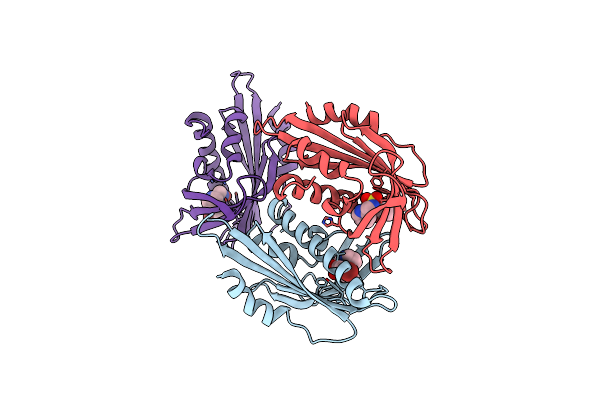
Crystal Structure Of Pyrabactin-Bound Abscisic Acid Receptor Pyl1 In Complex With Type 2C Protein Phosphatase Abi1
Organism: Arabidopsis thaliana
Method: X-RAY DIFFRACTION Resolution:2.15 Å Release Date: 2010-08-25 Classification: PROTEIN BINDING Ligands: PYV, MG |
Organism: Arabidopsis thaliana
Method: X-RAY DIFFRACTION
Release Date: 2010-08-25
Ligands: PYV, MG
 |
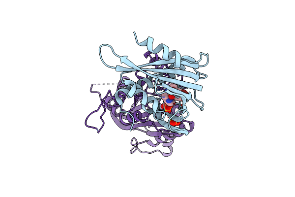
Crystal Structure Of The Abscisic Receptor Pyl2 Mutant A93F In Complex With Pyrabactin
Organism: Arabidopsis thaliana
Method: X-RAY DIFFRACTION Resolution:2.10 Å Release Date: 2010-08-25 Classification: hormone binding protein Ligands: PYV |
Organism: Arabidopsis thaliana
Method: X-RAY DIFFRACTION
Release Date: 2010-08-25
Ligands: PYV
 |
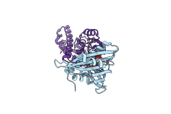
Crystal Structure Of Pyrabactin Bound Abscisic Acid Receptor Pyl2 Mutant A93F In Complex With Type 2C Protein Phosphatase Hab1
Organism: Arabidopsis thaliana
Method: X-RAY DIFFRACTION Resolution:2.56 Å Release Date: 2010-08-25 Classification: PROTEIN BINDING Ligands: PYV, MG, SO4 |
Organism: Arabidopsis thaliana
Method: X-RAY DIFFRACTION
Release Date: 2010-08-25
Ligands: PYV, MG, SO4
 |
Crystal Structure Of Pyrabactin-Bound Abscisic Acid Receptor Pyl2 Mutant A93F In Complex With Type 2C Protein Phosphatase Abi2
Organism: Arabidopsis thaliana
Method: X-RAY DIFFRACTION Resolution:2.10 Å Release Date: 2010-08-25 Classification: PROTEIN BINDING Ligands: PYV, MG |
Organism: Arabidopsis thaliana
Method: X-RAY DIFFRACTION
Release Date: 2010-08-25
Ligands: PYV, MG
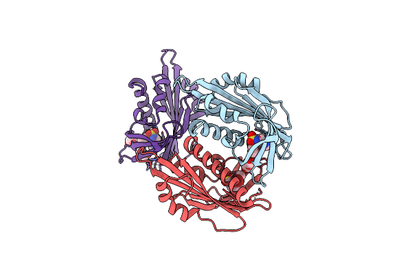 |
X-Ray Crystal Structure Of The Pyl2-Pyrabactin A Complex
Organism: Arabidopsis thaliana
Method: X-RAY DIFFRACTION Resolution:1.89 Å Release Date: 2010-08-18 Classification: HORMONE RECEPTOR Ligands: PYV, PEG, GOL |
Organism: Arabidopsis thaliana
Method: X-RAY DIFFRACTION
Release Date: 2010-08-18
Ligands: PYV, PEG, GOL