

Search Count: 31
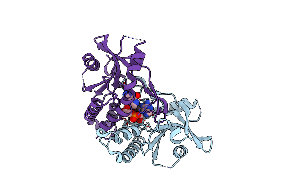 |
Human Sting G230A/R293Q Variant Bound To Cgamp
Organism: Homo sapiens
Method: X-RAY DIFFRACTION Resolution:2.22 Å Release Date: 2025-06-18 Classification: MEMBRANE PROTEIN Ligands: 1SY |
Organism: Homo sapiens
Method: X-RAY DIFFRACTION
Release Date: 2025-06-18
Ligands: 1SY
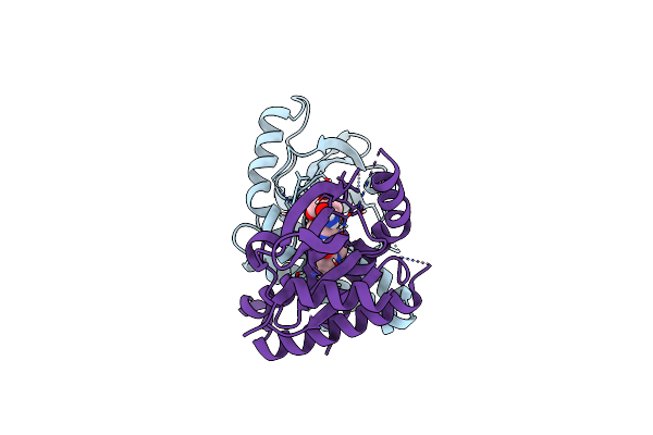 |
Human Sting G230A/R293Q Variant Bound To Diabzi-A1
Organism: Homo sapiens
Method: X-RAY DIFFRACTION Resolution:1.87 Å Release Date: 2025-06-18 Classification: MEMBRANE PROTEIN Ligands: A1A3A |
Organism: Homo sapiens
Method: X-RAY DIFFRACTION
Release Date: 2025-06-18
Ligands: A1A3A
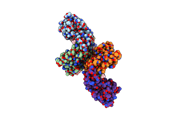 |
Human Sting G230A/R293Q Variant Bound To Thiqi
Organism: Homo sapiens
Method: X-RAY DIFFRACTION Resolution:2.10 Å Release Date: 2025-06-18 Classification: MEMBRANE PROTEIN Ligands: A1A4R |
Organism: Homo sapiens
Method: X-RAY DIFFRACTION
Release Date: 2025-06-18
Ligands: A1A4R
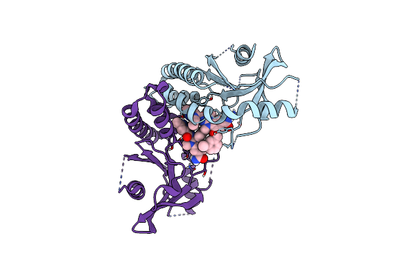 |
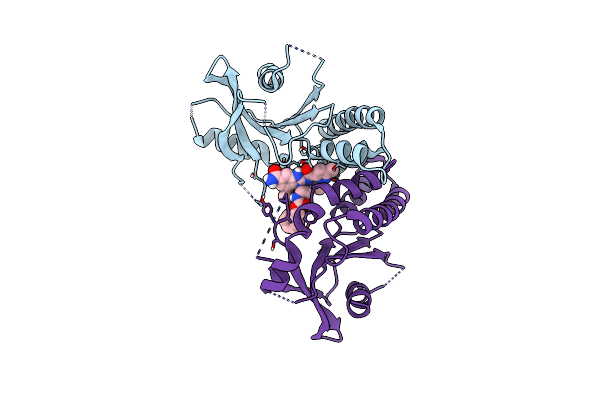
Human Sting G230A/R293Q Variant Bound To Diabzi-I
Organism: Homo sapiens
Method: X-RAY DIFFRACTION Resolution:1.53 Å Release Date: 2025-06-18 Classification: MEMBRANE PROTEIN Ligands: A1A4W |
Organism: Homo sapiens
Method: X-RAY DIFFRACTION
Release Date: 2025-06-18
Ligands: A1A4W
 |
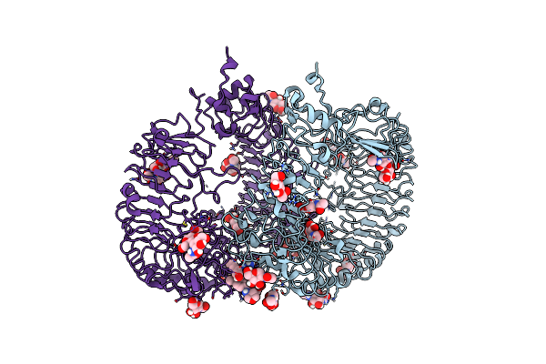
Human Sting H232R Variant Bound To Abzi
Organism: Homo sapiens
Method: X-RAY DIFFRACTION Resolution:1.95 Å Release Date: 2025-06-18 Classification: MEMBRANE PROTEIN Ligands: A1A4X |
Organism: Homo sapiens
Method: X-RAY DIFFRACTION
Release Date: 2025-06-18
Ligands: A1A4X
 |
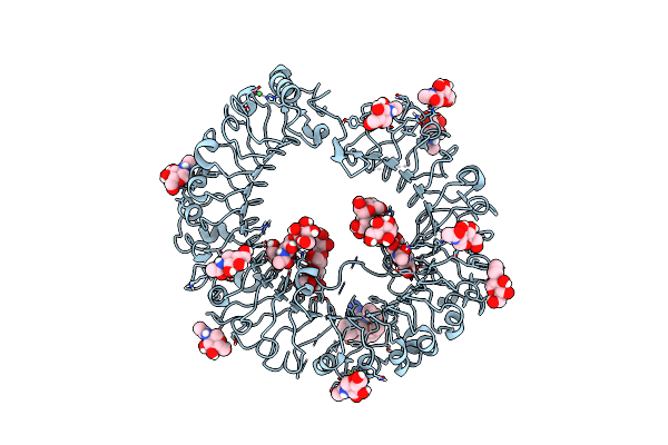
Monkey Tlr7 Ectodomain With Small Molecule Agonist 9
Organism: Macaca mulatta
Method: X-RAY DIFFRACTION Resolution:2.45 Å Release Date: 2025-03-05 Classification: IMMUNE SYSTEM Ligands: NAG, CL, A1BLT, CIT |
Organism: Macaca mulatta
Method: X-RAY DIFFRACTION
Release Date: 2025-03-05
Ligands: NAG, CL, A1BLT, CIT
 |
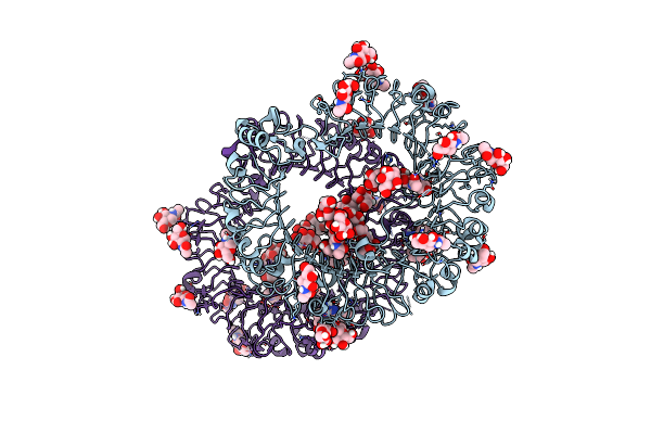
Human Tlr8 Ectodomain With Small Molecule Agonist 9
Organism: Homo sapiens
Method: X-RAY DIFFRACTION Resolution:1.52 Å Release Date: 2025-03-05 Classification: IMMUNE SYSTEM Ligands: NAG, NI, A1BLT |
Organism: Homo sapiens
Method: X-RAY DIFFRACTION
Release Date: 2025-03-05
Ligands: NAG, NI, A1BLT
 |
Human Tlr8 Ectodomain With Small Molecule Agonist 7
Organism: Homo sapiens
Method: X-RAY DIFFRACTION Resolution:2.13 Å Release Date: 2025-03-05 Classification: IMMUNE SYSTEM Ligands: NAG, A1BLO |
Organism: Homo sapiens
Method: X-RAY DIFFRACTION
Release Date: 2025-03-05
Ligands: NAG, A1BLO
 |
Human Tlr8 Ectodomain With Small Molecule Agonist 1
Organism: Homo sapiens
Method: X-RAY DIFFRACTION Resolution:1.66 Å Release Date: 2025-03-05 Classification: IMMUNE SYSTEM Ligands: NAG, SO4, A1BLN |
Organism: Homo sapiens
Method: X-RAY DIFFRACTION
Release Date: 2025-03-05
Ligands: NAG, SO4, A1BLN
 |
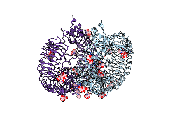
Crystal Structure Of Monkey Tlr7 Ectodomain With Compound 14
Organism: Macaca mulatta
Method: X-RAY DIFFRACTION Resolution:2.44 Å Release Date: 2024-02-07 Classification: IMMUNE SYSTEM Ligands: NAG, ZWV, SO4 |
Organism: Macaca mulatta
Method: X-RAY DIFFRACTION
Release Date: 2024-02-07
Ligands: NAG, ZWV, SO4
 |
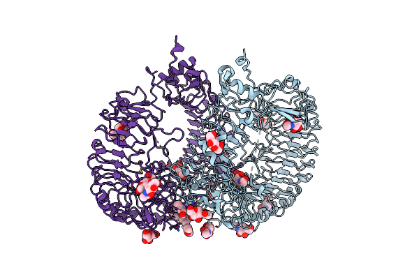
Crystal Structure Of Monkey Tlr7 Ectodomain With Compound 5
Organism: Macaca mulatta
Method: X-RAY DIFFRACTION Resolution:2.10 Å Release Date: 2024-02-07 Classification: IMMUNE SYSTEM Ligands: NAG, QEC, SO4 |
Organism: Macaca mulatta
Method: X-RAY DIFFRACTION
Release Date: 2024-02-07
Ligands: NAG, QEC, SO4
 |
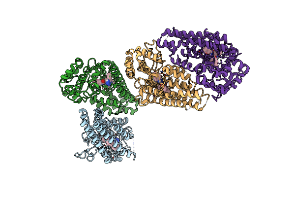
Crystal Structure Of Monkey Tlr7 Ectodomain With Compound 20
Organism: Macaca mulatta
Method: X-RAY DIFFRACTION Resolution:3.14 Å Release Date: 2024-02-07 Classification: IMMUNE SYSTEM Ligands: NAG, SO4, QCX |
Organism: Macaca mulatta
Method: X-RAY DIFFRACTION
Release Date: 2024-02-07
Ligands: NAG, SO4, QCX
 |
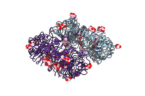
Crystal Structure Of Human Ido1 With Compound 11
Organism: Homo sapiens
Method: X-RAY DIFFRACTION Resolution:2.29 Å Release Date: 2023-04-26 Classification: OXIDOREDUCTASE/INHIBITOR Ligands: Y9X |
Organism: Homo sapiens
Method: X-RAY DIFFRACTION
Release Date: 2023-04-26
Ligands: Y9X
 |
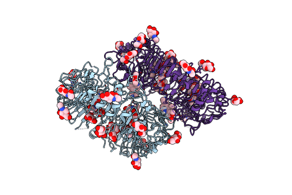
Crystal Structure Of Human Tlr8 Ectodomain Bound To Small Molecule Antagonist 21
Organism: Homo sapiens
Method: X-RAY DIFFRACTION Resolution:2.76 Å Release Date: 2022-05-04 Classification: RNA BINDING PROTEIN Ligands: NAG, RQY |
Organism: Homo sapiens
Method: X-RAY DIFFRACTION
Release Date: 2022-05-04
Ligands: NAG, RQY
 |
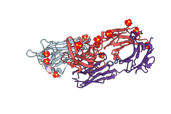
Crystal Structure Of Human Tlr8 Ectodomain Bound To Small Molecule Antagonist 14C
Organism: Homo sapiens
Method: X-RAY DIFFRACTION Resolution:2.65 Å Release Date: 2020-08-12 Classification: RNA BINDING PROTEIN Ligands: NAG, QSM |
Organism: Homo sapiens
Method: X-RAY DIFFRACTION
Release Date: 2020-08-12
Ligands: NAG, QSM
 |
Crystal Structure Of Vista Bound To A Ph-Selective Antibody Fab Fragment
Organism: Homo sapiens
Method: X-RAY DIFFRACTION Resolution:1.61 Å Release Date: 2019-10-23 Classification: IMMUNE SYSTEM Ligands: NAG, SO4 |
Organism: Homo sapiens
Method: X-RAY DIFFRACTION
Release Date: 2019-10-23
Ligands: NAG, SO4
 |
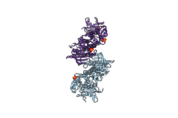
Tyrosine-Protein Kinase Lck Bound To Compound 11
Organism: Homo sapiens
Method: X-RAY DIFFRACTION Resolution:1.81 Å Release Date: 2019-10-09 Classification: SIGNALING PROTEIN/INHIBITOR Ligands: ODJ, CXS, SO4, NI |
Organism: Homo sapiens
Method: X-RAY DIFFRACTION
Release Date: 2019-10-09
Ligands: ODJ, CXS, SO4, NI
 |
Oxidized Shp2 Forms A Disulfide Bond Between Cys367 And Cys459
Organism: Homo sapiens
Method: X-RAY DIFFRACTION Resolution:2.50 Å Release Date: 2018-07-11 Classification: HYDROLASE Ligands: PO4 |
Organism: Homo sapiens
Method: X-RAY DIFFRACTION
Release Date: 2018-07-11
Ligands: PO4
 |
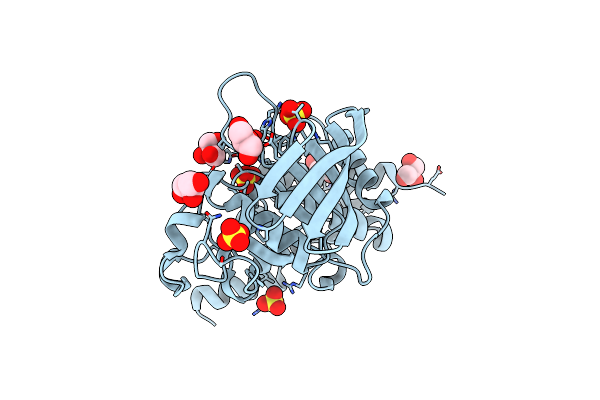
Crystal Structure Of A Braf Kinase Domain Monomer
Organism: Homo sapiens
Method: X-RAY DIFFRACTION Resolution:2.83 Å Release Date: 2014-12-03 Classification: TRANSFERASE/TRANSFERASE Inhibitor Ligands: 324 |
Organism: Homo sapiens
Method: X-RAY DIFFRACTION
Release Date: 2014-12-03
Ligands: 324
 |
Crystal Structure Of Heptp With A Closed Wpd Loop And An Ordered E-Loop
Organism: Homo sapiens
Method: X-RAY DIFFRACTION Resolution:1.90 Å Release Date: 2010-11-24 Classification: HYDROLASE Ligands: SO4, GOL |
Organism: Homo sapiens
Method: X-RAY DIFFRACTION
Release Date: 2010-11-24
Ligands: SO4, GOL