

Search Count: 137
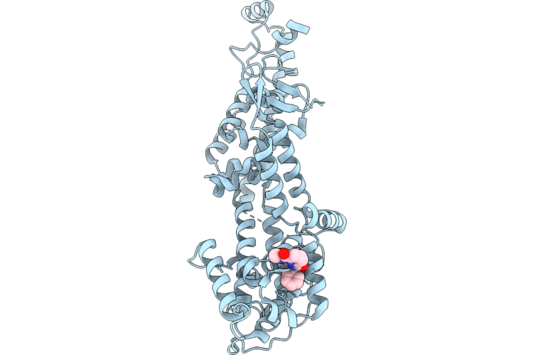 |
Sos1 In Complex With Compound 3
Organism: Homo sapiens
Method: X-RAY DIFFRACTION Resolution:1.92 Å Release Date: 2026-06-24 Classification: SIGNALING PROTEIN Ligands: A1JWK |
Organism: Homo sapiens
Method: X-RAY DIFFRACTION
Release Date: 2026-06-24
Ligands: A1JWK
 |
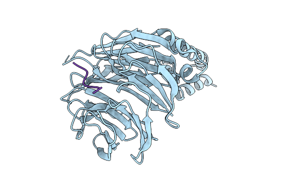
Crystal Structure Of Beta-Trcp Bound By Monophosphorylated Nrf2 Degron Peptide.
Organism: Homo sapiens
Method: X-RAY DIFFRACTION Resolution:1.80 Å Release Date: 2026-04-08 Classification: LIGASE |
Organism: Homo sapiens
Method: X-RAY DIFFRACTION
Release Date: 2026-04-08
 |
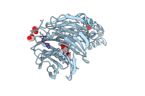
Crystal Structure Of Beta-Trcp Bound By Diphosphorylated Nrf2 Degron Peptide.
Organism: Homo sapiens
Method: X-RAY DIFFRACTION Resolution:2.15 Å Release Date: 2026-04-08 Classification: LIGASE |
Organism: Homo sapiens
Method: X-RAY DIFFRACTION
Release Date: 2026-04-08
 |
Crystal Structure Of Beta-Trcp Bound By Diphosphorylated I-Kappa-B-Alpha Degron Peptide
Organism: Homo sapiens
Method: X-RAY DIFFRACTION Resolution:1.16 Å Release Date: 2026-04-08 Classification: TRANSFERASE Ligands: EDO |
Organism: Homo sapiens
Method: X-RAY DIFFRACTION
Release Date: 2026-04-08
Ligands: EDO
 |
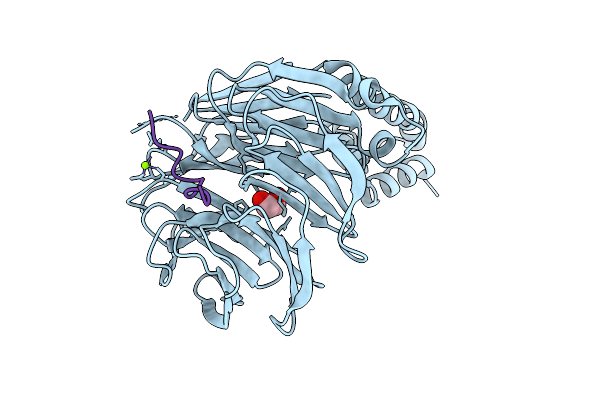
Crystal Structure Of Beta-Trcp Bound By Diphosphorylated Claspin Degron Peptide
Organism: Homo sapiens
Method: X-RAY DIFFRACTION Resolution:1.35 Å Release Date: 2026-04-08 Classification: TRANSFERASE Ligands: EDO, MG |
Organism: Homo sapiens
Method: X-RAY DIFFRACTION
Release Date: 2026-04-08
Ligands: EDO, MG
 |
Crystal Structure Of Beta-Trcp Bound By Diphosphorylated Pdcd4 Degron Peptide
Organism: Homo sapiens
Method: X-RAY DIFFRACTION Resolution:1.22 Å Release Date: 2026-04-08 Classification: TRANSFERASE Ligands: MG, EDO |
Organism: Homo sapiens
Method: X-RAY DIFFRACTION
Release Date: 2026-04-08
Ligands: MG, EDO
 |
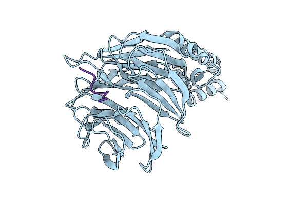
Crystal Structure Of Beta-Trcp Bound By Monophosphorylated Atf4 Degron Peptide
Organism: Homo sapiens
Method: X-RAY DIFFRACTION Resolution:2.00 Å Release Date: 2026-04-08 Classification: TRANSFERASE |
Organism: Homo sapiens
Method: X-RAY DIFFRACTION
Release Date: 2026-04-08
 |
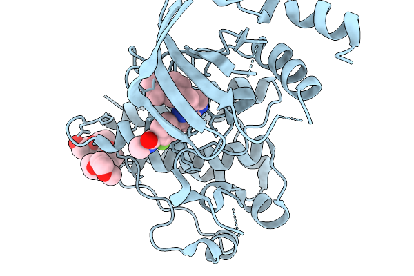
Crystal Structure Of Beta-Trcp Bound By Monophosphorylated Wee1 Degron Peptide
Organism: Homo sapiens
Method: X-RAY DIFFRACTION Resolution:1.68 Å Release Date: 2026-04-08 Classification: TRANSFERASE |
Organism: Homo sapiens
Method: X-RAY DIFFRACTION
Release Date: 2026-04-08
 |
Crystal Structure Of Wild-Type C-Met Bound By Capmatinib.
Organism: Homo sapiens
Method: X-RAY DIFFRACTION Resolution:1.32 Å Release Date: 2026-02-18 Classification: TRANSFERASE Ligands: A1JRF, 12P |
Organism: Homo sapiens
Method: X-RAY DIFFRACTION
Release Date: 2026-02-18
Ligands: A1JRF, 12P
 |
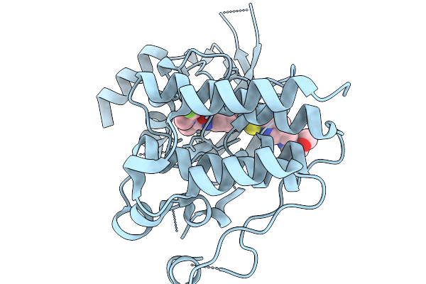
Crystal Structure Of Y1230H C-Met Bound By Capmatinib.
Organism: Homo sapiens
Method: X-RAY DIFFRACTION Resolution:2.29 Å Release Date: 2026-02-18 Classification: TRANSFERASE Ligands: A1JRF |
Organism: Homo sapiens
Method: X-RAY DIFFRACTION
Release Date: 2026-02-18
Ligands: A1JRF
 |
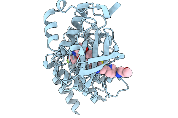
Crystal Structure Of Wild-Type C-Met Bound By Sitravatinib.
Organism: Homo sapiens
Method: X-RAY DIFFRACTION Resolution:1.46 Å Release Date: 2026-02-18 Classification: TRANSFERASE Ligands: A1JSO |
Organism: Homo sapiens
Method: X-RAY DIFFRACTION
Release Date: 2026-02-18
Ligands: A1JSO
 |
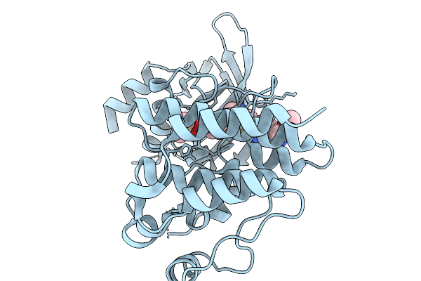
Crystal Structure Of D1228V C-Met Bound By Sitravatinib.
Organism: Homo sapiens
Method: X-RAY DIFFRACTION Resolution:1.54 Å Release Date: 2026-02-18 Classification: TRANSFERASE Ligands: A1JSO |
Organism: Homo sapiens
Method: X-RAY DIFFRACTION
Release Date: 2026-02-18
Ligands: A1JSO
 |
Crystal Structure Of Wild-Type C-Met Bound By Glesatinib
Organism: Homo sapiens
Method: X-RAY DIFFRACTION Resolution:1.20 Å Release Date: 2026-02-18 Classification: TRANSFERASE Ligands: A1JSR |
Organism: Homo sapiens
Method: X-RAY DIFFRACTION
Release Date: 2026-02-18
Ligands: A1JSR
 |
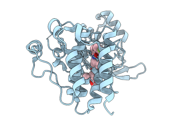
Crystal Structure Of D1228V C-Met Bound By Glesatinib.
Organism: Homo sapiens
Method: X-RAY DIFFRACTION Resolution:1.94 Å Release Date: 2026-02-18 Classification: TRANSFERASE Ligands: A1JSR |
Organism: Homo sapiens
Method: X-RAY DIFFRACTION
Release Date: 2026-02-18
Ligands: A1JSR
 |
Crystal Structure Of Wild-Type C-Met Bound By Cabozantinib.
Organism: Homo sapiens
Method: X-RAY DIFFRACTION Resolution:1.67 Å Release Date: 2026-02-18 Classification: TRANSFERASE Ligands: A1JS8 |
Organism: Homo sapiens
Method: X-RAY DIFFRACTION
Release Date: 2026-02-18
Ligands: A1JS8
 |
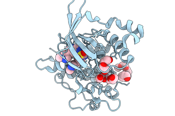
Crystal Structure Of D1228V C-Met Bound By Cabozantinib.
Organism: Homo sapiens
Method: X-RAY DIFFRACTION Resolution:1.63 Å Release Date: 2026-02-18 Classification: TRANSFERASE Ligands: A1JS8 |
Organism: Homo sapiens
Method: X-RAY DIFFRACTION
Release Date: 2026-02-18
Ligands: A1JS8
 |
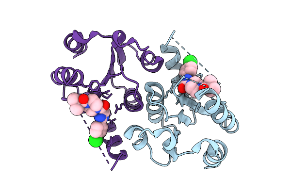
Crystal Structure Of Wild-Type C-Met Bound By Glumetinib.
Organism: Homo sapiens
Method: X-RAY DIFFRACTION Resolution:1.13 Å Release Date: 2026-02-18 Classification: TRANSFERASE Ligands: A1JT6, 15P, PEG |
Organism: Homo sapiens
Method: X-RAY DIFFRACTION
Release Date: 2026-02-18
Ligands: A1JT6, 15P, PEG
 |
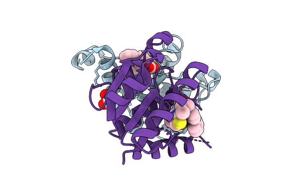
Crystal Structure Of Dnph1 Bound By Compound 3
Organism: Homo sapiens
Method: X-RAY DIFFRACTION Resolution:2.02 Å Release Date: 2025-12-24 Classification: HYDROLASE Ligands: A1JH5 |
Organism: Homo sapiens
Method: X-RAY DIFFRACTION
Release Date: 2025-12-24
Ligands: A1JH5
 |
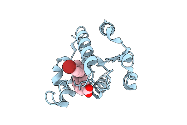
Crystal Structure Of Dnph1 Bound By Compound 6
Organism: Homo sapiens
Method: X-RAY DIFFRACTION Resolution:1.51 Å Release Date: 2025-12-24 Classification: HYDROLASE Ligands: A1JKT, EDO |
Organism: Homo sapiens
Method: X-RAY DIFFRACTION
Release Date: 2025-12-24
Ligands: A1JKT, EDO
 |
Crystal Structure Of Dnph1 Bound By Compound 19
Organism: Homo sapiens
Method: X-RAY DIFFRACTION Resolution:1.50 Å Release Date: 2025-12-24 Classification: HYDROLASE Ligands: A1JKZ |
Organism: Homo sapiens
Method: X-RAY DIFFRACTION
Release Date: 2025-12-24
Ligands: A1JKZ