

Search Count: 15
 |
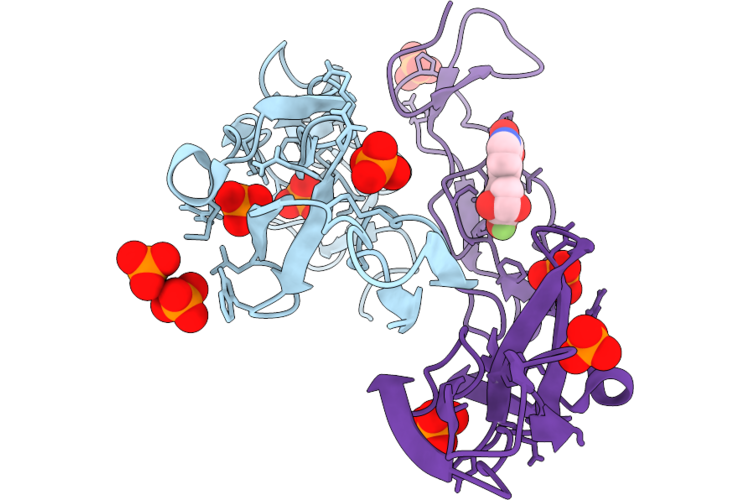
Crystal Structure Of The Human Tumor Necrosis Factor Receptor 1 Extracellular Domain At 1.64A Resolution
Organism: Homo sapiens
Method: X-RAY DIFFRACTION Resolution:1.64 Å Release Date: 2026-07-01 Classification: SIGNALING PROTEIN Ligands: PO4 |
 |
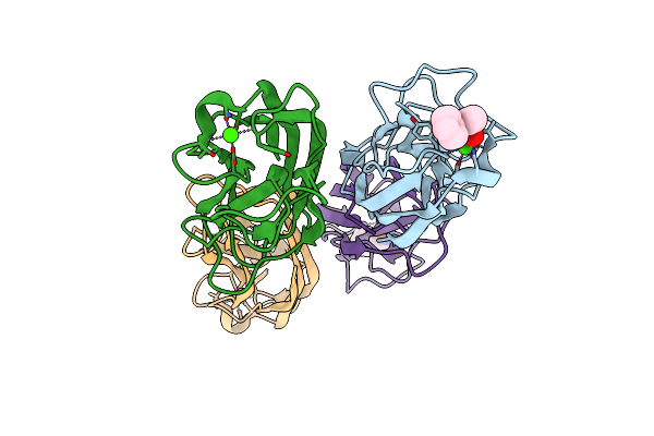
Crystal Structure Of The Human Tumor Necrosis Factor Receptor 1 Extracellular Domain In Complex With A Picolinic Acid Based Ligand At 1.77A Resolution
Organism: Homo sapiens
Method: X-RAY DIFFRACTION Resolution:1.77 Å Release Date: 2026-07-01 Classification: SIGNALING PROTEIN Ligands: PO4, A1JLJ |
 |
Leca From Pseudomonas Aeruginosa In Complex With 4-Phenylbutyryl Hydroxamic Acid (Cas: 32153-46-1)
Organism: Pseudomonas aeruginosa
Method: X-RAY DIFFRACTION Resolution:1.79 Å Release Date: 2022-06-08 Classification: SUGAR BINDING PROTEIN Ligands: 4R9, CA |
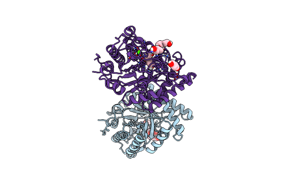 |
Ignisphaera Aggregans Gh53 Catalytic Domain
Organism: Ignisphaera aggregans (strain dsm 17230 / jcm 13409 / aq1.s1)
Method: X-RAY DIFFRACTION Resolution:2.65 Å Release Date: 2021-09-29 Classification: HYDROLASE Ligands: CA, HEZ, CL |
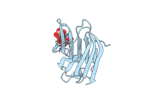 |
Crystal Structure Of Galectin-3 Crd In Complex With Galactopentaose
Organism: Homo sapiens
Method: X-RAY DIFFRACTION Resolution:1.41 Å Release Date: 2018-12-19 Classification: SUGAR BINDING PROTEIN |
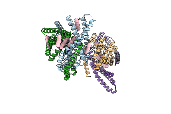 |
Crystal Structure Of Human Two Pore Domain Potassium Ion Channel Trek2 (K2P10.1) In An Alternate Conformation (Form 2)
Organism: Homo sapiens
Method: X-RAY DIFFRACTION Resolution:3.80 Å Release Date: 2015-03-18 Classification: TRANSPORT PROTEIN Ligands: K, PC1, TRD |
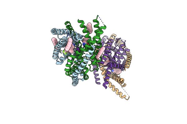 |
Crystal Structure Of Human Two Pore Domain Potassium Ion Channel Trek2 (K2P10.1) In Complex With Norfluoxetine
Organism: Homo sapiens
Method: X-RAY DIFFRACTION Resolution:3.60 Å Release Date: 2015-03-18 Classification: TRANSPORT PROTEIN Ligands: K, PC1, 408, 405 |
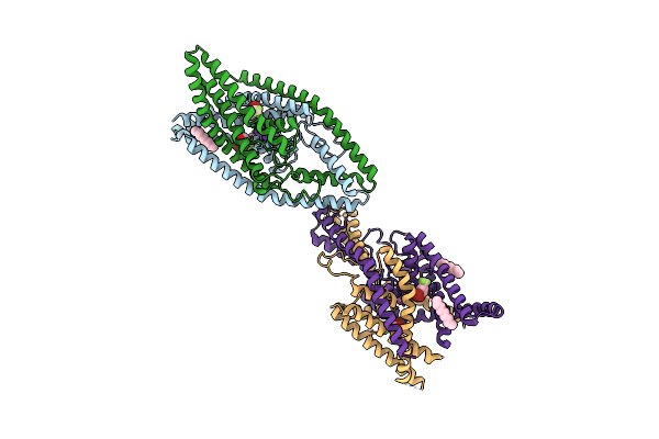 |
Crystal Structure Of Human Two Pore Domain Potassium Ion Channel Trek2 (K2P10.1) In Complex With A Brominated Fluoxetine Derivative.
Organism: Homo sapiens
Method: X-RAY DIFFRACTION Resolution:3.50 Å Release Date: 2015-03-18 Classification: TRANSPORT PROTEIN Ligands: K, PC1, CD, 40D |
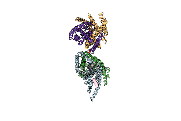 |
Crystal Structure Of Human Two Pore Domain Potassium Ion Channel Trek2 (K2P10.1)
Organism: Homo sapiens
Method: X-RAY DIFFRACTION Resolution:3.20 Å Release Date: 2013-07-31 Classification: TRANSPORT PROTEIN Ligands: K, CD, PC1 |
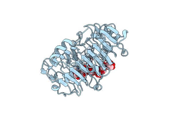 |
Structural Insights Into Substrate Specificity And The Anti Beta-Elimination Mechanism Of Pectate Lyase
Organism: Bacillus subtilis
Method: X-RAY DIFFRACTION Resolution:1.90 Å Release Date: 2010-02-09 Classification: LYASE Ligands: CO, GOL |
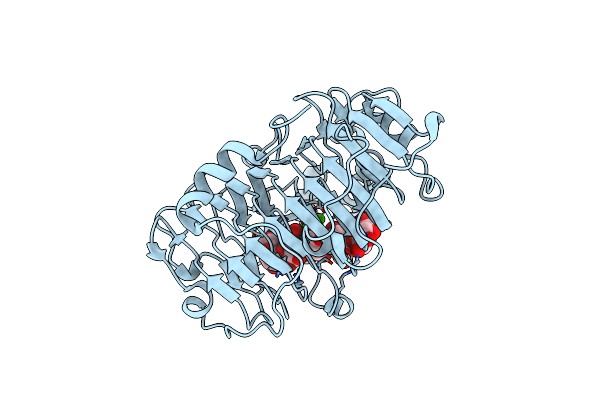 |
Hexasaccharide I Bound To Bacillus Subtilis Pectate Lyase
Organism: Bacillus subtilis
Method: X-RAY DIFFRACTION Resolution:1.80 Å Release Date: 2007-11-20 Classification: LYASE Ligands: CA |
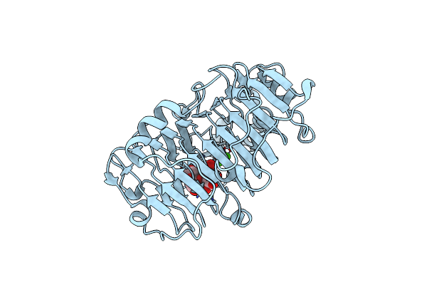 |
Pectate Lyase Bound To Hexasaccharide Compound Ii
Organism: Bacillus subtilis
Method: X-RAY DIFFRACTION Resolution:1.70 Å Release Date: 2007-11-20 Classification: LYASE Ligands: CA |
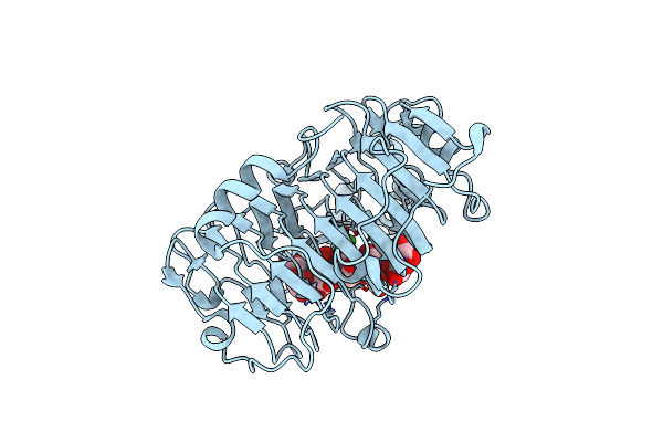 |
Pectate Lyase Bound To Hexasaccharide Compound Iii
Organism: Bacillus subtilis
Method: X-RAY DIFFRACTION Resolution:1.90 Å Release Date: 2007-11-20 Classification: LYASE Ligands: CA |
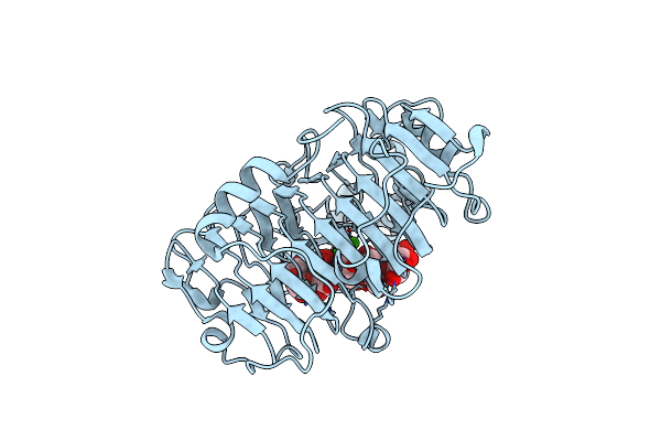 |
Pectate Lyase Bound To Hexasaccharide
Organism: Bacillus subtilis
Method: X-RAY DIFFRACTION Resolution:2.30 Å Release Date: 2007-11-20 Classification: LYASE Ligands: CA |
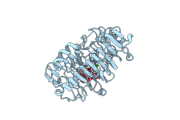 |
Pectate Lyase Bound To Trisaccharide
Organism: Bacillus subtilis
Method: X-RAY DIFFRACTION Resolution:2.00 Å Release Date: 2007-11-20 Classification: LYASE Ligands: CA |