

Search Count: 44
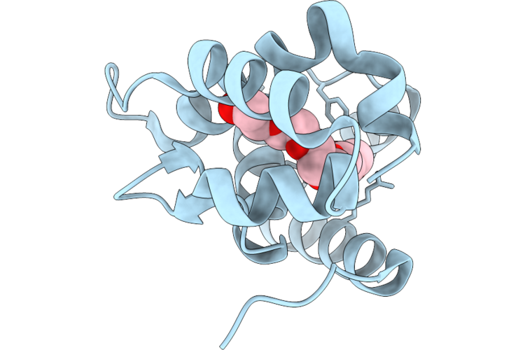 |
Structure Of Anopheles Gambiae Obp9 In Complex With Peg
Organism: Anopheles gambiae
Method: X-RAY DIFFRACTION Resolution:1.20 Å Release Date: 2026-06-24 Classification: TRANSPORT PROTEIN Ligands: P6G |
Organism: Anopheles gambiae
Method: X-RAY DIFFRACTION
Release Date: 2026-06-24
Ligands: P6G
 |
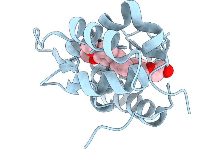
Structure Of Anopheles Gambiae Obp9 In Complex With N-Butyl Cinnamate
Organism: Anopheles gambiae
Method: X-RAY DIFFRACTION Resolution:1.20 Å Release Date: 2026-06-24 Classification: TRANSPORT PROTEIN Ligands: MRD, A1J28, MG |
Organism: Anopheles gambiae
Method: X-RAY DIFFRACTION
Release Date: 2026-06-24
Ligands: MRD, A1J28, MG
 |
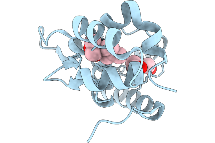
Structure Of Anopheles Gambiae Obp9 In Complex With Methyl Eugenol
Organism: Anopheles gambiae
Method: X-RAY DIFFRACTION Resolution:1.20 Å Release Date: 2026-06-24 Classification: TRANSPORT PROTEIN Ligands: A1J27, PG4 |
Organism: Anopheles gambiae
Method: X-RAY DIFFRACTION
Release Date: 2026-06-24
Ligands: A1J27, PG4
 |
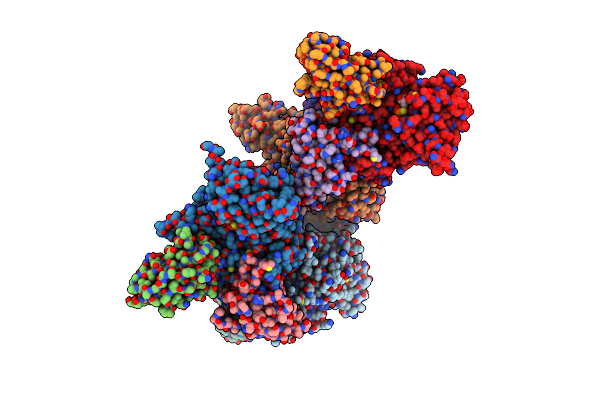
Structure Of Anopheles Gambiae Obp9 In Complex With Pmd (P-Menthane-3,8-Diol)
Organism: Anopheles gambiae
Method: X-RAY DIFFRACTION Resolution:1.20 Å Release Date: 2026-06-24 Classification: TRANSPORT PROTEIN Ligands: A1J26, PGE |
Organism: Anopheles gambiae
Method: X-RAY DIFFRACTION
Release Date: 2026-06-24
Ligands: A1J26, PGE
 |
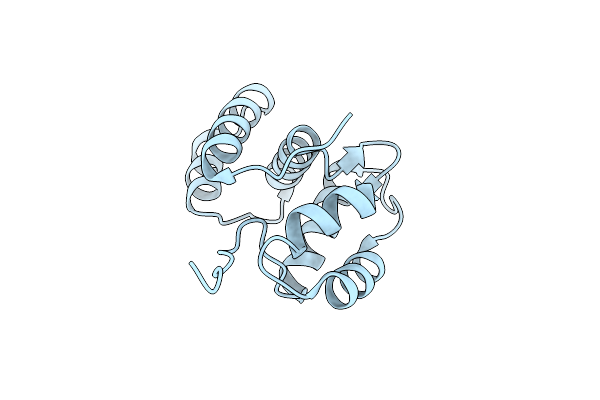
P. Falciparum Fikk13 In Complex With Atpgammas
Organism: Plasmodium falciparum, Lama glama
Method: X-RAY DIFFRACTION Resolution:2.81 Å Release Date: 2025-03-26 Classification: PEPTIDE BINDING PROTEIN Ligands: AGS, PE4 |
Organism: Plasmodium falciparum, Lama glama
Method: X-RAY DIFFRACTION
Release Date: 2025-03-26
Ligands: AGS, PE4
 |
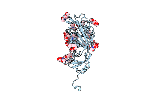
Crystal Structure Of Odorant Binding Protein 1 From Aedes Albopictus (Asian Tiger Mosquito)
Organism: Aedes albopictus
Method: X-RAY DIFFRACTION Resolution:2.00 Å Release Date: 2023-10-18 Classification: TRANSPORT PROTEIN |
Organism: Aedes albopictus
Method: X-RAY DIFFRACTION
Release Date: 2023-10-18
 |
Crystal Structure Of Sars-Cov-2 Spike Protein N-Terminal Domain In Complex With Biliverdin
Organism: Severe acute respiratory syndrome coronavirus 2
Method: X-RAY DIFFRACTION Resolution:1.82 Å Release Date: 2021-04-28 Classification: VIRAL PROTEIN Ligands: BLA, NAG, PG4, PEG, PGE, 1PE |
Organism: Severe acute respiratory syndrome coronavirus 2
Method: X-RAY DIFFRACTION
Release Date: 2021-04-28
Ligands: BLA, NAG, PG4, PEG, PGE, 1PE
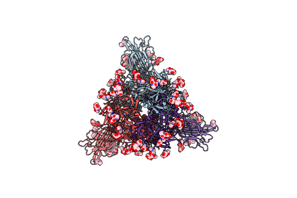 |
Trimeric Sars-Cov-2 Spike Ectodomain In Complex With Biliverdin (Closed Conformation)
Organism: Severe acute respiratory syndrome coronavirus 2
Method: ELECTRON MICROSCOPY Release Date: 2021-04-28 Classification: VIRAL PROTEIN Ligands: BLA, NAG |
Organism: Severe acute respiratory syndrome coronavirus 2
Method: ELECTRON MICROSCOPY
Release Date: 2021-04-28
Ligands: BLA, NAG
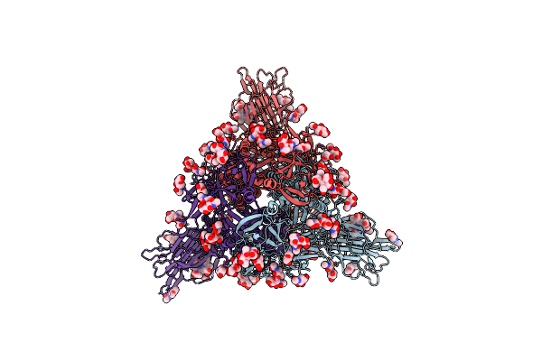 |
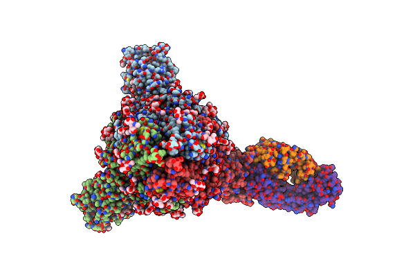
Trimeric Sars-Cov-2 Spike Ectodomain In Complex With Biliverdin (One Rbd Erect)
Organism: Severe acute respiratory syndrome coronavirus 2
Method: ELECTRON MICROSCOPY Release Date: 2021-04-28 Classification: VIRAL PROTEIN Ligands: BLA, NAG |
Organism: Severe acute respiratory syndrome coronavirus 2
Method: ELECTRON MICROSCOPY
Release Date: 2021-04-28
Ligands: BLA, NAG
 |
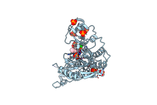
Trimeric Sars-Cov-2 Spike Ectodomain Bound To P008_056 Fab
Organism: Severe acute respiratory syndrome coronavirus 2, Homo sapiens
Method: ELECTRON MICROSCOPY Release Date: 2021-04-28 Classification: VIRAL PROTEIN Ligands: BLA, NAG |
Organism: Severe acute respiratory syndrome coronavirus 2, Homo sapiens
Method: ELECTRON MICROSCOPY
Release Date: 2021-04-28
Ligands: BLA, NAG
 |
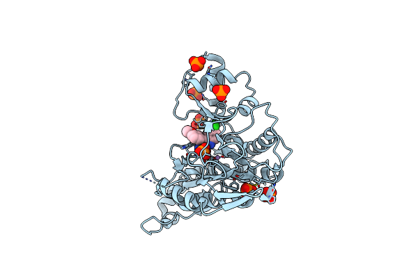
Crystal Structure Of Human Methionine Aminopeptidase-2 In Complex With An Inhibitor Gw557358X (Compound 9)
Organism: Homo sapiens
Method: X-RAY DIFFRACTION Resolution:2.00 Å Release Date: 2020-09-23 Classification: HYDROLASE Ligands: PO4, QVK, MN |
Organism: Homo sapiens
Method: X-RAY DIFFRACTION
Release Date: 2020-09-23
Ligands: PO4, QVK, MN
 |
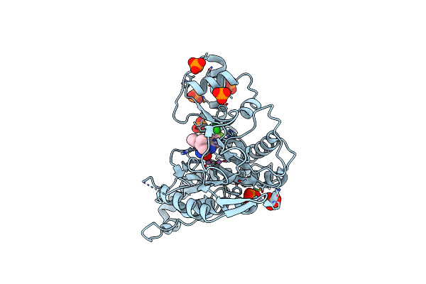
Crystal Structure Of Human Methionine Aminopeptidase-2 In Complex With An Inhibitor Gsk1978537A (Compound 27)
Organism: Homo sapiens
Method: X-RAY DIFFRACTION Resolution:2.04 Å Release Date: 2020-09-23 Classification: HYDROLASE Ligands: PO4, QW5, MN |
Organism: Homo sapiens
Method: X-RAY DIFFRACTION
Release Date: 2020-09-23
Ligands: PO4, QW5, MN
 |
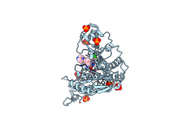
Crystal Structure Of Human Methionine Aminopeptidase-2 In Complex With An Inhibitor Gsk2218325A (Compound 32)
Organism: Homo sapiens
Method: X-RAY DIFFRACTION Resolution:2.14 Å Release Date: 2020-09-23 Classification: HYDROLASE Ligands: PO4, QVB, MN |
Organism: Homo sapiens
Method: X-RAY DIFFRACTION
Release Date: 2020-09-23
Ligands: PO4, QVB, MN
 |
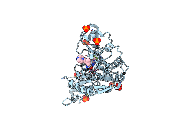
Crystal Structure Of Human Methionine Aminopeptidase-2 In Complex With An Inhibitor Gsk2224863A (Compound 42)
Organism: Homo sapiens
Method: X-RAY DIFFRACTION Resolution:2.15 Å Release Date: 2020-09-23 Classification: HYDROLASE Ligands: PO4, QV5, MN |
Organism: Homo sapiens
Method: X-RAY DIFFRACTION
Release Date: 2020-09-23
Ligands: PO4, QV5, MN
 |
Crystal Structure Of Human Methionine Aminopeptidase-2 In Complex With An Inhibitor Gsk2229238A (Compound 43)
Organism: Homo sapiens
Method: X-RAY DIFFRACTION Resolution:1.90 Å Release Date: 2020-09-23 Classification: HYDROLASE Ligands: PO4, HZE, MN |
Organism: Homo sapiens
Method: X-RAY DIFFRACTION
Release Date: 2020-09-23
Ligands: PO4, HZE, MN
 |
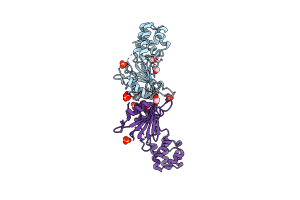
Solution Structure Of The Ashh2 Cw Domain With The N-Terminal Histone H3 Tail Mimicking Peptide Monomethylated On Lysine 4
Organism: Arabidopsis thaliana, Homo sapiens
Method: SOLUTION NMR Release Date: 2019-12-04 Classification: TRANSFERASE Ligands: ZN |
Organism: Arabidopsis thaliana, Homo sapiens
Method: SOLUTION NMR
Release Date: 2019-12-04
Ligands: ZN
 |
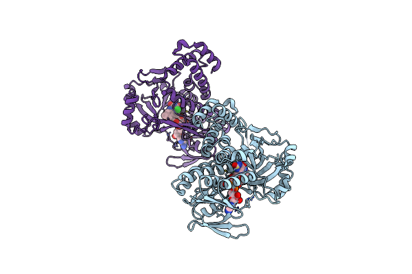
A Cryptic Rna-Binding Domain Mediates Syncrip Recognition And Exosomal Partitioning Of Mirna Targets
Organism: Drosophila melanogaster
Method: X-RAY DIFFRACTION Resolution:2.20 Å Release Date: 2018-03-07 Classification: RNA Ligands: SO4, EDO |
Organism: Drosophila melanogaster
Method: X-RAY DIFFRACTION
Release Date: 2018-03-07
Ligands: SO4, EDO
 |
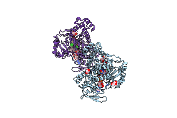
Pseudomonas Fluorescens Kynurenine 3-Monooxygenase (Kmo) Apo Structure
Organism: Pseudomonas fluorescens
Method: X-RAY DIFFRACTION Resolution:1.94 Å Release Date: 2017-06-21 Classification: OXIDOREDUCTASE Ligands: FAD, CL, GOL |
Organism: Pseudomonas fluorescens
Method: X-RAY DIFFRACTION
Release Date: 2017-06-21
Ligands: FAD, CL, GOL
 |
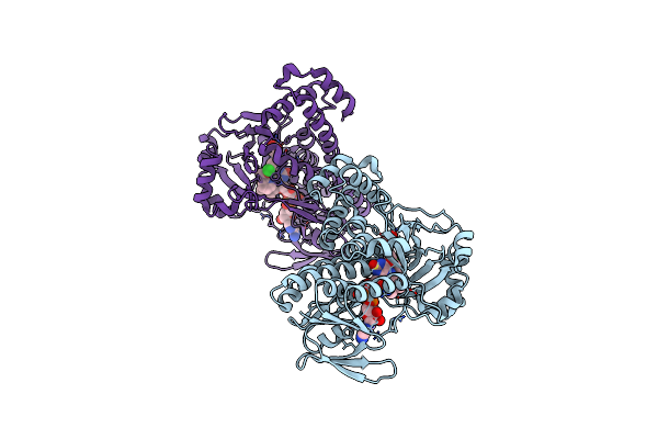
Pseudomonas Fluorescens Kynurenine 3-Monooxygenase (Kmo) In Complex With 3-(5-Chloro-6-Methyl-2-Oxo-2,3-Dihydro-1,3-Benzoxazol-3-Yl)Propanoic Acid
Organism: Pseudomonas fluorescens
Method: X-RAY DIFFRACTION Resolution:1.63 Å Release Date: 2017-06-21 Classification: OXIDOREDUCTASE Ligands: FAD, CL, 8RK, GOL |
Organism: Pseudomonas fluorescens
Method: X-RAY DIFFRACTION
Release Date: 2017-06-21
Ligands: FAD, CL, 8RK, GOL
 |
Pseudomonas Fluorescens Kynurenine 3-Monooxygenase (Kmo) In Complex With 3-{5-Chloro-2-Oxo-6-[(1R)-1-(Pyridin-2-Yl)Ethoxy]-2,3-Dihydro-1,3-Benzoxazol-3-Yl}Propanoic Acid
Organism: Pseudomonas fluorescens
Method: X-RAY DIFFRACTION Resolution:1.76 Å Release Date: 2017-06-21 Classification: OXIDOREDUCTASE Ligands: FAD, 8R8 |
Organism: Pseudomonas fluorescens
Method: X-RAY DIFFRACTION
Release Date: 2017-06-21
Ligands: FAD, 8R8