

Search Count: 100
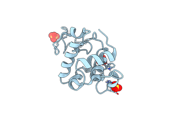 |
Structure Of The W64R Amyloidogenic Variant Of Human Lysozyme
Organism: Homo sapiens
Method: X-RAY DIFFRACTION Resolution:1.15 Å Release Date: 2021-08-25 Classification: HYDROLASE Ligands: SO4 |
Organism: Homo sapiens
Method: X-RAY DIFFRACTION
Release Date: 2021-08-25
Ligands: SO4
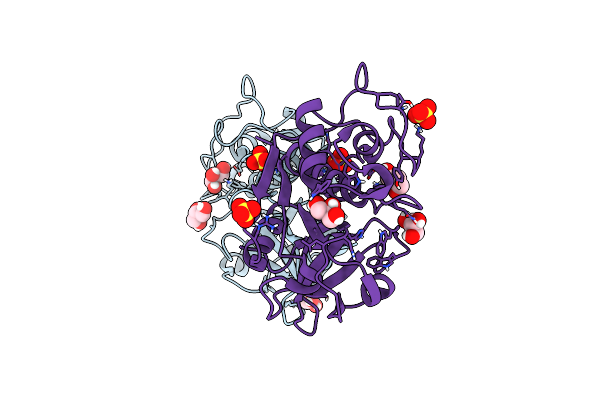 |
Structure The Ananain Protease From Ananas Comosus With A Thiomethylated Catalytic Cysteine
Organism: Ananas comosus
Method: X-RAY DIFFRACTION Resolution:1.30 Å Release Date: 2020-11-25 Classification: HYDROLASE Ligands: SO4, GOL |
Organism: Ananas comosus
Method: X-RAY DIFFRACTION
Release Date: 2020-11-25
Ligands: SO4, GOL
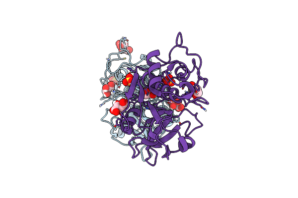 |
Structure The Ananain Protease From Ananas Comosus Covalently Bound To With The E64 Inhibitor
Organism: Ananas comosus
Method: X-RAY DIFFRACTION Resolution:1.26 Å Release Date: 2020-11-25 Classification: HYDROLASE Ligands: SO4, GOL, TLA |
Organism: Ananas comosus
Method: X-RAY DIFFRACTION
Release Date: 2020-11-25
Ligands: SO4, GOL, TLA
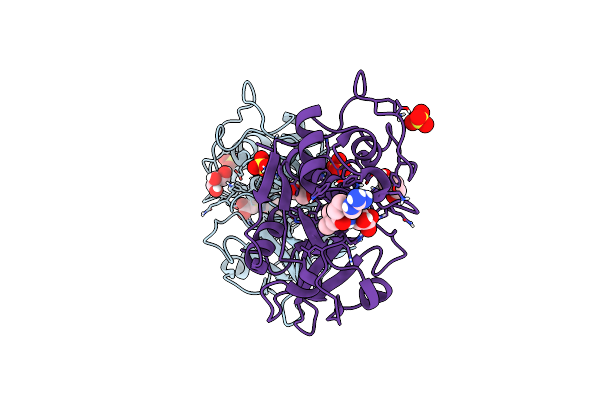 |
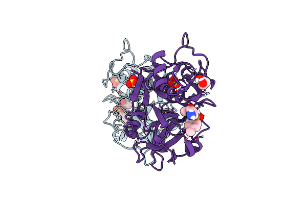
Structure The Ananain Protease From Ananas Comosus Covalently Bound To The E64 Inhibitor
Organism: Ananas comosus
Method: X-RAY DIFFRACTION Resolution:1.30 Å Release Date: 2020-11-25 Classification: HYDROLASE Ligands: E64, SO4, GOL |
Organism: Ananas comosus
Method: X-RAY DIFFRACTION
Release Date: 2020-11-25
Ligands: E64, SO4, GOL
 |
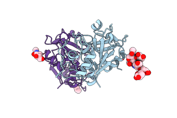
Structure The Ananain Protease From Ananas Comosus Covalently Bound To The Tlck Inhibitor
Organism: Ananas comosus
Method: X-RAY DIFFRACTION Resolution:1.35 Å Release Date: 2020-11-25 Classification: HYDROLASE Ligands: TCK, SO4, GOL |
Organism: Ananas comosus
Method: X-RAY DIFFRACTION
Release Date: 2020-11-25
Ligands: TCK, SO4, GOL
 |
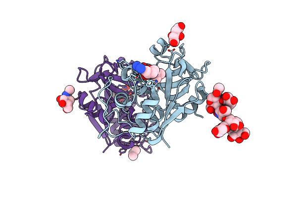
Structure The Bromelain Protease From Ananas Comosus With A Thiomethylated Active Cysteine
Organism: Ananas comosus
Method: X-RAY DIFFRACTION Resolution:1.80 Å Release Date: 2020-11-25 Classification: HYDROLASE Ligands: NAG, IPA |
Organism: Ananas comosus
Method: X-RAY DIFFRACTION
Release Date: 2020-11-25
Ligands: NAG, IPA
 |
Structure The Bromelain Protease From Ananas Comosus In Complex With The E64 Inhibitor
Organism: Ananas comosus
Method: X-RAY DIFFRACTION Resolution:1.85 Å Release Date: 2020-11-25 Classification: HYDROLASE Ligands: E64, CIT, NAG, IPA |
Organism: Ananas comosus
Method: X-RAY DIFFRACTION
Release Date: 2020-11-25
Ligands: E64, CIT, NAG, IPA
 |
Structure The Bromelain Protease From Ananas Comosus In Complex With The Tlck Inhibitor
Organism: Ananas comosus
Method: X-RAY DIFFRACTION Resolution:1.45 Å Release Date: 2020-11-25 Classification: HYDROLASE Ligands: IPA, CIT, TCK |
Organism: Ananas comosus
Method: X-RAY DIFFRACTION
Release Date: 2020-11-25
Ligands: IPA, CIT, TCK
 |
Crystal Structure Of Enterococcus Faecium D63R Penicillin-Binding Protein 5 (Pbp5Fm)
Organism: Enterococcus faecium
Method: X-RAY DIFFRACTION Resolution:3.30 Å Release Date: 2019-04-24 Classification: ANTIBIOTIC Ligands: RB6, SO4 |
Organism: Enterococcus faecium
Method: X-RAY DIFFRACTION
Release Date: 2019-04-24
Ligands: RB6, SO4
 |
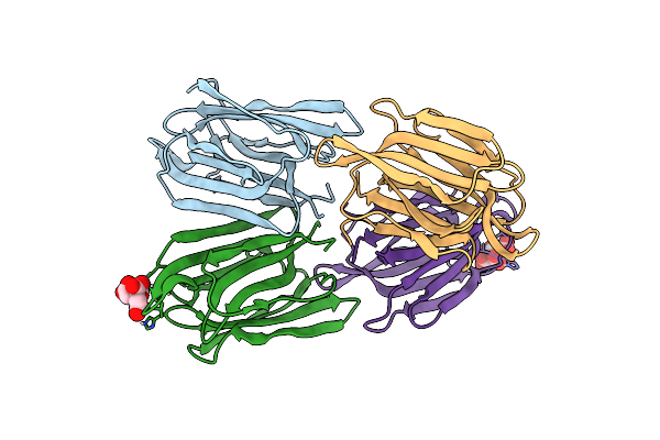
Crystal Structure Of Enterococcus Faecium D63R Penicillin-Binding Protein 5 (Pbp5Fm)
Organism: Enterococcus faecium
Method: X-RAY DIFFRACTION Resolution:2.90 Å Release Date: 2019-04-10 Classification: ANTIBIOTIC Ligands: SO4 |
Organism: Enterococcus faecium
Method: X-RAY DIFFRACTION
Release Date: 2019-04-10
Ligands: SO4
 |
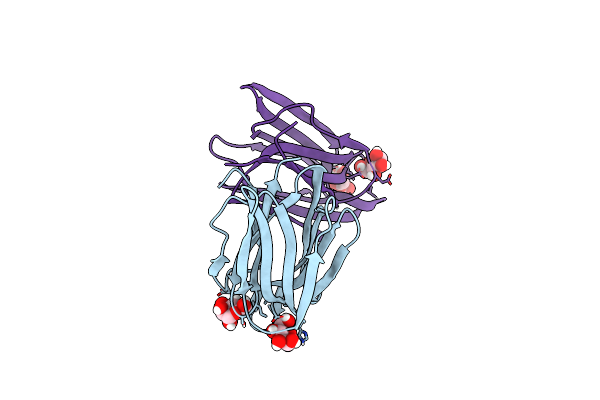
Structure Of Acmjrl, A Mannose Binding Jacalin Related Lectin From Ananas Comosus.
Organism: Ananas comosus
Method: X-RAY DIFFRACTION Resolution:1.80 Å Release Date: 2018-08-15 Classification: SUGAR BINDING PROTEIN Ligands: CIT |
Organism: Ananas comosus
Method: X-RAY DIFFRACTION
Release Date: 2018-08-15
Ligands: CIT
 |
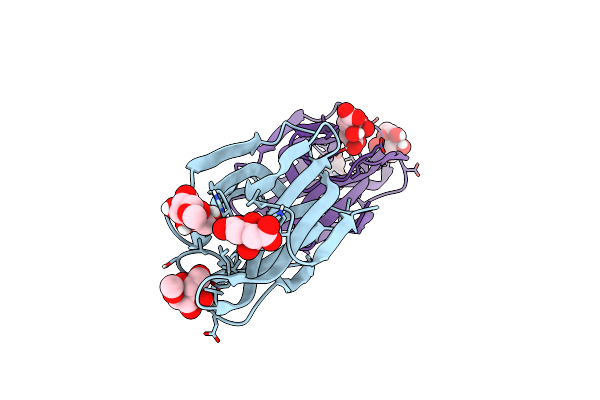
Structure Of Acmjrl, A Mannose Binding Jacalin Related Lectin From Ananas Comosus, In Complex With Mannose.
Organism: Ananas comosus
Method: X-RAY DIFFRACTION Resolution:2.75 Å Release Date: 2018-08-15 Classification: SUGAR BINDING PROTEIN Ligands: MAN |
Organism: Ananas comosus
Method: X-RAY DIFFRACTION
Release Date: 2018-08-15
Ligands: MAN
 |
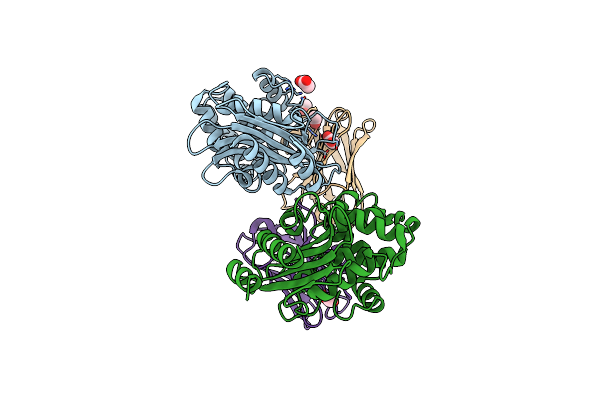
Structure Of Acmjrl, A Mannose Binding Jacalin Related Lectin From Ananas Comosus, In Complex With Methyl-Mannose.
Organism: Ananas comosus
Method: X-RAY DIFFRACTION Resolution:1.90 Å Release Date: 2018-08-15 Classification: SUGAR BINDING PROTEIN Ligands: MMA, CIT |
Organism: Ananas comosus
Method: X-RAY DIFFRACTION
Release Date: 2018-08-15
Ligands: MMA, CIT
 |
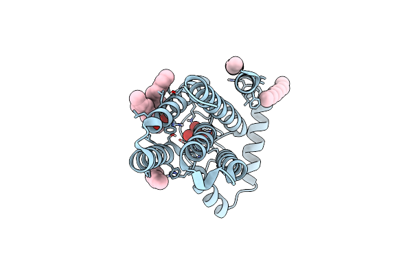
Structure Of A Single Domain Camelid Antibody Fragment Cab-G10S In Complex With The Blap Beta-Lactamase From Bacillus Licheniformis
Organism: Bacillus licheniformis, Lama glama
Method: X-RAY DIFFRACTION Resolution:2.56 Å Release Date: 2017-11-15 Classification: HYDROLASE Ligands: ACT |
Organism: Bacillus licheniformis, Lama glama
Method: X-RAY DIFFRACTION
Release Date: 2017-11-15
Ligands: ACT
 |
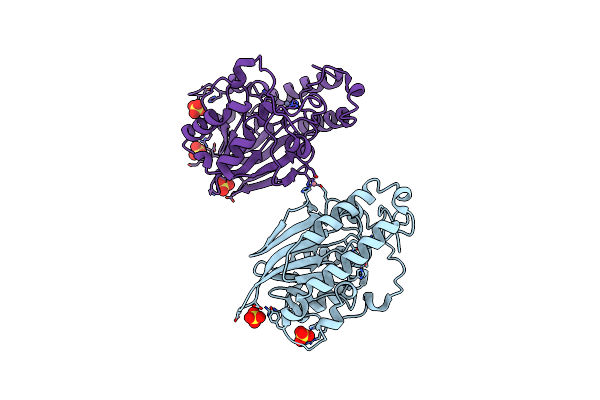
Crystal Structure Of The First Transmembrane Pap2 Type Phosphatidylglycerolphosphate Phosphatase From Bacillus Subtilis
Organism: Bacillus subtilis (strain 168)
Method: X-RAY DIFFRACTION Resolution:2.25 Å Release Date: 2017-02-22 Classification: HYDROLASE Ligands: WO4, OLC, UNL |
Organism: Bacillus subtilis (strain 168)
Method: X-RAY DIFFRACTION
Release Date: 2017-02-22
Ligands: WO4, OLC, UNL
 |
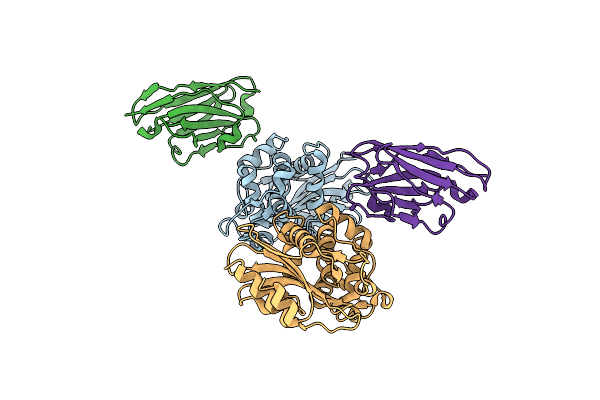
Crystal Structure Of The Class B3 Di-Zinc Metallo-Beta-Lactamase Lra- 12 From An Alaskan Soil Metagenome.
Organism: Uncultured bacterium blr12
Method: X-RAY DIFFRACTION Resolution:2.10 Å Release Date: 2015-09-16 Classification: HYDROLASE Ligands: ZN, SO4, CO |
Organism: Uncultured bacterium blr12
Method: X-RAY DIFFRACTION
Release Date: 2015-09-16
Ligands: ZN, SO4, CO
 |
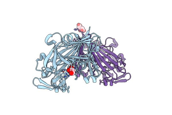
Structure Of A Single-Domain Camelid Antibody Fragment Cab-F11N In Complex With The Blap Beta-Lactamase From Bacillus Licheniformis
Organism: Bacillus licheniformis, Lama glama
Method: X-RAY DIFFRACTION Resolution:3.00 Å Release Date: 2014-10-08 Classification: HYDROLASE/IMMUNE SYSTEM |
Organism: Bacillus licheniformis, Lama glama
Method: X-RAY DIFFRACTION
Release Date: 2014-10-08
 |
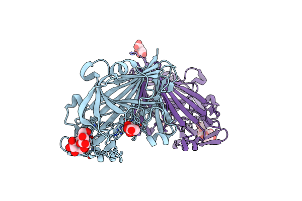
Crystal Structure Of Galactose Mutarotase Galm From Bacillus Subtilis
Organism: Bacillus subtilis subsp. subtilis str. 168
Method: X-RAY DIFFRACTION Resolution:2.00 Å Release Date: 2014-08-13 Classification: ISOMERASE Ligands: GOL, CIT |
Organism: Bacillus subtilis subsp. subtilis str. 168
Method: X-RAY DIFFRACTION
Release Date: 2014-08-13
Ligands: GOL, CIT
 |
Crystal Structure Of Galactose Mutarotase Galm From Bacillus Subtilis With Trehalose
Organism: Bacillus subtilis
Method: X-RAY DIFFRACTION Resolution:1.90 Å Release Date: 2014-08-13 Classification: ISOMERASE Ligands: PGE, CIT, ACT |
Organism: Bacillus subtilis
Method: X-RAY DIFFRACTION
Release Date: 2014-08-13
Ligands: PGE, CIT, ACT
 |
Crystal Structure Of Galactose Mutarotase Galm From Bacillus Subtilis In Complex With Maltose
Organism: Bacillus subtilis subsp. subtilis str. 168
Method: X-RAY DIFFRACTION Resolution:2.13 Å Release Date: 2014-08-13 Classification: ISOMERASE Ligands: CIT |
Organism: Bacillus subtilis subsp. subtilis str. 168
Method: X-RAY DIFFRACTION
Release Date: 2014-08-13
Ligands: CIT