

Search Count: 44
 |
Lectin/Toxin 2 From Coprinopsis Cinerea
Organism: Coprinopsis cinerea okayama7#130
Method: ELECTRON MICROSCOPY Resolution:3.16 Å Release Date: 2026-03-18 Classification: TOXIN |
Organism: Coprinopsis cinerea okayama7#130
Method: ELECTRON MICROSCOPY
Release Date: 2026-03-18
 |
Coprinopsis Cinerea Gh131 Protein Ccgh131B E161A In Complex With Cellobiose
Organism: Coprinopsis cinerea (strain okayama-7 / 130 / atcc mya-4618 / fgsc 9003)
Method: X-RAY DIFFRACTION Resolution:1.45 Å Release Date: 2024-11-06 Classification: HYDROLASE Ligands: MES, PEG, PGE |
Organism: Coprinopsis cinerea (strain okayama-7 / 130 / atcc mya-4618 / fgsc 9003)
Method: X-RAY DIFFRACTION
Release Date: 2024-11-06
Ligands: MES, PEG, PGE
 |
Coprinopsis Cinerea Gh131 Protein Ccgh131B
Organism: Coprinopsis cinerea (strain okayama-7 / 130 / atcc mya-4618 / fgsc 9003)
Method: X-RAY DIFFRACTION Resolution:1.90 Å Release Date: 2024-11-06 Classification: HYDROLASE Ligands: PEG |
Organism: Coprinopsis cinerea (strain okayama-7 / 130 / atcc mya-4618 / fgsc 9003)
Method: X-RAY DIFFRACTION
Release Date: 2024-11-06
Ligands: PEG
 |
Crystal Structure Of The Dioxygenase Cctet From Coprinopsis Cinerea Bound To 12Bp 5-Methylcytosine (5Mc) Containing Duplex Dna
Organism: Coprinopsis cinerea (strain okayama-7 / 130 / atcc mya-4618 / fgsc 9003), Homo sapiens
Method: X-RAY DIFFRACTION Resolution:2.30 Å Release Date: 2024-03-06 Classification: DNA BINDING PROTEIN/DNA Ligands: MN, OGA |
Organism: Coprinopsis cinerea (strain okayama-7 / 130 / atcc mya-4618 / fgsc 9003), Homo sapiens
Method: X-RAY DIFFRACTION
Release Date: 2024-03-06
Ligands: MN, OGA
 |
Crystal Structure Of Cocaprin 1, Inhibitor Of Cysteine And Aspartic Proteases From Coprinopsis Cinerea
Organism: Coprinopsis cinerea
Method: X-RAY DIFFRACTION Resolution:1.60 Å Release Date: 2022-06-01 Classification: PROTEIN BINDING |
Organism: Coprinopsis cinerea
Method: X-RAY DIFFRACTION
Release Date: 2022-06-01
 |
Crystal Structure Of The Dioxygenase Cctet From Coprinopsis Cinereain In Complex With Mn(Ii) And N-Oxalylglycine
Organism: Coprinopsis cinerea (strain okayama-7 / 130 / atcc mya-4618 / fgsc 9003)
Method: X-RAY DIFFRACTION Resolution:2.60 Å Release Date: 2022-03-30 Classification: DNA BINDING PROTEIN Ligands: MN, OGA |
Organism: Coprinopsis cinerea (strain okayama-7 / 130 / atcc mya-4618 / fgsc 9003)
Method: X-RAY DIFFRACTION
Release Date: 2022-03-30
Ligands: MN, OGA
 |
Crystal Structure Of The Dioxygenase Cctet From Coprinopsis Cinereain Bound To 12Bp N6-Methyldeoxyadenine (6Ma) Containing Duplex Dna
Organism: Coprinopsis cinerea, Synthetic construct
Method: X-RAY DIFFRACTION Resolution:2.30 Å Release Date: 2022-03-30 Classification: DNA BINDING PROTEIN/DNA Ligands: MN, OGA |
Organism: Coprinopsis cinerea, Synthetic construct
Method: X-RAY DIFFRACTION
Release Date: 2022-03-30
Ligands: MN, OGA
 |
Cml1 Crystal Structure In Complex With Lewis A Tetrasaccharide
Organism: Coprinopsis cinerea
Method: X-RAY DIFFRACTION Resolution:1.95 Å Release Date: 2021-08-04 Classification: SUGAR BINDING PROTEIN Ligands: SO4 |
Organism: Coprinopsis cinerea
Method: X-RAY DIFFRACTION
Release Date: 2021-08-04
Ligands: SO4
 |
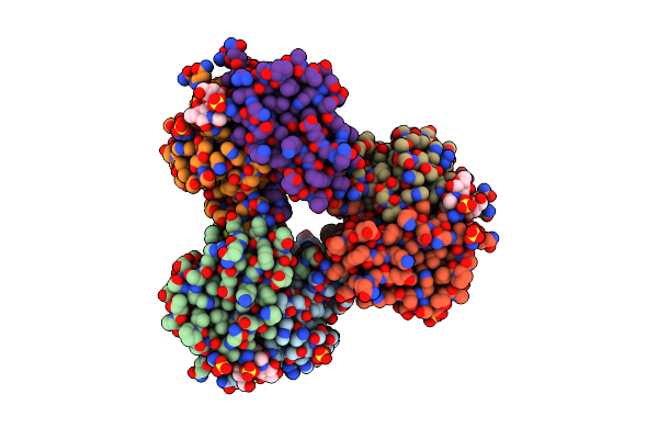
Crystal Structure Of The Fungal Lectin Cml1
Organism: Coprinopsis cinerea
Method: X-RAY DIFFRACTION Resolution:1.35 Å Release Date: 2021-07-28 Classification: CARBOHYDRATE Ligands: GOL, PC4, K, ACT |
Organism: Coprinopsis cinerea
Method: X-RAY DIFFRACTION
Release Date: 2021-07-28
Ligands: GOL, PC4, K, ACT
 |
Cml1 Crystal Structure In Complex With H-Type 1 Trisaccharide
Organism: Coprinopsis cinerea
Method: X-RAY DIFFRACTION Resolution:1.55 Å Release Date: 2021-07-28 Classification: SUGAR BINDING PROTEIN Ligands: SO4 |
Organism: Coprinopsis cinerea
Method: X-RAY DIFFRACTION
Release Date: 2021-07-28
Ligands: SO4
 |
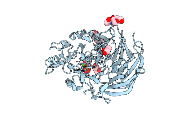
Crystal Structure Of Cytochrome B Domain Of Pyranose Dehydrogenase From Coprinopsis Cinerea
Organism: Coprinopsis cinerea
Method: X-RAY DIFFRACTION Resolution:2.00 Å Release Date: 2019-11-13 Classification: OXIDOREDUCTASE Ligands: NAG, HEM, MPD, ACT |
Organism: Coprinopsis cinerea
Method: X-RAY DIFFRACTION
Release Date: 2019-11-13
Ligands: NAG, HEM, MPD, ACT
 |
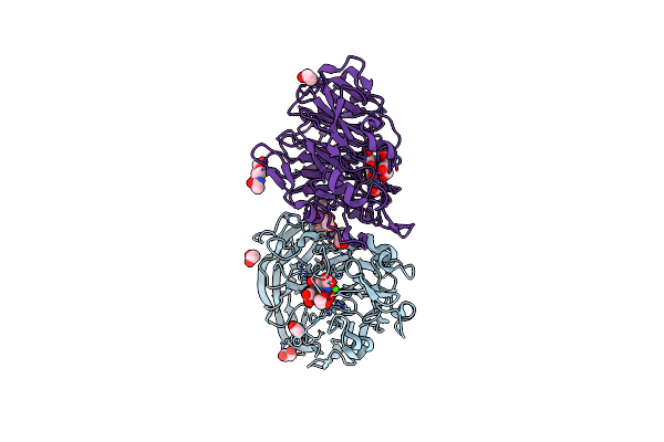
Crystal Structure Of Pqq Doamin Of Pyranose Dehydrogenase From Coprinopsis Cinerea: Apo-From
Organism: Coprinopsis cinerea
Method: X-RAY DIFFRACTION Resolution:1.50 Å Release Date: 2019-11-06 Classification: OXIDOREDUCTASE Ligands: NAG, CA, SO4, FMT, PGE, GOL |
Organism: Coprinopsis cinerea
Method: X-RAY DIFFRACTION
Release Date: 2019-11-06
Ligands: NAG, CA, SO4, FMT, PGE, GOL
 |
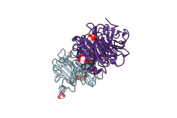
Holo Form Of Pyranose Dehydrogenase Pqq Domain From Coprinopsis Cinerea
Organism: Coprinopsis cinerea
Method: X-RAY DIFFRACTION Resolution:1.30 Å Release Date: 2019-11-06 Classification: OXIDOREDUCTASE Ligands: CA, PQQ, EDO, ACT, NAG |
Organism: Coprinopsis cinerea
Method: X-RAY DIFFRACTION
Release Date: 2019-11-06
Ligands: CA, PQQ, EDO, ACT, NAG
 |
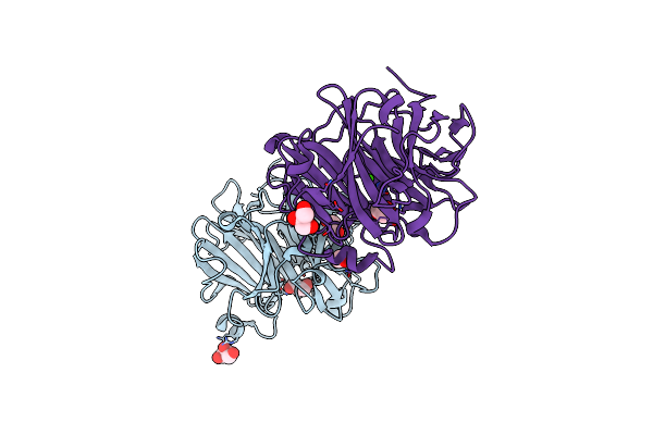
Catalytic Domain Of Coprinopsis Cinerea Gh62 Alpha-L-Arabinofuranosidase
Organism: Coprinopsis cinerea (strain okayama-7 / 130 / atcc mya-4618 / fgsc 9003)
Method: X-RAY DIFFRACTION Resolution:1.70 Å Release Date: 2016-09-07 Classification: HYDROLASE Ligands: CA, GOL |
Organism: Coprinopsis cinerea (strain okayama-7 / 130 / atcc mya-4618 / fgsc 9003)
Method: X-RAY DIFFRACTION
Release Date: 2016-09-07
Ligands: CA, GOL
 |
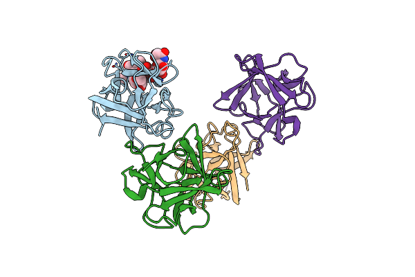
Catalytic Domain Of Coprinopsis Cinerea Gh62 Alpha-L-Arabinofuranosidase Complexed With Pb
Organism: Coprinopsis cinerea (strain okayama-7 / 130 / atcc mya-4618 / fgsc 9003)
Method: X-RAY DIFFRACTION Resolution:1.48 Å Release Date: 2016-09-07 Classification: HYDROLASE Ligands: PB, CA, GOL |
Organism: Coprinopsis cinerea (strain okayama-7 / 130 / atcc mya-4618 / fgsc 9003)
Method: X-RAY DIFFRACTION
Release Date: 2016-09-07
Ligands: PB, CA, GOL
 |
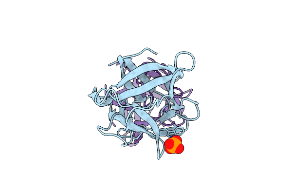
X-Ray Structure Of The Ccl2 Lectin In Complex With Sialyl Lewis X
Organism: Coprinopsis cinerea
Method: X-RAY DIFFRACTION Resolution:1.95 Å Release Date: 2015-07-22 Classification: SUGAR BINDING PROTEIN |
Organism: Coprinopsis cinerea
Method: X-RAY DIFFRACTION
Release Date: 2015-07-22
 |
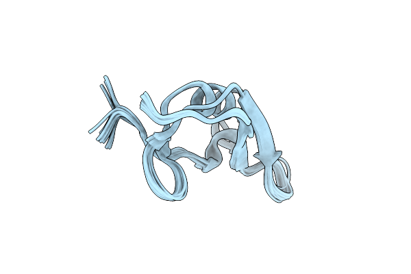
X-Ray Structure Of The Dimeric Ccl2 Lectin In Native Form
Organism: Coprinopsis cinerea
Method: X-RAY DIFFRACTION Resolution:2.25 Å Release Date: 2015-07-22 Classification: SUGAR BINDING PROTEIN Ligands: CL, PO4 |
Organism: Coprinopsis cinerea
Method: X-RAY DIFFRACTION
Release Date: 2015-07-22
Ligands: CL, PO4
 |
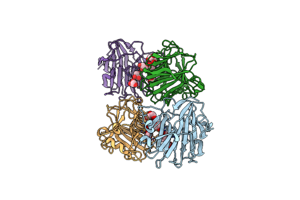
Nmr Structure Of Copsin
Organism: Coprinopsis cinerea
Method: SOLUTION NMR Release Date: 2014-10-29 Classification: ANTIMICROBIAL PROTEIN |
Organism: Coprinopsis cinerea
Method: SOLUTION NMR
Release Date: 2014-10-29
 |
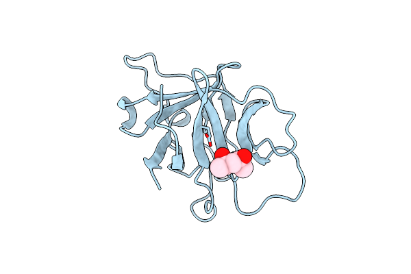
Crystal Structure Of The Catalytic Domain Of The Glycoside Hydrolase Family 131 Protein From Coprinopsis Cinerea
Organism: Coprinopsis cinerea
Method: X-RAY DIFFRACTION Resolution:1.99 Å Release Date: 2013-05-22 Classification: HYDROLASE Ligands: GOL |
Organism: Coprinopsis cinerea
Method: X-RAY DIFFRACTION
Release Date: 2013-05-22
Ligands: GOL
 |
High Resolution Structure Of Proteinase Inhibitor From Coprinopsis Cinerea
Organism: Coprinopsis cinerea
Method: X-RAY DIFFRACTION Resolution:1.50 Å Release Date: 2012-09-05 Classification: HYDROLASE INHIBITOR Ligands: MPD |
Organism: Coprinopsis cinerea
Method: X-RAY DIFFRACTION
Release Date: 2012-09-05
Ligands: MPD