

Search Count: 31
 |
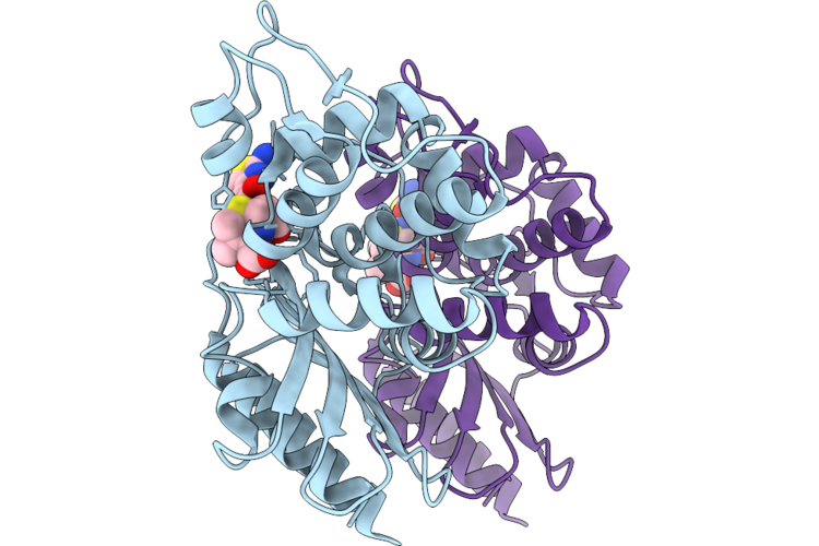
Crystal Structure Of The Transpeptidase Domain Of A Y422A Mutant Of Pbp2 From Neisseria Gonorrhoeae Strain H041 Acylated By Ceftriaxone
Organism: Neisseria gonorrhoeae
Method: X-RAY DIFFRACTION Resolution:2.40 Å Release Date: 2026-06-03 Classification: LIGASE Ligands: CEF, PGE, GOL |
Organism: Neisseria gonorrhoeae
Method: X-RAY DIFFRACTION
Release Date: 2026-06-03
Ligands: CEF, PGE, GOL
 |
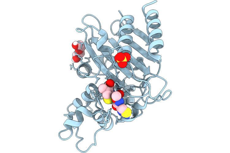
Crystal Structure Of Sme-1 E166A In Complex With Cefotaxime
Organism: Serratia marcescens
Method: X-RAY DIFFRACTION Resolution:2.40 Å Release Date: 2026-04-22 Classification: HORMONE Ligands: CEF |
Organism: Serratia marcescens
Method: X-RAY DIFFRACTION
Release Date: 2026-04-22
Ligands: CEF
 |
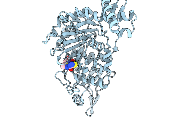
Crystal Structure Of Sme-1 E166A With Cefpirome
Organism: Serratia marcescens
Method: X-RAY DIFFRACTION Resolution:2.00 Å Release Date: 2026-04-22 Classification: HYDROLASE Ligands: CEF, PEG, SO4 |
Organism: Serratia marcescens
Method: X-RAY DIFFRACTION
Release Date: 2026-04-22
Ligands: CEF, PEG, SO4
 |
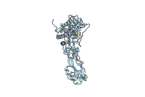
Penicillin-Binding Protein 1B (Pbp-1B) In Complex With Ceftriaxone - Streptococcus Pneumoniae R6
Organism: Streptococcus pneumoniae r6
Method: X-RAY DIFFRACTION Resolution:1.79 Å Release Date: 2026-02-18 Classification: TRANSFERASE Ligands: CEF, CL |
Organism: Streptococcus pneumoniae r6
Method: X-RAY DIFFRACTION
Release Date: 2026-02-18
Ligands: CEF, CL
 |
Pseudomonas Aeruginosa Penicillin Binding Protein 3 In Complex With Cefepime
Organism: Pseudomonas aeruginosa
Method: X-RAY DIFFRACTION Resolution:2.70 Å Release Date: 2024-10-09 Classification: HYDROLASE Ligands: CEF |
Organism: Pseudomonas aeruginosa
Method: X-RAY DIFFRACTION
Release Date: 2024-10-09
Ligands: CEF
 |
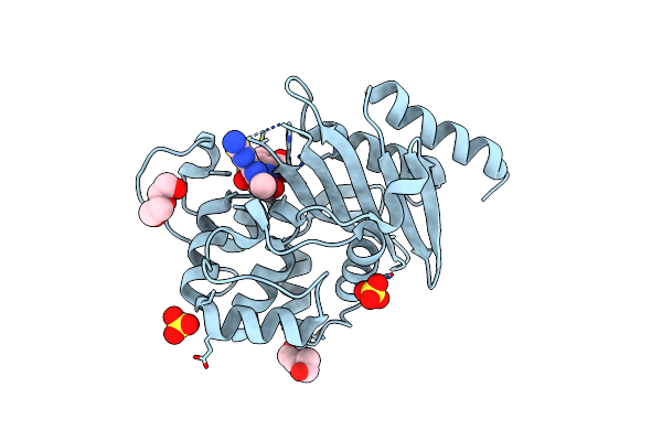
Crystal Structure Of Penicillin-Binding Protein 3 (Pbp3) From Staphylococcus Epidermidis In Complex With Cefotaxime
Organism: Staphylococcus epidermidis (strain atcc 35984 / rp62a)
Method: X-RAY DIFFRACTION Resolution:2.51 Å Release Date: 2023-12-13 Classification: PENICILLIN-BINDING PROTEIN Ligands: CEF |
Organism: Staphylococcus epidermidis (strain atcc 35984 / rp62a)
Method: X-RAY DIFFRACTION
Release Date: 2023-12-13
Ligands: CEF
 |
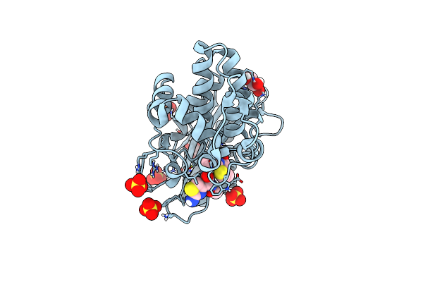
X-Ray Crystal Structure Of Oxa-24/40 K84D In Complex With Cefotaxime
Organism: Acinetobacter baumannii
Method: X-RAY DIFFRACTION Resolution:1.62 Å Release Date: 2022-07-06 Classification: HYDROLASE/Inhibitor Ligands: PEG, SO4, CEF |
Organism: Acinetobacter baumannii
Method: X-RAY DIFFRACTION
Release Date: 2022-07-06
Ligands: PEG, SO4, CEF
 |
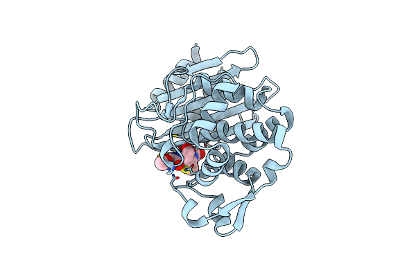
Acylenzyme Complex Of Cefotaxime Bound To Deacylation Mutant Kpc-2 (E166Q)
Organism: Klebsiella pneumoniae
Method: X-RAY DIFFRACTION Resolution:1.31 Å Release Date: 2020-12-23 Classification: ANTIMICROBIAL PROTEIN Ligands: GOL, SO4, CEF |
Organism: Klebsiella pneumoniae
Method: X-RAY DIFFRACTION
Release Date: 2020-12-23
Ligands: GOL, SO4, CEF
 |
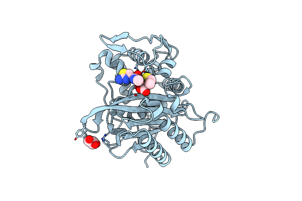
Crystal Structure Of Ctx-M-14 E166A/K234R Beta-Lactamase In Complex With Hydrolyzed Cefotaxime
Organism: Escherichia coli
Method: X-RAY DIFFRACTION Resolution:1.40 Å Release Date: 2020-11-04 Classification: HYDROLASE Ligands: CEF |
Organism: Escherichia coli
Method: X-RAY DIFFRACTION
Release Date: 2020-11-04
Ligands: CEF
 |
Crystal Structure Of Transpeptidase Domain Of Pbp2 From Neisseria Gonorrhoeae Cephalosporin-Resistant Strain H041 Acylated By Ceftriaxone
Organism: Neisseria gonorrhoeae
Method: X-RAY DIFFRACTION Resolution:1.80 Å Release Date: 2020-04-15 Classification: HYDROLASE/ANTIBIOTIC Ligands: CEF, GOL |
Organism: Neisseria gonorrhoeae
Method: X-RAY DIFFRACTION
Release Date: 2020-04-15
Ligands: CEF, GOL
 |
Crystal Structure Of Class D Beta-Lactamase Oxa-48 With Cefotaxime
Organism: Klebsiella pneumoniae
Method: X-RAY DIFFRACTION Resolution:2.05 Å Release Date: 2020-01-22 Classification: HYDROLASE Ligands: CEF, CL, ACT |
Organism: Klebsiella pneumoniae
Method: X-RAY DIFFRACTION
Release Date: 2020-01-22
Ligands: CEF, CL, ACT
 |
Crystal Structure Of A Class C Beta-Lactamase In Complex With Cefotaxime
Organism: Klebsiella pneumoniae
Method: X-RAY DIFFRACTION Resolution:1.46 Å Release Date: 2019-10-16 Classification: HYDROLASE Ligands: CEF |
Organism: Klebsiella pneumoniae
Method: X-RAY DIFFRACTION
Release Date: 2019-10-16
Ligands: CEF
 |
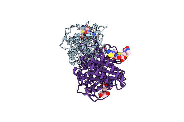
Structure Of Cefotaxime-Cdd-1 Beta-Lactamase Complex
Organism: Clostridioides difficile
Method: X-RAY DIFFRACTION Resolution:2.10 Å Release Date: 2019-10-09 Classification: HYDROLASE/HYDROLASE Inhibitor Ligands: CEF, SO4, MES, MPD |
Organism: Clostridioides difficile
Method: X-RAY DIFFRACTION
Release Date: 2019-10-09
Ligands: CEF, SO4, MES, MPD
 |
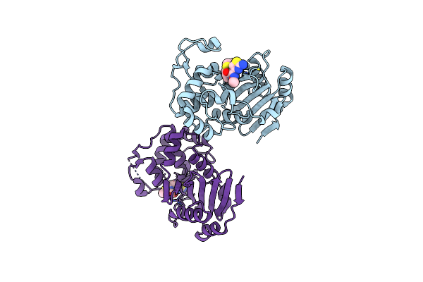
Crystal Structure Of Transpeptidase Domain Of Pbp2 From Neisseria Gonorrhoeae Acylated By Ceftriaxone
Organism: Neisseria gonorrhoeae
Method: X-RAY DIFFRACTION Resolution:1.83 Å Release Date: 2019-08-07 Classification: HYDROLASE/ANTIBIOTIC Ligands: NZV, CEF, 9F2, PEG |
Organism: Neisseria gonorrhoeae
Method: X-RAY DIFFRACTION
Release Date: 2019-08-07
Ligands: NZV, CEF, 9F2, PEG
 |
Crystal Structure Of Penicillin-Binding Protein D2 From Listeria Monocytogenes In The Cefotaxime Bound Form
Organism: Listeria monocytogenes egd-e
Method: X-RAY DIFFRACTION Resolution:1.89 Å Release Date: 2018-07-25 Classification: ANTIBIOTIC Ligands: CEF, GOL |
Organism: Listeria monocytogenes egd-e
Method: X-RAY DIFFRACTION
Release Date: 2018-07-25
Ligands: CEF, GOL
 |
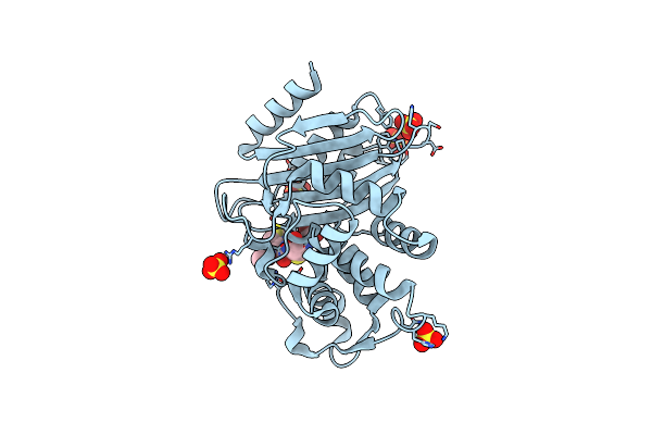
Structure Of Acinetobacter Baumannii Carbapenemase Oxa-239 K82D Bound To Cefotaxime
Organism: Acinetobacter sp. enrichment culture clone 8407
Method: X-RAY DIFFRACTION Resolution:1.81 Å Release Date: 2017-12-27 Classification: HYDROLASE Ligands: CEF |
Organism: Acinetobacter sp. enrichment culture clone 8407
Method: X-RAY DIFFRACTION
Release Date: 2017-12-27
Ligands: CEF
 |
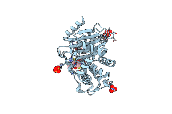
15K X-Ray Structure With Cefotaxime: Exploring The Mechanism Of Beta- Lactam Ring Protonation In The Class A Beta-Lactamase Acylation Mechanism Using Neutron And X-Ray Crystallography
Organism: Escherichia coli
Method: X-RAY DIFFRACTION Resolution:1.05 Å Release Date: 2015-12-16 Classification: HYDROLASE Ligands: SO4, CEF |
Organism: Escherichia coli
Method: X-RAY DIFFRACTION
Release Date: 2015-12-16
Ligands: SO4, CEF
 |
293K Joint X-Ray Neutron With Cefotaxime: Exploring The Mechanism Of Beta-Lactam Ring Protonation In The Class A Beta-Lactamase Acylation Mechanism Using Neutron And X-Ray Crystallography
Organism: Escherichia coli
Method: NEUTRON DIFFRACTION, X-RAY DIFFRACTION Resolution:1.598 Å, 2.200 Å Release Date: 2015-12-16 Classification: HYDROLASE Ligands: CEF, SO4 |
Organism: Escherichia coli
Method: NEUTRON DIFFRACTION, X-RAY DIFFRACTION
Release Date: 2015-12-16
Ligands: CEF, SO4
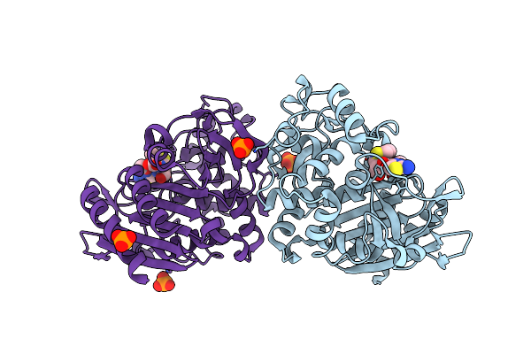 |
Crystal Structure Of Ampc Beta-Lactamase From E. Coli In Complex With Cefotaxime
Organism: Escherichia coli
Method: X-RAY DIFFRACTION Resolution:1.89 Å Release Date: 2014-10-29 Classification: Hydrolase/antibiotic Ligands: PO4, CEF |
Organism: Escherichia coli
Method: X-RAY DIFFRACTION
Release Date: 2014-10-29
Ligands: PO4, CEF
 |
Crystal Structure Of Ampc Beta-Lactamase N152G Mutant In Complex With Cefotaxime
Organism: Escherichia coli
Method: X-RAY DIFFRACTION Resolution:2.11 Å Release Date: 2014-10-29 Classification: Hydrolase/antibiotic Ligands: PO4, CEF |
Organism: Escherichia coli
Method: X-RAY DIFFRACTION
Release Date: 2014-10-29
Ligands: PO4, CEF