

Search Count: 12
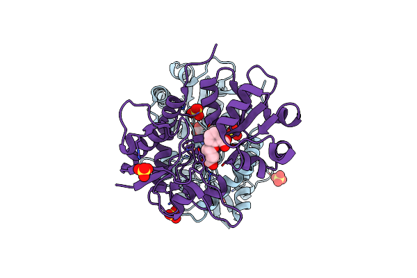 |
Structure Of Gluk1 Ligand-Binding Domain (S1S2) In Complex With (2S,4R)-4-(2-Carboxyphenoxy)Pyrrolidine-2-Carboxylic Acid At 3.18 A Resolution
Organism: Rattus norvegicus
Method: X-RAY DIFFRACTION Resolution:3.18 Å Release Date: 2017-01-11 Classification: MEMBRANE PROTEIN Ligands: 7E5, SO4, CL |
Organism: Rattus norvegicus
Method: X-RAY DIFFRACTION
Release Date: 2017-01-11
Ligands: 7E5, SO4, CL
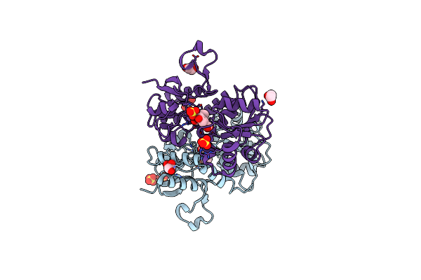 |
Structure Of The Ligand-Binding Domain Of Glua2 In Complex With The Antagonist Cng10109
Organism: Rattus norvegicus
Method: X-RAY DIFFRACTION Resolution:1.90 Å Release Date: 2015-08-05 Classification: SIGNALING PROTEIN Ligands: SO4, GOL, 4E5, EDO, ACT |
Organism: Rattus norvegicus
Method: X-RAY DIFFRACTION
Release Date: 2015-08-05
Ligands: SO4, GOL, 4E5, EDO, ACT
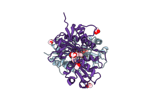 |
Structure Of The Ligand-Binding Domain Of Gluk1 In Complex With The Antagonist Cng10111
Organism: Rattus norvegicus
Method: X-RAY DIFFRACTION Resolution:1.93 Å Release Date: 2015-08-05 Classification: SIGNALING PROTEIN Ligands: 4E7, ACT, CL, PEG, EDO |
Organism: Rattus norvegicus
Method: X-RAY DIFFRACTION
Release Date: 2015-08-05
Ligands: 4E7, ACT, CL, PEG, EDO
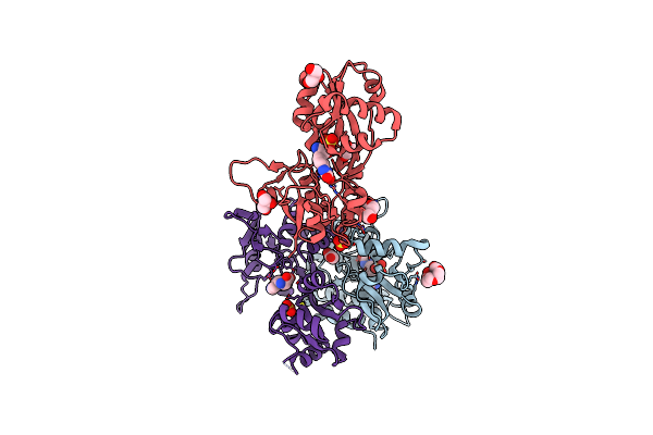 |
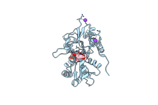
Structure Of Gluk1 Ligand-Binding Domain (S1S2) In Complex With (S)-2-Amino-4-(2,3-Dioxo-1,2,3,4-Tetrahydroquinoxalin-6-Yl)Butanoic Acid At 2.28 A Resolution
Organism: Rattus norvegicus
Method: X-RAY DIFFRACTION Resolution:2.28 Å Release Date: 2015-04-22 Classification: MEMBRANE PROTEIN Ligands: 35K, SO4, PG4, CL, GOL, ACT |
Organism: Rattus norvegicus
Method: X-RAY DIFFRACTION
Release Date: 2015-04-22
Ligands: 35K, SO4, PG4, CL, GOL, ACT
 |
Crystal Structure Of The Gluk3 Ligand-Binding Domain (S1S2) In Complex With The Agonist (2S,4R)-4-(3-Methoxy-3-Oxopropyl)Glutamic Acid At 2.01 A Resolution.
Organism: Rattus norvegicus
Method: X-RAY DIFFRACTION Resolution:2.01 Å Release Date: 2014-08-06 Classification: MEMBRANE PROTEIN, RECEPTOR/Agonist Ligands: K, PO4, CL, 2QE |
Organism: Rattus norvegicus
Method: X-RAY DIFFRACTION
Release Date: 2014-08-06
Ligands: K, PO4, CL, 2QE
 |
Crystal Structure Of The Kainate Receptor Gluk3 Ligand-Binding Domain In Complex With The Agonist (2S,4R)-4-(3-Methylamino-3-Oxopropyl)Glutamic Acid At 2.6 A Resolution
Organism: Rattus norvegicus
Method: X-RAY DIFFRACTION Resolution:2.60 Å Release Date: 2014-08-06 Classification: MEMBRANE PROTEIN/AGONIST Ligands: 2QD, K, CL |
Organism: Rattus norvegicus
Method: X-RAY DIFFRACTION
Release Date: 2014-08-06
Ligands: 2QD, K, CL
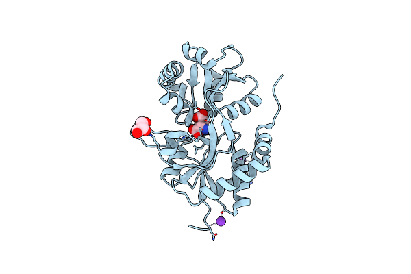 |
Crystal Structure Of The Kainate Receptor Gluk3 Ligand Binding Domain In Complex With (S)-Glutamate
Organism: Rattus norvegicus
Method: X-RAY DIFFRACTION Resolution:1.65 Å Release Date: 2013-10-16 Classification: Receptor/agonist Ligands: GLU, K, CL, GOL |
Organism: Rattus norvegicus
Method: X-RAY DIFFRACTION
Release Date: 2013-10-16
Ligands: GLU, K, CL, GOL
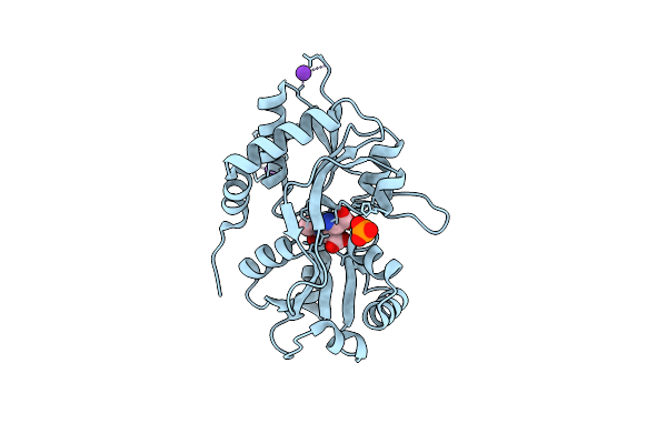 |
Crystal Structure Of The Kainate Receptor Gluk3 Ligand-Binding Domain In Complex With The Agonist Za302
Organism: Rattus norvegicus
Method: X-RAY DIFFRACTION Resolution:2.65 Å Release Date: 2013-03-06 Classification: MEMBRANE PROTEIN Ligands: 3ZA, K, CL, PO4 |
Organism: Rattus norvegicus
Method: X-RAY DIFFRACTION
Release Date: 2013-03-06
Ligands: 3ZA, K, CL, PO4
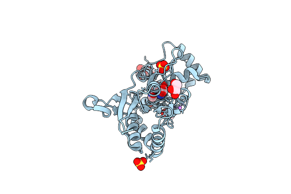 |
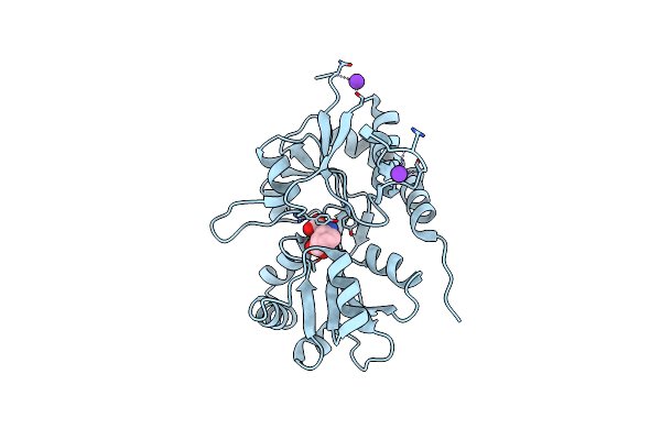
Crystal Structure Of The Glua2 Ligand-Binding Domain (S1S2J) In Complex With The Agonist Za302 At 1.24A Resolution
Organism: Rattus norvegicus
Method: X-RAY DIFFRACTION Resolution:1.24 Å Release Date: 2013-03-06 Classification: MEMBRANE PROTEIN Ligands: 3ZA, SO4, LI, GOL |
Organism: Rattus norvegicus
Method: X-RAY DIFFRACTION
Release Date: 2013-03-06
Ligands: 3ZA, SO4, LI, GOL
 |
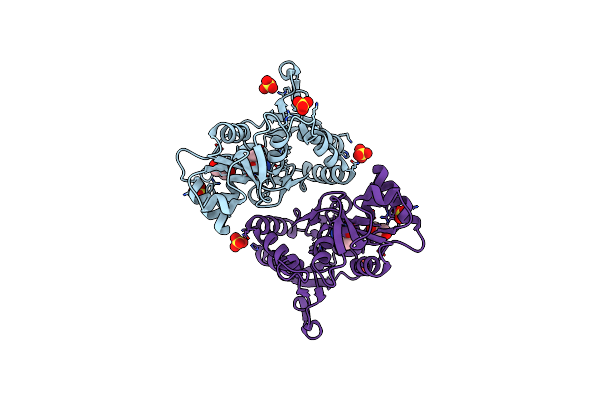
Crystal Structure Of The Kainate Receptor Gluk3 Ligand-Binding Domain In Complex With The Agonist G8M
Organism: Rattus norvegicus
Method: X-RAY DIFFRACTION Resolution:2.30 Å Release Date: 2012-08-29 Classification: MEMBRANE PROTEIN/Agonist Ligands: G8M, CL, K |
Organism: Rattus norvegicus
Method: X-RAY DIFFRACTION
Release Date: 2012-08-29
Ligands: G8M, CL, K
 |
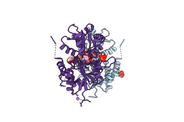
Crystal Structure Of The Glua2 Ligand-Binding Domain (S1S2J) In Complex With The Agonist Cbg-Iv At 2.05A Resolution
Organism: Rattus norvegicus
Method: X-RAY DIFFRACTION Resolution:2.05 Å Release Date: 2012-08-08 Classification: Membrane Protein/Agonist Ligands: G8M, SO4, CL, GOL |
Organism: Rattus norvegicus
Method: X-RAY DIFFRACTION
Release Date: 2012-08-08
Ligands: G8M, SO4, CL, GOL
 |
Crystal Structure Of The Kainate Receptor Gluk3 Ligand Binding Domain In Complex With (S)-Glutamate
Organism: Rattus norvegicus
Method: X-RAY DIFFRACTION Resolution:1.60 Å Release Date: 2011-09-28 Classification: MEMBRANE PROTEIN Ligands: GLU, GOL, CL, PO4, NA |
Organism: Rattus norvegicus
Method: X-RAY DIFFRACTION
Release Date: 2011-09-28
Ligands: GLU, GOL, CL, PO4, NA