

Search Count: 89
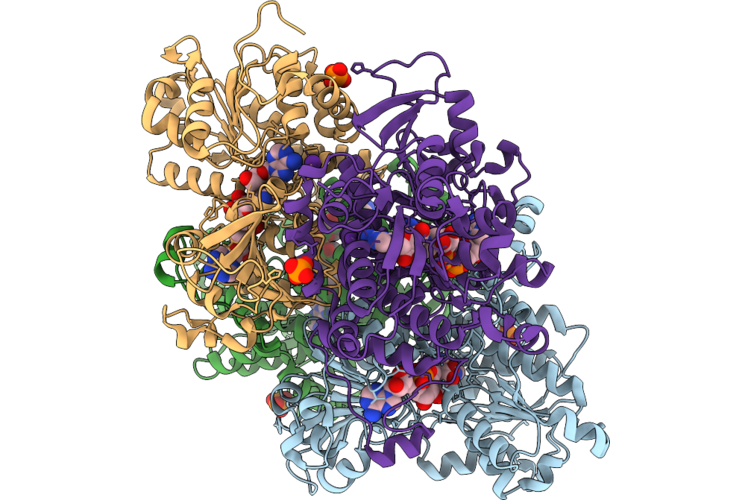 |
Crystal Structure Of S-Adenosyl-L-Homocysteine Hydrolase From P. Aeruginosa: F324A Mutant Complexed With Adenosine And Rubidium Ions Replacing Native Potassium
Organism: Pseudomonas aeruginosa pao1
Method: X-RAY DIFFRACTION Resolution:2.06 Å Release Date: 2026-07-08 Classification: HYDROLASE Ligands: NAD, ADN, PO4, RB |
Organism: Pseudomonas aeruginosa pao1
Method: X-RAY DIFFRACTION
Release Date: 2026-07-08
Ligands: NAD, ADN, PO4, RB
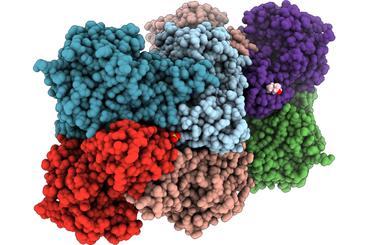 |
Crystal Structure Of Human S-Adenosyl-L-Homocysteine Hydrolase Complex With Adenosine
Organism: Homo sapiens
Method: X-RAY DIFFRACTION Resolution:1.59 Å Release Date: 2026-04-29 Classification: HYDROLASE Ligands: NAD, ADN, K, SO4, MPD |
Organism: Homo sapiens
Method: X-RAY DIFFRACTION
Release Date: 2026-04-29
Ligands: NAD, ADN, K, SO4, MPD
 |
Crystal Structure Of Human S-Adenosyl-L-Homocysteine Hydrolase In Complex With Adenosine And Cadmium Ions.
Organism: Homo sapiens
Method: X-RAY DIFFRACTION Resolution:1.93 Å Release Date: 2026-04-29 Classification: HYDROLASE Ligands: NAD, ADN, CD, K |
Organism: Homo sapiens
Method: X-RAY DIFFRACTION
Release Date: 2026-04-29
Ligands: NAD, ADN, CD, K
 |
Crystal Structure Of The S-Adenosyl-L-Homocysteine Hydrolase From P. Aeruginosa, Q65N Mutant Probed With Rubidium To Confirm Disruption Of A Potassium Binding Site.
Organism: Pseudomonas aeruginosa pao1
Method: X-RAY DIFFRACTION Resolution:1.91 Å Release Date: 2026-04-08 Classification: HYDROLASE Ligands: NAD, GOL, PO4 |
Organism: Pseudomonas aeruginosa pao1
Method: X-RAY DIFFRACTION
Release Date: 2026-04-08
Ligands: NAD, GOL, PO4
 |
Crystal Structure Of S-Adenosyl-L-Homocysteine Hydrolase From P. Aeruginosa: Q65A Mutant Soaked With Adenosine And Probed With Rubidium To Confirm Disruption Of A Potassium Binding Site.
Organism: Pseudomonas aeruginosa pao1
Method: X-RAY DIFFRACTION Resolution:2.40 Å Release Date: 2026-04-08 Classification: HYDROLASE Ligands: NAD, ADN, PO4 |
Organism: Pseudomonas aeruginosa pao1
Method: X-RAY DIFFRACTION
Release Date: 2026-04-08
Ligands: NAD, ADN, PO4
 |
Crystal Structure Of S-Adenosyl-L-Homocysteine Hydrolase From P. Aeruginosa, Q65N Mutant Soaked With Adenosine And Probed With Rubidium To Confirm Disruption Of A Potassium Binding Site.
Organism: Pseudomonas aeruginosa pao1
Method: X-RAY DIFFRACTION Resolution:2.34 Å Release Date: 2026-04-08 Classification: HYDROLASE Ligands: NAD, ADN, PO4 |
Organism: Pseudomonas aeruginosa pao1
Method: X-RAY DIFFRACTION
Release Date: 2026-04-08
Ligands: NAD, ADN, PO4
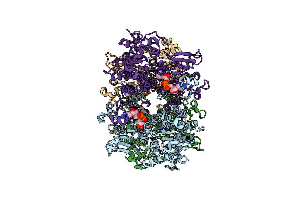 |
Cryo-Em Structure Of Pseudomonas Aeruginosa Tetrameric S-Adenosyl-L-Homocysteine Hydrolase In The Fully Open State
Organism: Pseudomonas aeruginosa pao1
Method: ELECTRON MICROSCOPY Release Date: 2025-02-19 Classification: HYDROLASE Ligands: NAD |
Organism: Pseudomonas aeruginosa pao1
Method: ELECTRON MICROSCOPY
Release Date: 2025-02-19
Ligands: NAD
 |
Cryo-Em Structure Of Pseudomonas Aeruginosa Tetrameric S-Adenosyl-L-Homocysteine Hydrolase With 3 Open And 1 Closed Subunits
Organism: Pseudomonas aeruginosa pao1
Method: ELECTRON MICROSCOPY Release Date: 2025-02-19 Classification: HYDROLASE Ligands: NAD, ADN, K |
Organism: Pseudomonas aeruginosa pao1
Method: ELECTRON MICROSCOPY
Release Date: 2025-02-19
Ligands: NAD, ADN, K
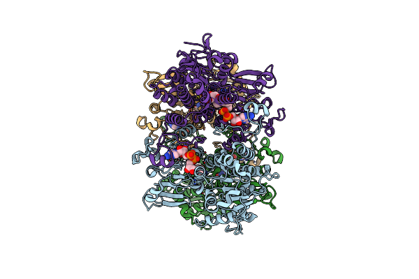 |
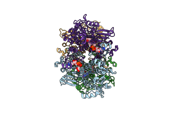
Cryo-Em Structure Of Pseudomonas Aeruginosa Tetrameric S-Adenosyl-L-Homocysteine Hydrolase With 1 Open And 3 Closed Subunits
Organism: Pseudomonas aeruginosa pao1
Method: ELECTRON MICROSCOPY Release Date: 2025-02-19 Classification: HYDROLASE Ligands: NAD, ADN, K |
Organism: Pseudomonas aeruginosa pao1
Method: ELECTRON MICROSCOPY
Release Date: 2025-02-19
Ligands: NAD, ADN, K
 |
Cryo-Em Structure Of Pseudomonas Aeruginosa Tetrameric S-Adenosyl-L-Homocysteine Hydrolase With 2 Open And 2 Closed Subunits
Organism: Pseudomonas aeruginosa pao1
Method: ELECTRON MICROSCOPY Release Date: 2025-02-19 Classification: HYDROLASE Ligands: NAD, ADN, K |
Organism: Pseudomonas aeruginosa pao1
Method: ELECTRON MICROSCOPY
Release Date: 2025-02-19
Ligands: NAD, ADN, K
 |
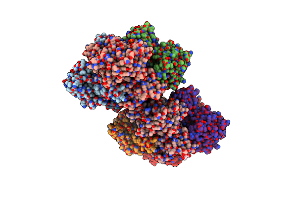
Cryo-Em Structure Of Pseudomonas Aeruginosa Tetrameric S-Adenosyl-L-Homocysteine Hydrolase With 2 Wide Open, 1 Open And 1 Closed Subunits
Organism: Pseudomonas aeruginosa
Method: ELECTRON MICROSCOPY Release Date: 2025-02-19 Classification: HYDROLASE Ligands: NAD, ADN |
Organism: Pseudomonas aeruginosa
Method: ELECTRON MICROSCOPY
Release Date: 2025-02-19
Ligands: NAD, ADN
 |
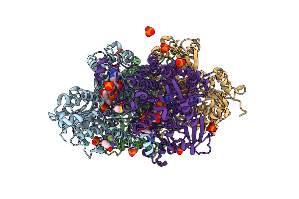
Crystal Structure Of S-Adenosyl-L-Homocysteine Hydrolase From P. Aeruginosa In Complex With F2X-Entry Library Fragment H09
Organism: Pseudomonas aeruginosa pao1
Method: X-RAY DIFFRACTION Resolution:1.77 Å Release Date: 2024-05-22 Classification: HYDROLASE Ligands: NAD, ADE, PO4, K, A1H8H, GOL |
Organism: Pseudomonas aeruginosa pao1
Method: X-RAY DIFFRACTION
Release Date: 2024-05-22
Ligands: NAD, ADE, PO4, K, A1H8H, GOL
 |
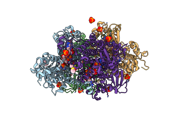
Crystal Structure Of S-Adenosyl-L-Homocysteine Hydrolase From P. Aeruginosa In Complex With F2X-Entry Library Fragment D02
Organism: Pseudomonas aeruginosa pao1
Method: X-RAY DIFFRACTION Resolution:2.07 Å Release Date: 2024-02-21 Classification: HYDROLASE Ligands: NAD, ADE, DMS, K, PO4, SY4 |
Organism: Pseudomonas aeruginosa pao1
Method: X-RAY DIFFRACTION
Release Date: 2024-02-21
Ligands: NAD, ADE, DMS, K, PO4, SY4
 |
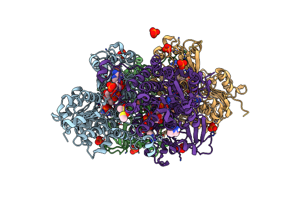
Crystal Structure Of S-Adenosyl-L-Homocysteine Hydrolase From P. Aeruginosa In Complex With F2X-Entry Library Fragment A01
Organism: Pseudomonas aeruginosa pao1
Method: X-RAY DIFFRACTION Resolution:1.64 Å Release Date: 2024-02-21 Classification: HYDROLASE Ligands: NAD, ADE, K, PO4, UHR, DMS |
Organism: Pseudomonas aeruginosa pao1
Method: X-RAY DIFFRACTION
Release Date: 2024-02-21
Ligands: NAD, ADE, K, PO4, UHR, DMS
 |
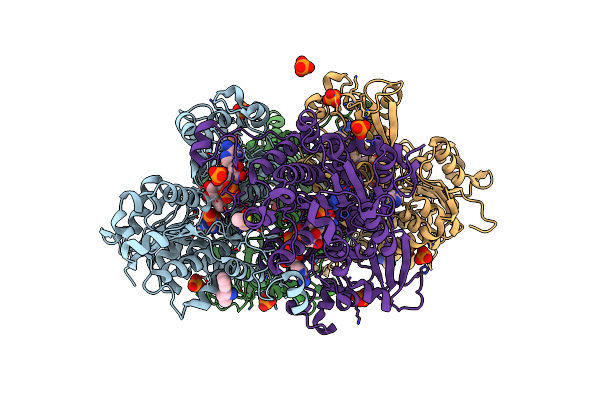
Crystal Structure Of S-Adenosyl-L-Homocysteine Hydrolase From P. Aeruginosa In Complex With F2X-Entry Library Fragment A07
Organism: Pseudomonas aeruginosa pao1
Method: X-RAY DIFFRACTION Resolution:1.88 Å Release Date: 2024-02-21 Classification: HYDROLASE Ligands: NAD, ADE, K, PO4, DMS, R8A |
Organism: Pseudomonas aeruginosa pao1
Method: X-RAY DIFFRACTION
Release Date: 2024-02-21
Ligands: NAD, ADE, K, PO4, DMS, R8A
 |
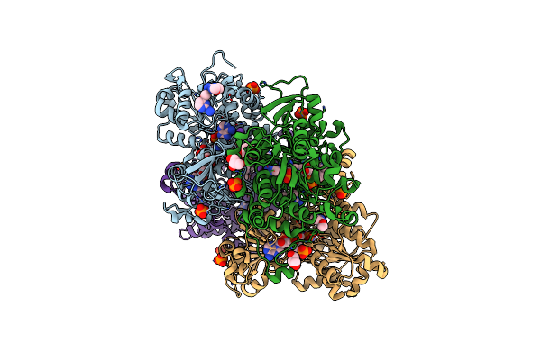
Crystal Structure Of S-Adenosyl-L-Homocysteine Hydrolase From P. Aeruginosa In Complex With F2X-Entry Library Fragment B03
Organism: Pseudomonas aeruginosa pao1
Method: X-RAY DIFFRACTION Resolution:1.80 Å Release Date: 2024-02-21 Classification: HYDROLASE Ligands: NAD, ADE, T9G, K, PO4, DMS |
Organism: Pseudomonas aeruginosa pao1
Method: X-RAY DIFFRACTION
Release Date: 2024-02-21
Ligands: NAD, ADE, T9G, K, PO4, DMS
 |
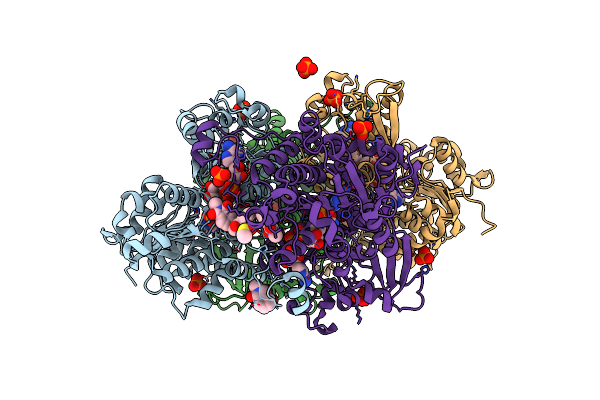
Crystal Structure Of S-Adenosyl-L-Homocysteine Hydrolase From P. Aeruginosa In Complex With F2X-Entry Library Fragment C04
Organism: Pseudomonas aeruginosa pao1
Method: X-RAY DIFFRACTION Resolution:1.73 Å Release Date: 2024-02-21 Classification: HYDROLASE Ligands: NAD, ADE, DMS, K, PO4, R8S, GOL |
Organism: Pseudomonas aeruginosa pao1
Method: X-RAY DIFFRACTION
Release Date: 2024-02-21
Ligands: NAD, ADE, DMS, K, PO4, R8S, GOL
 |
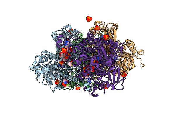
Crystal Structure Of S-Adenosyl-L-Homocysteine Hydrolase From P. Aeruginosa In Complex With F2X-Entry Library Fragment C08
Organism: Pseudomonas aeruginosa pao1
Method: X-RAY DIFFRACTION Resolution:1.60 Å Release Date: 2024-02-21 Classification: HYDROLASE Ligands: NAD, ADE, K, PO4, R8V, DMS |
Organism: Pseudomonas aeruginosa pao1
Method: X-RAY DIFFRACTION
Release Date: 2024-02-21
Ligands: NAD, ADE, K, PO4, R8V, DMS
 |
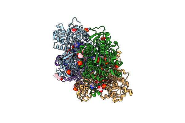
Crystal Structure Of S-Adenosyl-L-Homocysteine Hydrolase From P. Aeruginosa In Complex With F2X-Entry Library Fragment D07
Organism: Pseudomonas aeruginosa pao1
Method: X-RAY DIFFRACTION Resolution:2.02 Å Release Date: 2024-02-21 Classification: HYDROLASE Ligands: NAD, ADE, SYJ, K, PO4, DMS |
Organism: Pseudomonas aeruginosa pao1
Method: X-RAY DIFFRACTION
Release Date: 2024-02-21
Ligands: NAD, ADE, SYJ, K, PO4, DMS
 |
Crystal Structure Of S-Adenosyl-L-Homocysteine Hydrolase From P. Aeruginosa In Complex With F2X-Entry Library Fragment D10
Organism: Pseudomonas aeruginosa pao1
Method: X-RAY DIFFRACTION Resolution:1.64 Å Release Date: 2024-02-21 Classification: HYDROLASE Ligands: NAD, ADE, R9J, GOL, K, PO4, DMS |
Organism: Pseudomonas aeruginosa pao1
Method: X-RAY DIFFRACTION
Release Date: 2024-02-21
Ligands: NAD, ADE, R9J, GOL, K, PO4, DMS