

Search Count: 94
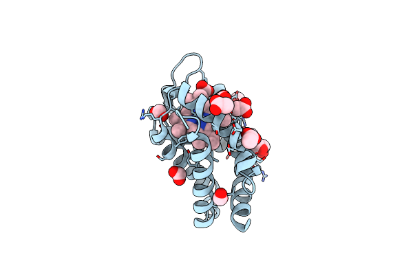 |
Crystal Structure Of Murine Neuroglobin Bound To Co At 40 K.
Organism: Mus musculus
Method: X-RAY DIFFRACTION Resolution:2.00 Å Release Date: 2019-09-11 Classification: OXYGEN TRANSPORT Ligands: HEM, CMO, FMT, ACT, GOL |
Organism: Mus musculus
Method: X-RAY DIFFRACTION
Release Date: 2019-09-11
Ligands: HEM, CMO, FMT, ACT, GOL
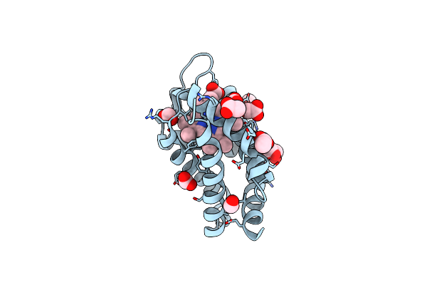 |
Crystal Structure Of Murine Neuroglobin Bound To Co At 15K Under Illumination Using Optical Fiber
Organism: Mus musculus
Method: X-RAY DIFFRACTION Resolution:1.90 Å Release Date: 2019-09-11 Classification: OXYGEN TRANSPORT Ligands: HEM, CMO, FMT, ACT, GOL |
Organism: Mus musculus
Method: X-RAY DIFFRACTION
Release Date: 2019-09-11
Ligands: HEM, CMO, FMT, ACT, GOL
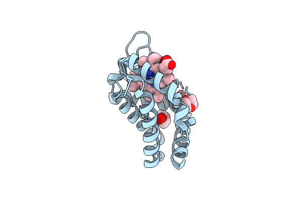 |
Metneuroglobin Under Oxygen At 50 Bar
Organism: Mus musculus
Method: X-RAY DIFFRACTION Resolution:1.62 Å Release Date: 2017-12-20 Classification: TRANSPORT PROTEIN Ligands: HEM, DIO, SO4, OXY |
Organism: Mus musculus
Method: X-RAY DIFFRACTION
Release Date: 2017-12-20
Ligands: HEM, DIO, SO4, OXY
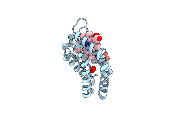 |
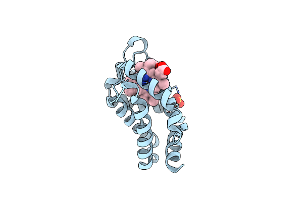
Metngb Under Oxygen At 80 Bar
Organism: Mus musculus
Method: X-RAY DIFFRACTION Resolution:1.70 Å Release Date: 2017-12-20 Classification: TRANSPORT PROTEIN Ligands: HEM, SO4, OXY |
Organism: Mus musculus
Method: X-RAY DIFFRACTION
Release Date: 2017-12-20
Ligands: HEM, SO4, OXY
 |
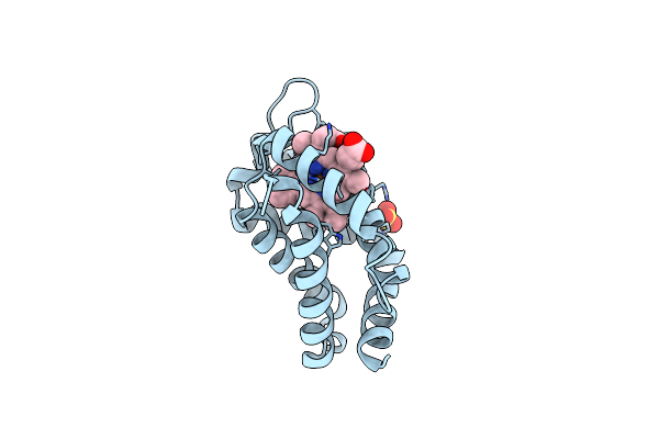
Crystal Structure Of Murine Neuroglobin Mutant M144W
Organism: Mus musculus
Method: X-RAY DIFFRACTION Resolution:1.80 Å Release Date: 2014-06-18 Classification: TRANSPORT PROTEIN Ligands: HEM, SO4 |
Organism: Mus musculus
Method: X-RAY DIFFRACTION
Release Date: 2014-06-18
Ligands: HEM, SO4
 |
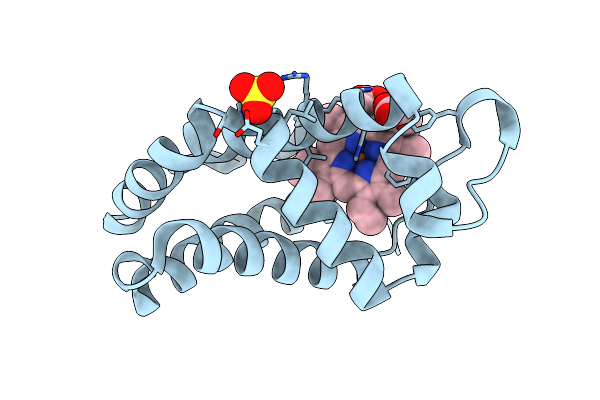
Crystal Structure Of Murine Neuroglobin Mutant V140W
Organism: Mus musculus
Method: X-RAY DIFFRACTION Resolution:2.10 Å Release Date: 2014-06-18 Classification: TRANSPORT PROTEIN Ligands: HEM, SO4 |
Organism: Mus musculus
Method: X-RAY DIFFRACTION
Release Date: 2014-06-18
Ligands: HEM, SO4
 |
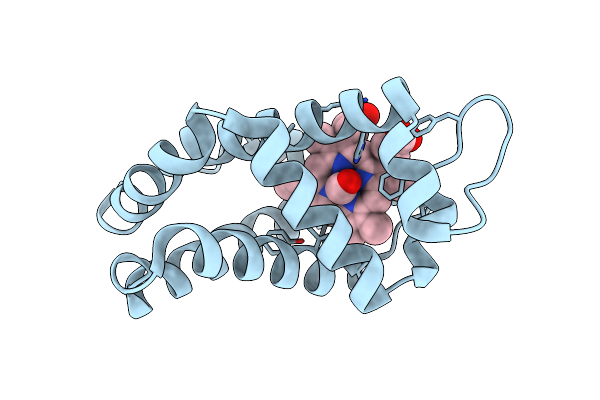
Crystal Structure Of Murine Neuroglobin Mutant F106W
Organism: Mus musculus
Method: X-RAY DIFFRACTION Resolution:1.60 Å Release Date: 2014-06-18 Classification: TRANSPORT PROTEIN Ligands: HEM, SO4 |
Organism: Mus musculus
Method: X-RAY DIFFRACTION
Release Date: 2014-06-18
Ligands: HEM, SO4
 |
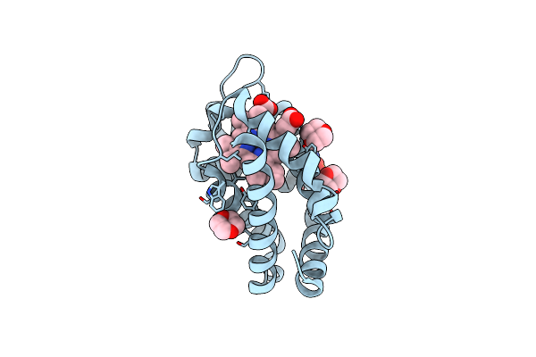
Crystal Structure Of Carbomonoxy Murine Neuroglobin Mutant V140W
Organism: Mus musculus
Method: X-RAY DIFFRACTION Resolution:2.70 Å Release Date: 2014-06-18 Classification: TRANSPORT PROTEIN Ligands: HEM, CMO |
Organism: Mus musculus
Method: X-RAY DIFFRACTION
Release Date: 2014-06-18
Ligands: HEM, CMO
 |
Crystal Structure Of Carbomonoxy Murine Neuroglobin Mutant F106W
Organism: Mus musculus
Method: X-RAY DIFFRACTION Resolution:1.80 Å Release Date: 2014-06-18 Classification: TRANSPORT PROTEIN Ligands: HEM, CMO, SO4, DIO |
Organism: Mus musculus
Method: X-RAY DIFFRACTION
Release Date: 2014-06-18
Ligands: HEM, CMO, SO4, DIO
 |
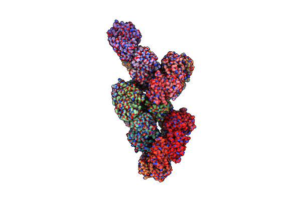
Crystal Structure Of Schistosoma Mansoni Peroxiredoxin I C48S Mutant With One Decamer In The Asu
Organism: Schistosoma mansoni
Method: X-RAY DIFFRACTION Resolution:2.49 Å Release Date: 2013-09-11 Classification: OXIDOREDUCTASE Ligands: SO4, PEG |
Organism: Schistosoma mansoni
Method: X-RAY DIFFRACTION
Release Date: 2013-09-11
Ligands: SO4, PEG
 |
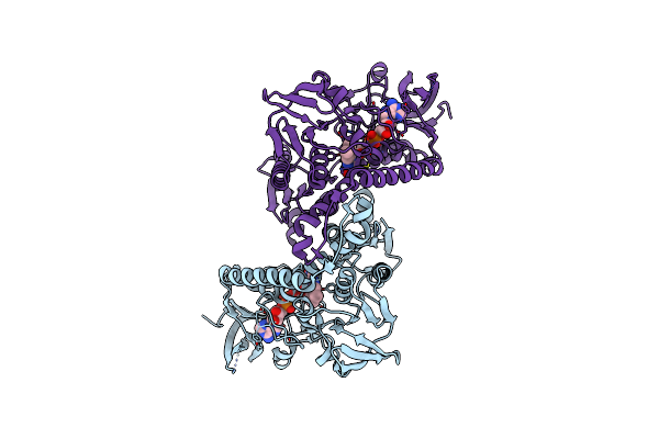
Crystal Structure Of Schistosoma Mansoni Peroxiredoxin 1 C48P Mutant Form With Four Decamers In The Asymmetric Unit
Organism: Schistosoma mansoni
Method: X-RAY DIFFRACTION Resolution:3.52 Å Release Date: 2013-09-11 Classification: OXIDOREDUCTASE |
Organism: Schistosoma mansoni
Method: X-RAY DIFFRACTION
Release Date: 2013-09-11
 |
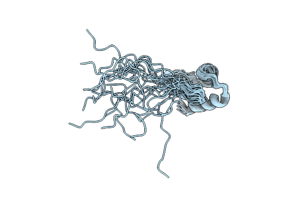
Crystal Structure Of Plasmodium Falciparum Oxidised Thioredoxin Reductase At 2.9 Angstrom
Organism: Plasmodium falciparum
Method: X-RAY DIFFRACTION Resolution:2.90 Å Release Date: 2012-08-29 Classification: OXIDOREDUCTASE Ligands: FAD |
Organism: Plasmodium falciparum
Method: X-RAY DIFFRACTION
Release Date: 2012-08-29
Ligands: FAD
 |
Nmr Structure Of Npm1_C70
Organism: Homo sapiens
Method: SOLUTION NMR Release Date: 2012-06-27 Classification: DNA BINDING PROTEIN, CHAPERONE |
Organism: Homo sapiens
Method: SOLUTION NMR
Release Date: 2012-06-27
 |
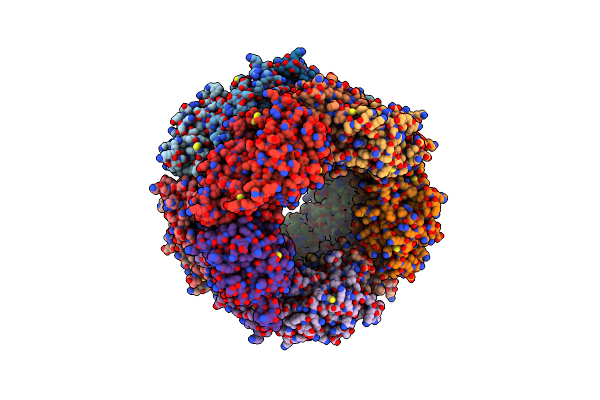
Crystal Structure Of Decameric Form Of Peroxiredoxin I From Schistosoma Mansoni
Organism: Schistosoma mansoni
Method: X-RAY DIFFRACTION Resolution:3.00 Å Release Date: 2012-03-21 Classification: OXIDOREDUCTASE |
Organism: Schistosoma mansoni
Method: X-RAY DIFFRACTION
Release Date: 2012-03-21
 |
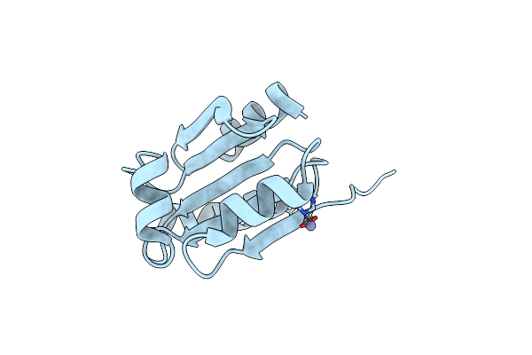
Crystal Structure Of High Molecular Weight (Hmw) Form Of Peroxiredoxin I From Schistosoma Mansoni
Organism: Schistosoma mansoni
Method: X-RAY DIFFRACTION Resolution:3.00 Å Release Date: 2012-03-21 Classification: OXIDOREDUCTASE |
Organism: Schistosoma mansoni
Method: X-RAY DIFFRACTION
Release Date: 2012-03-21
 |
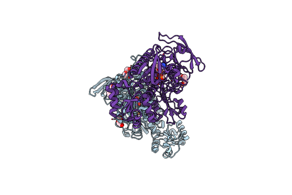
Crystal Structure Of Oxidized Schistosoma Mansoni Thioredoxin Pre- Protein At 1.6 Angstrom
Organism: Schistosoma mansoni
Method: X-RAY DIFFRACTION Resolution:1.56 Å Release Date: 2010-08-11 Classification: OXIDOREDUCTASE Ligands: CA, ZN |
Organism: Schistosoma mansoni
Method: X-RAY DIFFRACTION
Release Date: 2010-08-11
Ligands: CA, ZN
 |
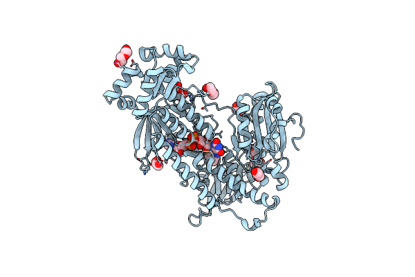
Thioredoxin Glutathione Reductase From Schistosoma Mansoni With The Reduced C-Terminal End
Organism: Schistosoma mansoni
Method: X-RAY DIFFRACTION Resolution:3.10 Å Release Date: 2010-07-21 Classification: OXIDOREDUCTASE/FLAVOPROTEIN Ligands: FAD, PGE, PEG |
Organism: Schistosoma mansoni
Method: X-RAY DIFFRACTION
Release Date: 2010-07-21
Ligands: FAD, PGE, PEG
 |
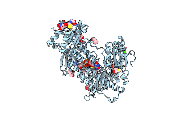
Oxidized Thioredoxin Glutathione Reductase From Schistosoma Mansoni
Organism: Schistosoma mansoni
Method: X-RAY DIFFRACTION Resolution:1.90 Å Release Date: 2010-07-21 Classification: OXIDOREDUCTASE Ligands: FAD, PEG, GOL, PG4 |
Organism: Schistosoma mansoni
Method: X-RAY DIFFRACTION
Release Date: 2010-07-21
Ligands: FAD, PEG, GOL, PG4
 |
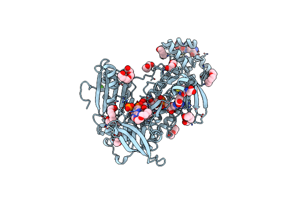
Thioredoxin Glutathione Reductase From Schistosoma Mansoni In Complex With Gsh
Organism: Schistosoma mansoni
Method: X-RAY DIFFRACTION Resolution:1.90 Å Release Date: 2010-07-21 Classification: OXIDOREDUCTASE Ligands: FAD, GSH, BME, PGE, PG4, CA, PEG |
Organism: Schistosoma mansoni
Method: X-RAY DIFFRACTION
Release Date: 2010-07-21
Ligands: FAD, GSH, BME, PGE, PG4, CA, PEG
 |
Thioredoxin Glutathione Reductase From Schistosoma Mansoni In Complex With Nadph
Organism: Schistosoma mansoni
Method: X-RAY DIFFRACTION Resolution:2.30 Å Release Date: 2010-07-21 Classification: OXIDOREDUCTASE Ligands: FAD, NDP, GSH, CA, PEG, PG4 |
Organism: Schistosoma mansoni
Method: X-RAY DIFFRACTION
Release Date: 2010-07-21
Ligands: FAD, NDP, GSH, CA, PEG, PG4