

Search Count: 28
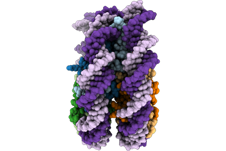 |
Nucleosome Containing An 8Oxog:A At Shl-6
Organism: Homo sapiens, Synthetic construct
Method: ELECTRON MICROSCOPY Resolution:2.80 Å Release Date: 2026-07-22 Classification: DNA BINDING PROTEIN |
Organism: Homo sapiens, Synthetic construct
Method: ELECTRON MICROSCOPY
Release Date: 2026-07-22
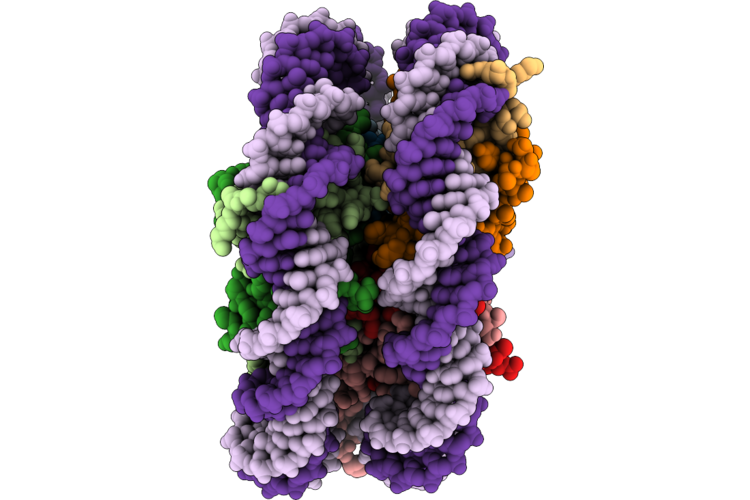 |
Nucleosome Containing An 8Oxog:A At Shl+4
Organism: Homo sapiens, Synthetic construct
Method: ELECTRON MICROSCOPY Resolution:2.70 Å Release Date: 2026-07-22 Classification: DNA BINDING PROTEIN |
Organism: Homo sapiens, Synthetic construct
Method: ELECTRON MICROSCOPY
Release Date: 2026-07-22
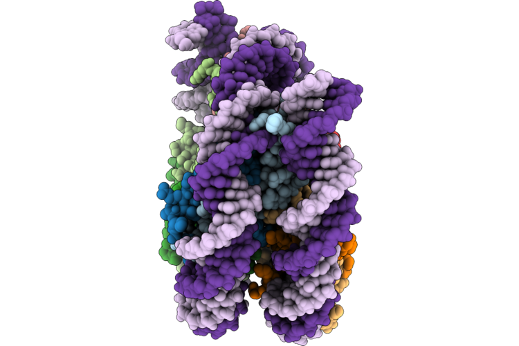 |
Nucleosome Containing An 8Oxog:A At Shl+3
Organism: Homo sapiens, Synthetic construct
Method: ELECTRON MICROSCOPY Resolution:2.50 Å Release Date: 2026-07-22 Classification: DNA BINDING PROTEIN |
Organism: Homo sapiens, Synthetic construct
Method: ELECTRON MICROSCOPY
Release Date: 2026-07-22
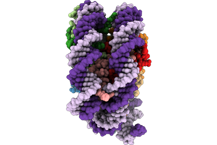 |
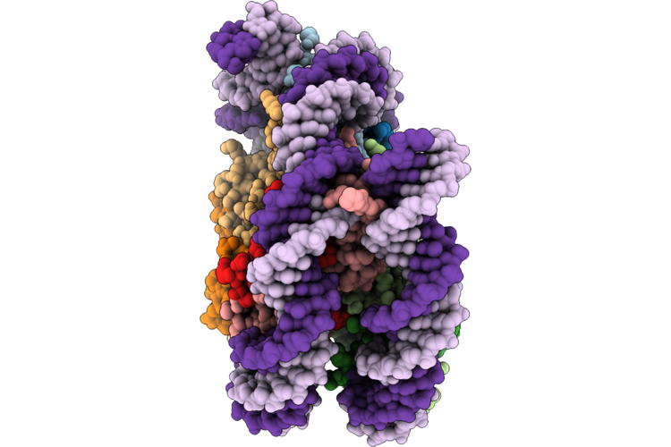
Nucleosome Containing An 8Oxog:A At Shl+2
Organism: Homo sapiens, Synthetic construct
Method: ELECTRON MICROSCOPY Resolution:2.50 Å Release Date: 2026-07-22 Classification: DNA BINDING PROTEIN |
Organism: Homo sapiens, Synthetic construct
Method: ELECTRON MICROSCOPY
Release Date: 2026-07-22
 |
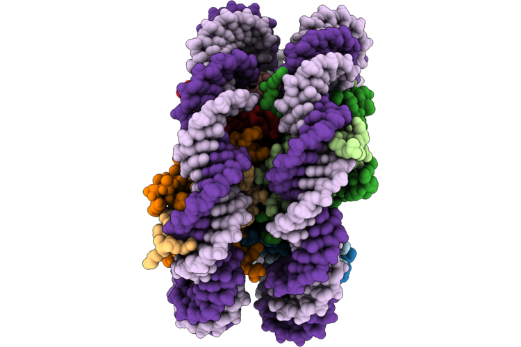
Nucleosome Containing An 8Oxog At Shl+3
Organism: Homo sapiens, Synthetic construct
Method: ELECTRON MICROSCOPY Resolution:3.00 Å Release Date: 2026-07-22 Classification: DNA BINDING PROTEIN |
Organism: Homo sapiens, Synthetic construct
Method: ELECTRON MICROSCOPY
Release Date: 2026-07-22
 |
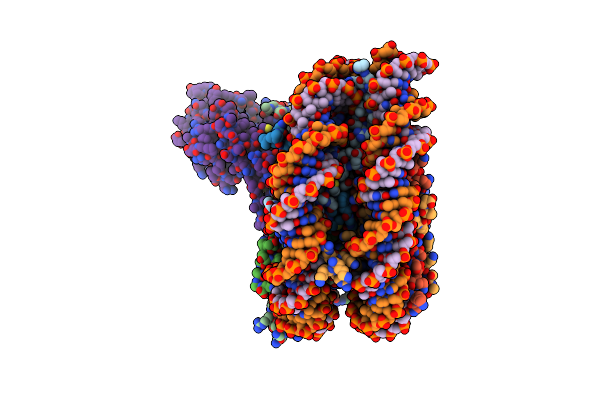
Nucleosome Containing An 8Oxog At Shl+2
Organism: Homo sapiens, Synthetic construct
Method: ELECTRON MICROSCOPY Resolution:2.80 Å Release Date: 2026-07-22 Classification: DNA BINDING PROTEIN |
Organism: Homo sapiens, Synthetic construct
Method: ELECTRON MICROSCOPY
Release Date: 2026-07-22
 |
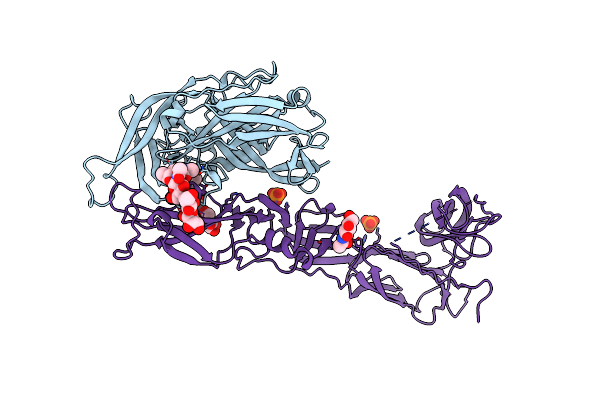
Nucleosome Bound Human Sirt6 (Composite)
Organism: Xenopus laevis, Homo sapiens
Method: ELECTRON MICROSCOPY Release Date: 2023-08-09 Classification: GENE REGULATION Ligands: ZN |
Organism: Xenopus laevis, Homo sapiens
Method: ELECTRON MICROSCOPY
Release Date: 2023-08-09
Ligands: ZN
 |
Crystal Structure Of The Envelope Glycoprotein Prefusion Complex Of Andes Virus - Mutant H953F
Organism: Andes orthohantavirus
Method: X-RAY DIFFRACTION Resolution:3.20 Å Release Date: 2020-10-14 Classification: VIRAL PROTEIN Ligands: NAG, PO4 |
Organism: Andes orthohantavirus
Method: X-RAY DIFFRACTION
Release Date: 2020-10-14
Ligands: NAG, PO4
 |
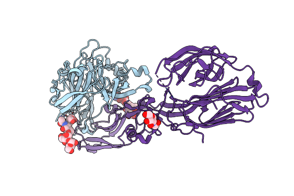
Crystal Structure Of The Envelope Glycoprotein Complex Of Andes Virus In A Near Postfusion Conformation
Organism: Andes orthohantavirus
Method: X-RAY DIFFRACTION Resolution:2.55 Å Release Date: 2020-10-14 Classification: VIRAL PROTEIN |
Organism: Andes orthohantavirus
Method: X-RAY DIFFRACTION
Release Date: 2020-10-14
 |
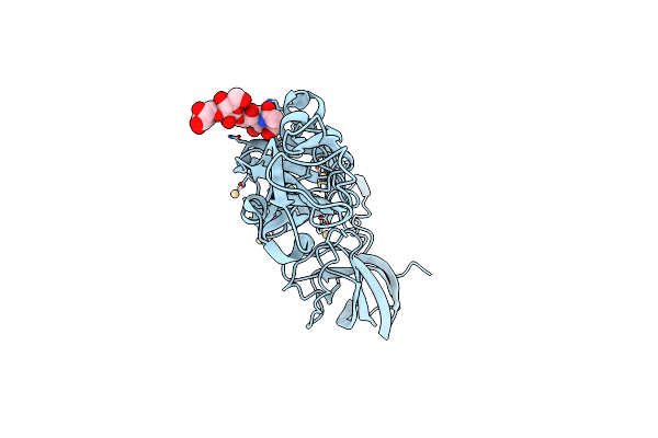
Crystal Structure Of The Envelope Glycoprotein Complex Of Maporal Virus In A Prefusion Conformation
Organism: Maporal virus
Method: X-RAY DIFFRACTION Resolution:2.20 Å Release Date: 2020-10-14 Classification: VIRAL PROTEIN Ligands: NAG |
Organism: Maporal virus
Method: X-RAY DIFFRACTION
Release Date: 2020-10-14
Ligands: NAG
 |
Structure Of Maporal Virus Envelope Glycoprotein Gc In Postfusion Conformation
Organism: Maporal virus
Method: X-RAY DIFFRACTION Resolution:2.40 Å Release Date: 2020-10-14 Classification: VIRAL PROTEIN Ligands: CD |
Organism: Maporal virus
Method: X-RAY DIFFRACTION
Release Date: 2020-10-14
Ligands: CD
 |
Structure Of Hantaan Virus Envelope Glycoprotein Gn
Organism: Hantaan orthohantavirus
Method: X-RAY DIFFRACTION Resolution:1.94 Å Release Date: 2020-10-14 Classification: VIRAL PROTEIN |
Organism: Hantaan orthohantavirus
Method: X-RAY DIFFRACTION
Release Date: 2020-10-14
 |
Structure Of Andes Virus Envelope Glycoprotein Gc In Postfusion Conformation
Organism: Andes orthohantavirus
Method: X-RAY DIFFRACTION Resolution:2.70 Å Release Date: 2020-10-14 Classification: VIRAL PROTEIN Ligands: SO4 |
Organism: Andes orthohantavirus
Method: X-RAY DIFFRACTION
Release Date: 2020-10-14
Ligands: SO4
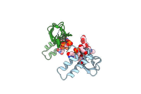 |
Crystal Structure Of The Tetramerization Domain Of The Glycoprotein Gn (Andes Virus) At Ph 7.5
Organism: Andes orthohantavirus, Drosophila melanogaster
Method: X-RAY DIFFRACTION Resolution:2.35 Å Release Date: 2020-10-14 Classification: VIRAL PROTEIN Ligands: IOD |
Organism: Andes orthohantavirus, Drosophila melanogaster
Method: X-RAY DIFFRACTION
Release Date: 2020-10-14
Ligands: IOD
 |
Crystal Structure Of The Tetramerization Domain Of The Glycoprotein Gn (Andes Virus) At Ph 4.6
Organism: Andes orthohantavirus, Drosophila melanogaster
Method: X-RAY DIFFRACTION Resolution:1.90 Å Release Date: 2020-10-14 Classification: VIRAL PROTEIN Ligands: NAG, MPD |
Organism: Andes orthohantavirus, Drosophila melanogaster
Method: X-RAY DIFFRACTION
Release Date: 2020-10-14
Ligands: NAG, MPD
 |
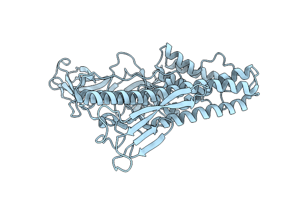
Atomic Model Of Andes Virus Glycoprotein Spike Tetramer Generated By Fitting Into A Tula Virus Reconstruction
Organism: Tula orthohantavirus
Method: ELECTRON MICROSCOPY Release Date: 2020-10-14 Classification: VIRAL PROTEIN Ligands: NAG |
Organism: Tula orthohantavirus
Method: ELECTRON MICROSCOPY
Release Date: 2020-10-14
Ligands: NAG
 |
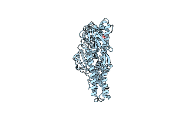
Structure Of Hrpv6 Vp5 Fitted In The Cryoem Density Of The Spike
Organism: Halorubrum pleomorphic virus 6
Method: ELECTRON MICROSCOPY Release Date: 2019-03-06 Classification: VIRAL PROTEIN |
Organism: Halorubrum pleomorphic virus 6
Method: ELECTRON MICROSCOPY
Release Date: 2019-03-06
 |
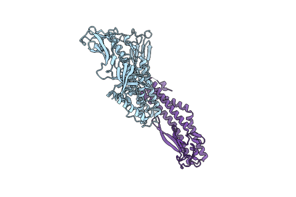
Crystal Structure Of Vp5 From Haloarchaeal Pleomorphic Virus 2
Organism: Halorubrum pleomorphic virus 2
Method: X-RAY DIFFRACTION Resolution:2.46 Å Release Date: 2019-02-27 Classification: VIRAL PROTEIN Ligands: NAG, CL |
Organism: Halorubrum pleomorphic virus 2
Method: X-RAY DIFFRACTION
Release Date: 2019-02-27
Ligands: NAG, CL
 |
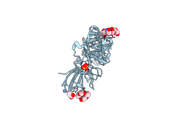
Crystal Structure Of Vp5 From Haloarchaeal Pleomorphic Virus 6
Organism: Halorubrum pleomorphic virus 6
Method: X-RAY DIFFRACTION Resolution:2.69 Å Release Date: 2019-02-27 Classification: VIRAL PROTEIN Ligands: BR |
Organism: Halorubrum pleomorphic virus 6
Method: X-RAY DIFFRACTION
Release Date: 2019-02-27
Ligands: BR
 |
Crystal Structure Of Glycoprotein C From Puumala Virus In The Post-Fusion Conformation (Ph 6.0)
Organism: Puumala virus (strain p360)
Method: X-RAY DIFFRACTION Resolution:1.80 Å Release Date: 2016-10-05 Classification: VIRAL PROTEIN Ligands: P33, SO4, ACT |
Organism: Puumala virus (strain p360)
Method: X-RAY DIFFRACTION
Release Date: 2016-10-05
Ligands: P33, SO4, ACT