

Search Count: 18
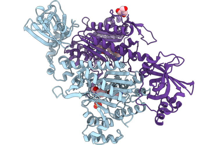 |
Cpkrs In Complex With Inhibitor Ddd01038762
Organism: Cryptosporidium parvum iowa ii
Method: X-RAY DIFFRACTION Resolution:1.84 Å Release Date: 2026-06-10 Classification: LIGASE Ligands: LYS, A1JCS, SO4, TRS |
Organism: Cryptosporidium parvum iowa ii
Method: X-RAY DIFFRACTION
Release Date: 2026-06-10
Ligands: LYS, A1JCS, SO4, TRS
 |
Crystal Structure Of Lysyl-Trna Synthetase From Cryptosporidium Parvum Complexed With L-Lysine And Inhibitor Ddd01887015
Organism: Cryptosporidium parvum iowa ii
Method: X-RAY DIFFRACTION Resolution:1.90 Å Release Date: 2026-06-10 Classification: LIGASE Ligands: A1JCT, EDO, TRS, GOL, LYS, SO4 |
Organism: Cryptosporidium parvum iowa ii
Method: X-RAY DIFFRACTION
Release Date: 2026-06-10
Ligands: A1JCT, EDO, TRS, GOL, LYS, SO4
 |
Crystal Structure Of Lysyl-Trna Synthetase From Cryptosporidium Parvum Complexed With L-Lysine And Inhibitor Ddd01932549
Organism: Cryptosporidium parvum iowa ii
Method: X-RAY DIFFRACTION Resolution:2.30 Å Release Date: 2026-06-10 Classification: LIGASE Ligands: LYS, A1JC7, SO4 |
Organism: Cryptosporidium parvum iowa ii
Method: X-RAY DIFFRACTION
Release Date: 2026-06-10
Ligands: LYS, A1JC7, SO4
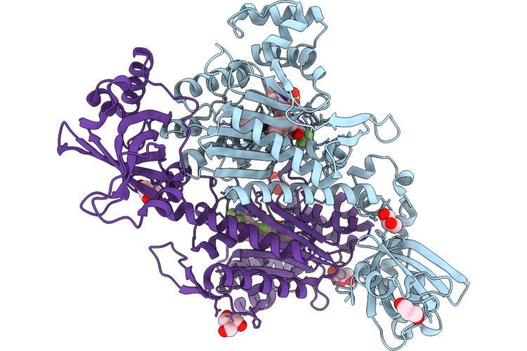 |
Crystal Structure Of Lysyl-Trna Synthetase From Cryptosporidium Parvum / Plasmodium Falciparum Chimera Complexed With L-Lysine And Inhibitor Ddd02174286
Organism: Cryptosporidium parvum iowa ii
Method: X-RAY DIFFRACTION Resolution:1.90 Å Release Date: 2026-06-10 Classification: LIGASE Ligands: LYS, A1JC4, GOL, SO4, TRS |
Organism: Cryptosporidium parvum iowa ii
Method: X-RAY DIFFRACTION
Release Date: 2026-06-10
Ligands: LYS, A1JC4, GOL, SO4, TRS
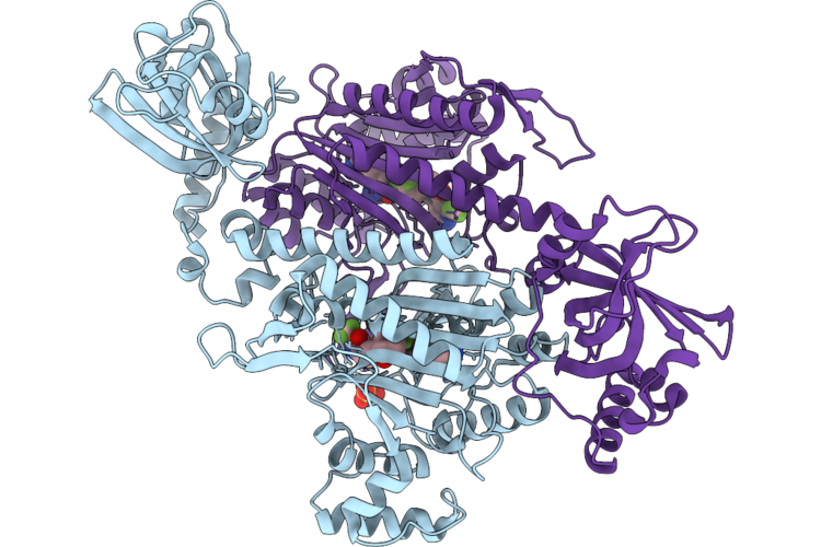 |
Crystal Structure Of Lysyl-Trna Synthetase From Cryptosporidium Parvum Complexed With L-Lysine And Inhibitor Ddd01827593
Organism: Cryptosporidium parvum iowa ii
Method: X-RAY DIFFRACTION Resolution:1.50 Å Release Date: 2026-06-10 Classification: LIGASE Ligands: LYS, A1JC9, SO4 |
Organism: Cryptosporidium parvum iowa ii
Method: X-RAY DIFFRACTION
Release Date: 2026-06-10
Ligands: LYS, A1JC9, SO4
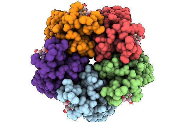 |
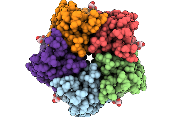
Structure Of The Honeybee Gabaa Rdl Receptor With Gaba And Abamectin
Organism: Apis mellifera
Method: ELECTRON MICROSCOPY Release Date: 2026-02-18 Classification: MEMBRANE PROTEIN Ligands: ABU, NAG, A1JNZ |
Organism: Apis mellifera
Method: ELECTRON MICROSCOPY
Release Date: 2026-02-18
Ligands: ABU, NAG, A1JNZ
 |
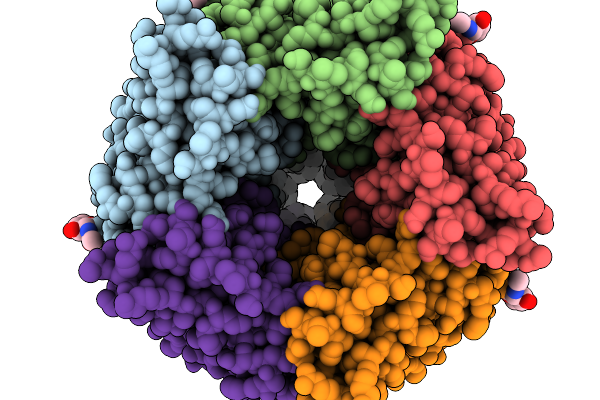
Structure Of The Honeybee Gabaa Rdl Receptor With Gaba
Organism: Apis mellifera
Method: ELECTRON MICROSCOPY Release Date: 2026-02-11 Classification: MEMBRANE PROTEIN Ligands: NAG, ABU |
Organism: Apis mellifera
Method: ELECTRON MICROSCOPY
Release Date: 2026-02-11
Ligands: NAG, ABU
 |
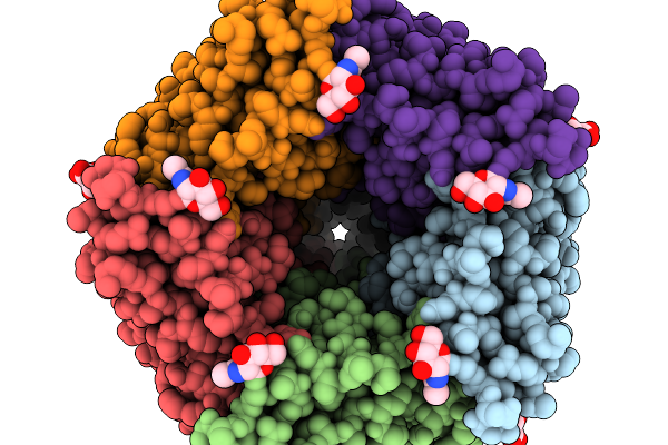
Structure Of The Honeybee Gabaa Rdl Receptor Apo State
Organism: Apis mellifera
Method: ELECTRON MICROSCOPY Release Date: 2026-02-11 Classification: MEMBRANE PROTEIN Ligands: NAG |
Organism: Apis mellifera
Method: ELECTRON MICROSCOPY
Release Date: 2026-02-11
Ligands: NAG
 |
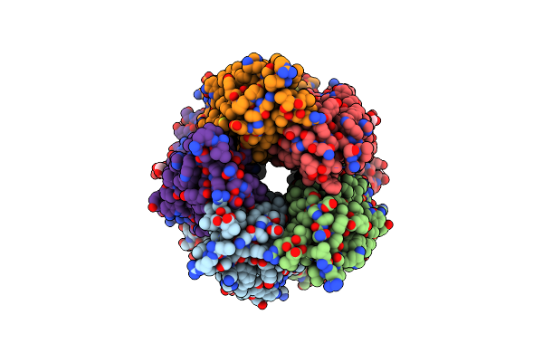
Structure Of The Honeybee Gabaa Rdl Receptor With Chrodrimanin B
Organism: Apis mellifera
Method: ELECTRON MICROSCOPY Release Date: 2026-02-11 Classification: MEMBRANE PROTEIN Ligands: NAG, PT5, A1JN7 |
Organism: Apis mellifera
Method: ELECTRON MICROSCOPY
Release Date: 2026-02-11
Ligands: NAG, PT5, A1JN7
 |
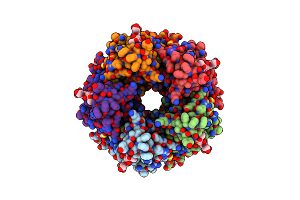
Human Serotonin 5-Ht3A Receptor (Apo, Active/Distorted Conformation)
Organism: Homo sapiens
Method: ELECTRON MICROSCOPY Release Date: 2024-05-15 Classification: MEMBRANE PROTEIN Ligands: NAG |
Organism: Homo sapiens
Method: ELECTRON MICROSCOPY
Release Date: 2024-05-15
Ligands: NAG
 |
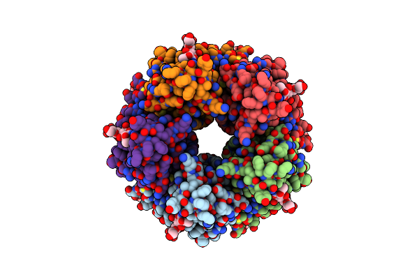
Human Serotonin 5-Ht3A Receptor In Complex With Vortioxetine (Detergent, Ecd Only, Active/Distorted Conformation)
Organism: Homo sapiens
Method: ELECTRON MICROSCOPY Release Date: 2024-05-15 Classification: MEMBRANE PROTEIN Ligands: NAG, VTX |
Organism: Homo sapiens
Method: ELECTRON MICROSCOPY
Release Date: 2024-05-15
Ligands: NAG, VTX
 |
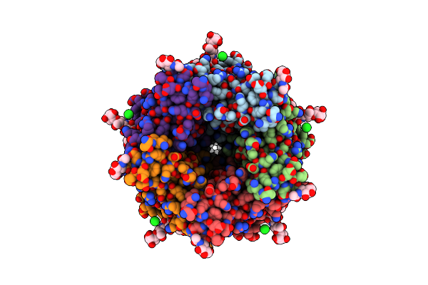
Human Serotonin 5-Ht3A Receptor In Complex With Vortioxetine (Nanodiscs, Ecd, Active/Distorted Conformation)
Organism: Homo sapiens
Method: ELECTRON MICROSCOPY Release Date: 2024-05-15 Classification: MEMBRANE PROTEIN Ligands: NAG, VTX |
Organism: Homo sapiens
Method: ELECTRON MICROSCOPY
Release Date: 2024-05-15
Ligands: NAG, VTX
 |
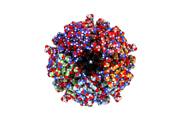
Mouse Serotonin 5-Ht3A Receptor In Complex With Vortioxetine
Organism: Mus musculus
Method: ELECTRON MICROSCOPY Release Date: 2024-03-06 Classification: MEMBRANE PROTEIN Ligands: NAG, CL, VTX |
Organism: Mus musculus
Method: ELECTRON MICROSCOPY
Release Date: 2024-03-06
Ligands: NAG, CL, VTX
 |
Human Serotonin 5-Ht3A Receptor (Apo, Resting Conformation)
Organism: Homo sapiens
Method: ELECTRON MICROSCOPY Release Date: 2024-03-06 Classification: MEMBRANE PROTEIN Ligands: NAG |
Organism: Homo sapiens
Method: ELECTRON MICROSCOPY
Release Date: 2024-03-06
Ligands: NAG
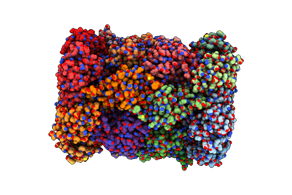 |
Structure Of Plasmodium Falciparum 20S Proteasome With Bound Bortezomib
Organism: Plasmodium falciparum (isolate 3d7), Plasmodium falciparum
Method: ELECTRON MICROSCOPY Release Date: 2021-09-22 Classification: HYDROLASE Ligands: BO2 |
Organism: Plasmodium falciparum (isolate 3d7), Plasmodium falciparum
Method: ELECTRON MICROSCOPY
Release Date: 2021-09-22
Ligands: BO2
 |
Structure Of Plasmodium Falciparum 20S Proteasome With Bound Mpi-5
Organism: Plasmodium falciparum (isolate 3d7)
Method: ELECTRON MICROSCOPY Release Date: 2021-09-22 Classification: HYDROLASE Ligands: YHD |
Organism: Plasmodium falciparum (isolate 3d7)
Method: ELECTRON MICROSCOPY
Release Date: 2021-09-22
Ligands: YHD
 |
Structure Of Human 20S Proteasome With Bound Mpi-5
Organism: Homo sapiens
Method: ELECTRON MICROSCOPY Release Date: 2021-09-22 Classification: HYDROLASE Ligands: YHD |
Organism: Homo sapiens
Method: ELECTRON MICROSCOPY
Release Date: 2021-09-22
Ligands: YHD
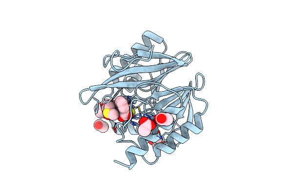 |
Crystal Structure Of The Vim-2 Acquired Metallo-Beta-Lactamase In Complex With Jmv-4690 (Cpd 31)
Organism: Pseudomonas aeruginosa
Method: X-RAY DIFFRACTION Resolution:1.95 Å Release Date: 2020-09-30 Classification: HYDROLASE Ligands: ZN, PJB, EDO, ACT, DMS |
Organism: Pseudomonas aeruginosa
Method: X-RAY DIFFRACTION
Release Date: 2020-09-30
Ligands: ZN, PJB, EDO, ACT, DMS