

Search Count: 72
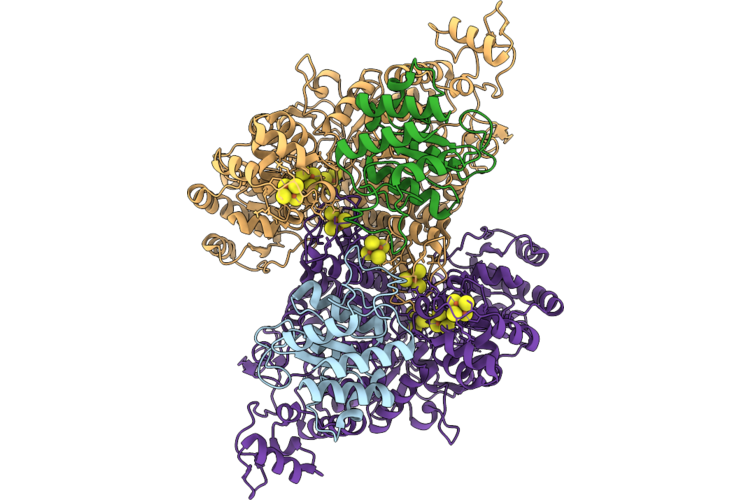 |
Cryo-Em Structure Of The Co Dehydrogenase (Codh) Subcomplex From Methanosarcina Acetivorans
Organism: Methanosarcina acetivorans
Method: ELECTRON MICROSCOPY Release Date: 2026-07-29 Classification: METAL BINDING PROTEIN Ligands: SF4, RQM |
Organism: Methanosarcina acetivorans
Method: ELECTRON MICROSCOPY
Release Date: 2026-07-29
Ligands: SF4, RQM
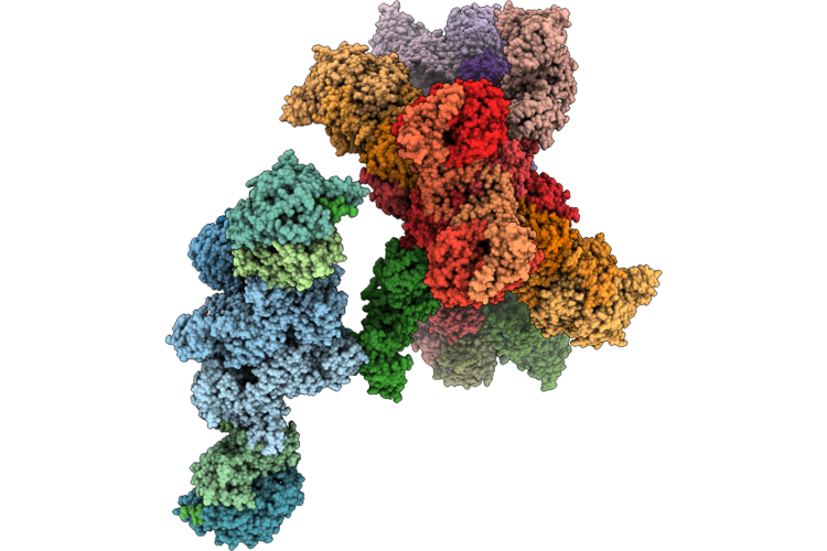 |
Heterodisulfide Reductase-Formylmethanofuran Dehydrogenase Super-Assembly
Organism: Methanococcus maripaludis, Methanococcus maripaludis deltae
Method: ELECTRON MICROSCOPY Resolution:4.00 Å Release Date: 2026-07-01 Classification: ELECTRON TRANSPORT Ligands: SF4, FAD, 9S8, FES, ZN, MGD, W, NFU |
Organism: Methanococcus maripaludis, Methanococcus maripaludis deltae
Method: ELECTRON MICROSCOPY
Release Date: 2026-07-01
Ligands: SF4, FAD, 9S8, FES, ZN, MGD, W, NFU
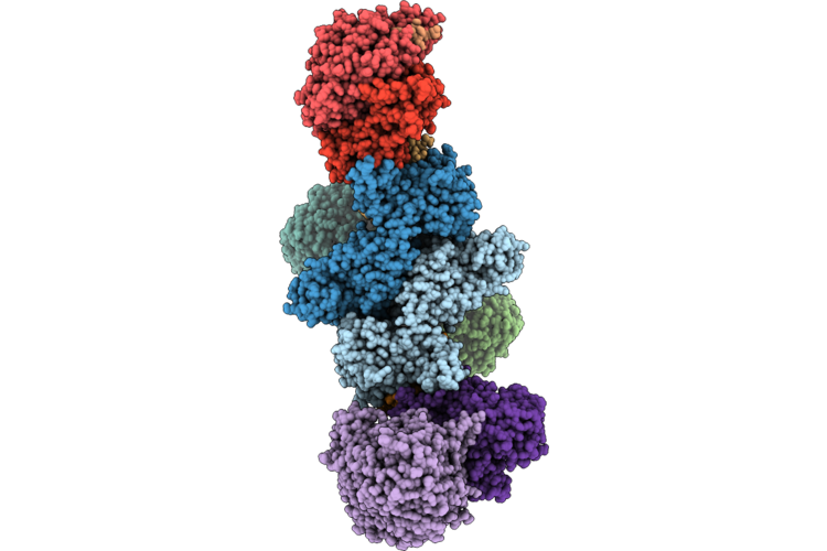 |
Heterodisulfide-Hydrogenase-Formate Dehydrogenase Dimer
Organism: Methanococcus maripaludis
Method: ELECTRON MICROSCOPY Resolution:4.00 Å Release Date: 2026-07-01 Classification: ELECTRON TRANSPORT Ligands: SF4, FAD, 9S8, NFU, FES, W, MGD |
Organism: Methanococcus maripaludis
Method: ELECTRON MICROSCOPY
Release Date: 2026-07-01
Ligands: SF4, FAD, 9S8, NFU, FES, W, MGD
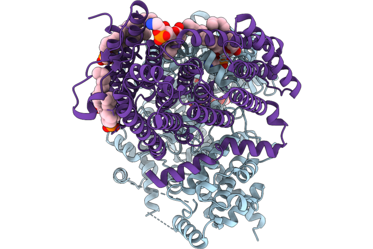 |
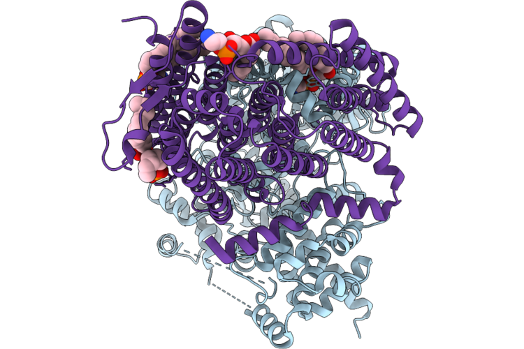
Structure Of Daba2B2 Complex Solved Under Ambient Condition
Organism: Halothiobacillus neapolitanus c2
Method: ELECTRON MICROSCOPY Release Date: 2026-04-29 Classification: TRANSPORT PROTEIN Ligands: ZN, CO2, 6OU |
Organism: Halothiobacillus neapolitanus c2
Method: ELECTRON MICROSCOPY
Release Date: 2026-04-29
Ligands: ZN, CO2, 6OU
 |
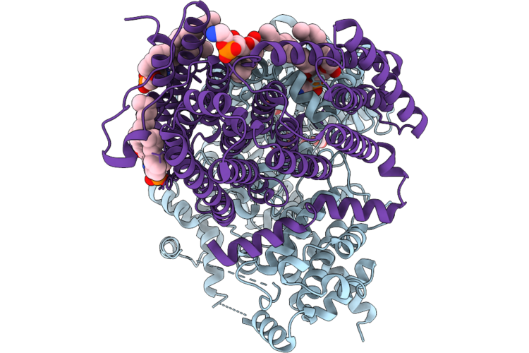
Structure Of Daba2B2 Complex Under 0.1M Bicarbonate
Organism: Halothiobacillus neapolitanus c2
Method: ELECTRON MICROSCOPY Release Date: 2026-04-29 Classification: TRANSPORT PROTEIN Ligands: ZN, BCT, CO2, 6OU |
Organism: Halothiobacillus neapolitanus c2
Method: ELECTRON MICROSCOPY
Release Date: 2026-04-29
Ligands: ZN, BCT, CO2, 6OU
 |
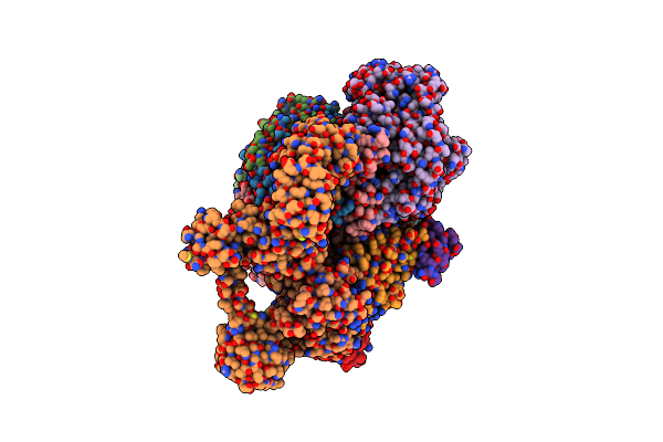
Structure Of Daba2B2 Complex Under 17 Mm Co2
Organism: Halothiobacillus neapolitanus c2
Method: ELECTRON MICROSCOPY Release Date: 2026-04-29 Classification: TRANSPORT PROTEIN Ligands: ZN, CO2, 6OU |
Organism: Halothiobacillus neapolitanus c2
Method: ELECTRON MICROSCOPY
Release Date: 2026-04-29
Ligands: ZN, CO2, 6OU
 |
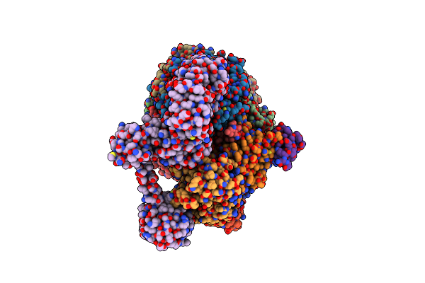
Methyl-Coenzyme M Reductase Activation Complex Binding To The A2 Component
Organism: Methanococcus maripaludis
Method: ELECTRON MICROSCOPY Release Date: 2025-02-26 Classification: OXIDOREDUCTASE Ligands: SHT, TP7, COM, F43, S5Q, ZN, ATP, MG |
Organism: Methanococcus maripaludis
Method: ELECTRON MICROSCOPY
Release Date: 2025-02-26
Ligands: SHT, TP7, COM, F43, S5Q, ZN, ATP, MG
 |
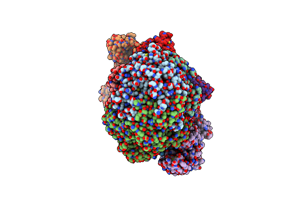
Methyl-Coenzyme M Reductase Activation Complex Without The A2 Component
Organism: Methanococcus maripaludis
Method: ELECTRON MICROSCOPY Release Date: 2025-02-26 Classification: OXIDOREDUCTASE Ligands: F43, SHT, COM, TP7, S5Q |
Organism: Methanococcus maripaludis
Method: ELECTRON MICROSCOPY
Release Date: 2025-02-26
Ligands: F43, SHT, COM, TP7, S5Q
 |
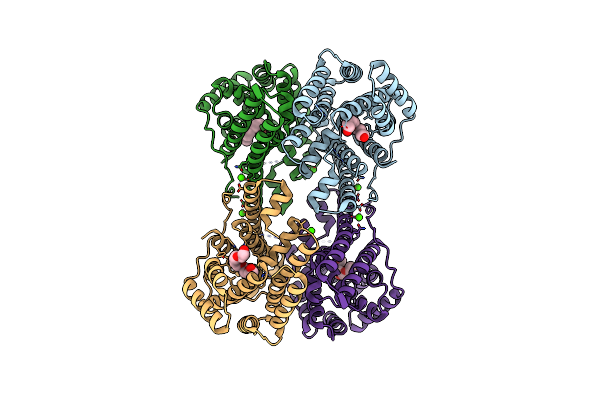
Methyl-Coenzyme M Reductase Activation Complex Binding To The A2 Component After Incubation With Atp
Organism: Methanococcus maripaludis
Method: ELECTRON MICROSCOPY Release Date: 2025-02-26 Classification: OXIDOREDUCTASE Ligands: F43, COM, TP7, SHT, S5Q, ZN, ATP, MG |
Organism: Methanococcus maripaludis
Method: ELECTRON MICROSCOPY
Release Date: 2025-02-26
Ligands: F43, COM, TP7, SHT, S5Q, ZN, ATP, MG
 |
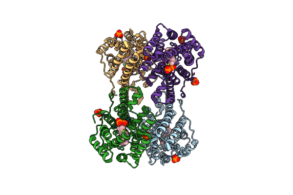
Crystal Structure Of Caryolan-1-Ol Synthase From S. Griseus With Peg Molecule In The Active Site
Organism: Streptomyces griseus
Method: X-RAY DIFFRACTION Resolution:2.33 Å Release Date: 2024-11-06 Classification: METAL BINDING PROTEIN Ligands: PG4, CA, 1PE |
Organism: Streptomyces griseus
Method: X-RAY DIFFRACTION
Release Date: 2024-11-06
Ligands: PG4, CA, 1PE
 |
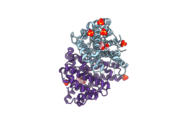
Crystal Structure Of Caryolan-1-Ol Synthase Complexed With 2-Fluorofarnesyl Diphosphate
Organism: Streptomyces griseus
Method: X-RAY DIFFRACTION Resolution:2.65 Å Release Date: 2024-11-06 Classification: METAL BINDING PROTEIN Ligands: FPF, PO4, MG, DPO |
Organism: Streptomyces griseus
Method: X-RAY DIFFRACTION
Release Date: 2024-11-06
Ligands: FPF, PO4, MG, DPO
 |
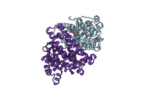
Crystal Structure Of Pentalenene Synthase Variant F76A With Peg Molecule In The Active Site
Organism: Streptomyces exfoliatus
Method: X-RAY DIFFRACTION Resolution:2.50 Å Release Date: 2024-11-06 Classification: METAL BINDING PROTEIN Ligands: PG4, SO4 |
Organism: Streptomyces exfoliatus
Method: X-RAY DIFFRACTION
Release Date: 2024-11-06
Ligands: PG4, SO4
 |
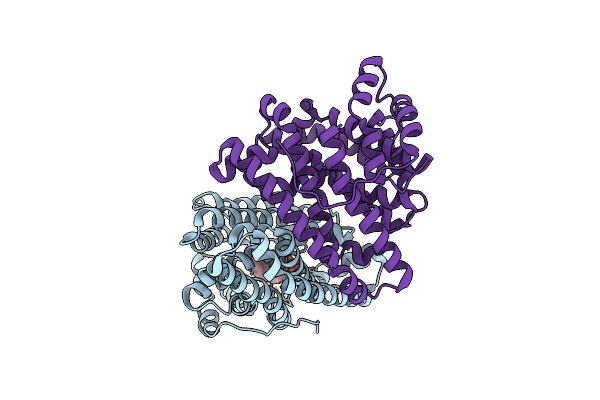
Crystal Structure Of Pentalenene Synthase Variant F76A Complexed With 2-Fluorofarnesyl Diphosphate
Organism: Streptomyces exfoliatus
Method: X-RAY DIFFRACTION Resolution:2.20 Å Release Date: 2024-11-06 Classification: METAL BINDING PROTEIN Ligands: FPF, MG |
Organism: Streptomyces exfoliatus
Method: X-RAY DIFFRACTION
Release Date: 2024-11-06
Ligands: FPF, MG
 |
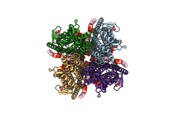
Crystal Structure Of Pentalenene Synthase Variant F76A Complexed With 12,13-Difluorofarnesyl Diphosphate
Organism: Streptomyces exfoliatus
Method: X-RAY DIFFRACTION Resolution:2.65 Å Release Date: 2024-11-06 Classification: METAL BINDING PROTEIN Ligands: FDF, MG |
Organism: Streptomyces exfoliatus
Method: X-RAY DIFFRACTION
Release Date: 2024-11-06
Ligands: FDF, MG
 |
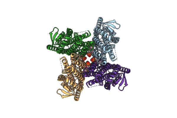
Cryoem Structure Of Insect Gustatory Receptor Bmgr9
Organism: Bombyx mori
Method: ELECTRON MICROSCOPY Release Date: 2024-01-10 Classification: MEMBRANE PROTEIN Ligands: PSC, PC7, 9Z9 |
Organism: Bombyx mori
Method: ELECTRON MICROSCOPY
Release Date: 2024-01-10
Ligands: PSC, PC7, 9Z9
 |
Cryoem Structure Of Insect Gustatory Receptor Bmgr9 In The Presence Of Fructose
Organism: Bombyx mori
Method: ELECTRON MICROSCOPY Release Date: 2024-01-10 Classification: MEMBRANE PROTEIN Ligands: PSC |
Organism: Bombyx mori
Method: ELECTRON MICROSCOPY
Release Date: 2024-01-10
Ligands: PSC
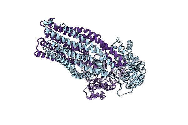 |
If(Apo/As Isolated) Conformation Of Cyddc (Dataset-1)
Organism: Escherichia coli k-12
Method: ELECTRON MICROSCOPY Release Date: 2023-04-19 Classification: MEMBRANE PROTEIN |
Organism: Escherichia coli k-12
Method: ELECTRON MICROSCOPY
Release Date: 2023-04-19
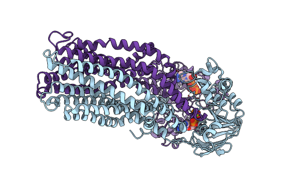 |
If(Apo/Asym) Conformation Of Cyddc In Adp+Pi(Cydc)/Atp(Cydd) Bound State (Dataset-2)
Organism: Escherichia coli k-12
Method: ELECTRON MICROSCOPY Release Date: 2023-04-19 Classification: MEMBRANE PROTEIN Ligands: MG, ADP, PO3, ATP |
Organism: Escherichia coli k-12
Method: ELECTRON MICROSCOPY
Release Date: 2023-04-19
Ligands: MG, ADP, PO3, ATP
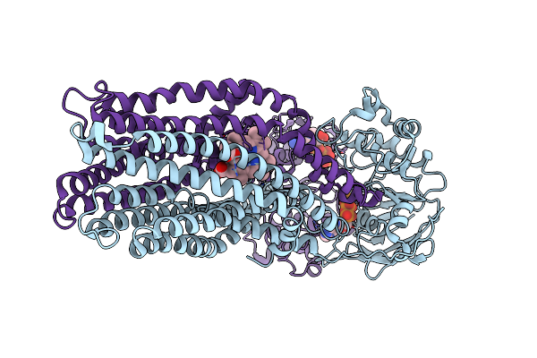 |
If(Heme/Bound) Conformation Of Cyddc In Adp+Pi(Cydc)/Atp(Cydd) Bound State (Dataset-2)
Organism: Escherichia coli k-12
Method: ELECTRON MICROSCOPY Release Date: 2023-04-19 Classification: MEMBRANE PROTEIN Ligands: HEB, MG, ADP, PO3, ATP |
Organism: Escherichia coli k-12
Method: ELECTRON MICROSCOPY
Release Date: 2023-04-19
Ligands: HEB, MG, ADP, PO3, ATP
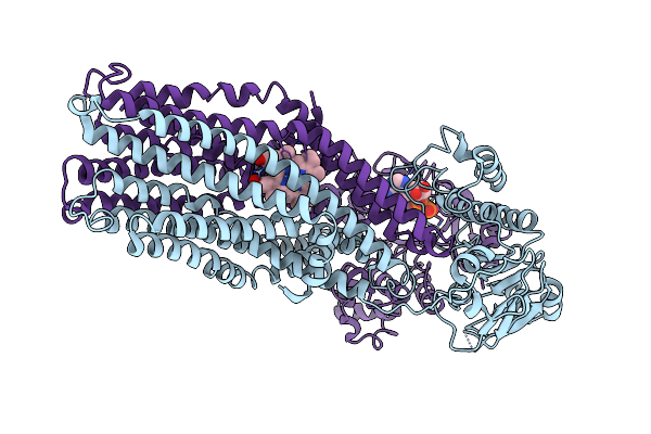 |
If(Heme/Confined) Conformation Of Cyddc In Adp(Cydd) Bound State (Dataset-3)
Organism: Escherichia coli k-12
Method: ELECTRON MICROSCOPY Release Date: 2023-04-19 Classification: MEMBRANE PROTEIN Ligands: HEB, MG, ADP |
Organism: Escherichia coli k-12
Method: ELECTRON MICROSCOPY
Release Date: 2023-04-19
Ligands: HEB, MG, ADP