

Search Count: 36
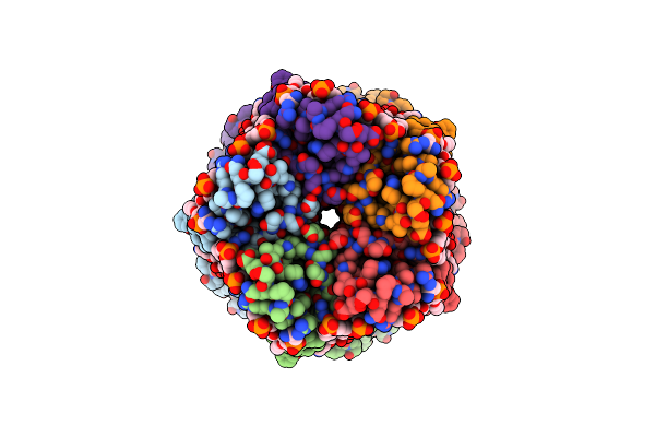 |
Cryo-Em Structure Of Nanodisc (Pe:Ps:Pc) Reconstituted Glic At Ph 5.5
Organism: Gloeobacter violaceus
Method: ELECTRON MICROSCOPY Release Date: 2024-04-17 Classification: MEMBRANE PROTEIN Ligands: PEE |
Organism: Gloeobacter violaceus
Method: ELECTRON MICROSCOPY
Release Date: 2024-04-17
Ligands: PEE
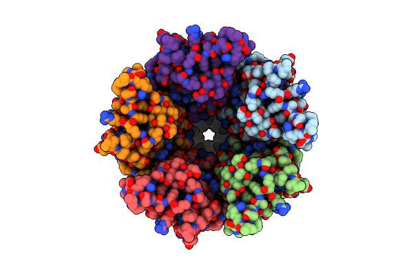 |
Cryo-Em Structure Of Nanodisc (Pe:Ps:Pc) Reconstituted Glic At Ph 4 In Closed State
Organism: Gloeobacter violaceus
Method: ELECTRON MICROSCOPY Release Date: 2024-04-17 Classification: MEMBRANE PROTEIN Ligands: CL, PEE |
Organism: Gloeobacter violaceus
Method: ELECTRON MICROSCOPY
Release Date: 2024-04-17
Ligands: CL, PEE
 |
Cryo-Em Structure Of Nanodisc (Pe:Ps:Pc) Reconstituted Glic At Ph 2.5
Organism: Gloeobacter violaceus
Method: ELECTRON MICROSCOPY Release Date: 2024-04-17 Classification: MEMBRANE PROTEIN Ligands: CL, PEE |
Organism: Gloeobacter violaceus
Method: ELECTRON MICROSCOPY
Release Date: 2024-04-17
Ligands: CL, PEE
 |
Cryo-Em Structure Of Nanodisc (Pe:Ps:Pc) Reconstituted Glic At Ph 4 In Intermediate State
Organism: Gloeobacter violaceus
Method: ELECTRON MICROSCOPY Release Date: 2024-04-17 Classification: MEMBRANE PROTEIN Ligands: CL, PEE |
Organism: Gloeobacter violaceus
Method: ELECTRON MICROSCOPY
Release Date: 2024-04-17
Ligands: CL, PEE
 |
Cryo-Em Structure Of Nanodisc (Pe:Ps:Pc) Reconstituted Glic At Ph 4 In Open State
Organism: Gloeobacter violaceus
Method: ELECTRON MICROSCOPY Release Date: 2024-04-17 Classification: MEMBRANE PROTEIN Ligands: CL, PEE |
Organism: Gloeobacter violaceus
Method: ELECTRON MICROSCOPY
Release Date: 2024-04-17
Ligands: CL, PEE
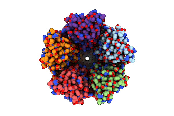 |
Cryo-Em Structure Of Nanodisc (Asolectin) Reconstituted Glic At Ph 7.5
Organism: Gloeobacter violaceus
Method: ELECTRON MICROSCOPY Release Date: 2024-04-10 Classification: MEMBRANE PROTEIN Ligands: PLC |
Organism: Gloeobacter violaceus
Method: ELECTRON MICROSCOPY
Release Date: 2024-04-10
Ligands: PLC
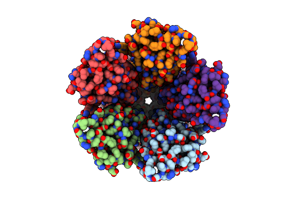 |
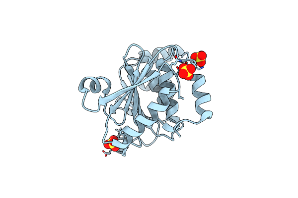
Cryo-Em Structure Of Nanodisc (Pe:Ps:Pc) Reconstituted Glic At Ph 7.5
Organism: Gloeobacter violaceus
Method: ELECTRON MICROSCOPY Release Date: 2024-04-10 Classification: MEMBRANE PROTEIN Ligands: PEE |
Organism: Gloeobacter violaceus
Method: ELECTRON MICROSCOPY
Release Date: 2024-04-10
Ligands: PEE
 |
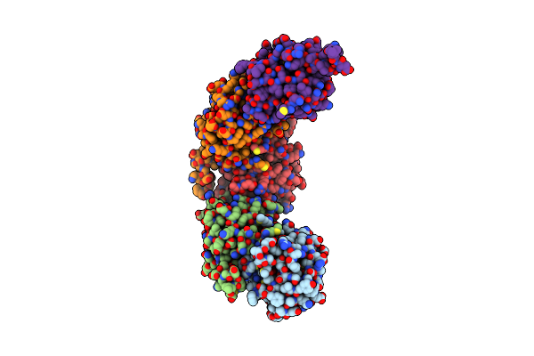
Structure Of Enterococcus Faecalis (V583) Alkylhydroperoxide Reductase Subunit F (Ahpf) C503A Mutant
Organism: Enterococcus faecalis v583
Method: X-RAY DIFFRACTION Resolution:2.50 Å Release Date: 2019-05-22 Classification: OXIDOREDUCTASE Ligands: SO4 |
Organism: Enterococcus faecalis v583
Method: X-RAY DIFFRACTION
Release Date: 2019-05-22
Ligands: SO4
 |
Crystal Structure Of Enterococcus Faecalis Ahpc
Organism: Enterococcus faecalis (strain atcc 700802 / v583)
Method: X-RAY DIFFRACTION Resolution:2.87 Å Release Date: 2017-12-27 Classification: OXIDOREDUCTASE |
Organism: Enterococcus faecalis (strain atcc 700802 / v583)
Method: X-RAY DIFFRACTION
Release Date: 2017-12-27
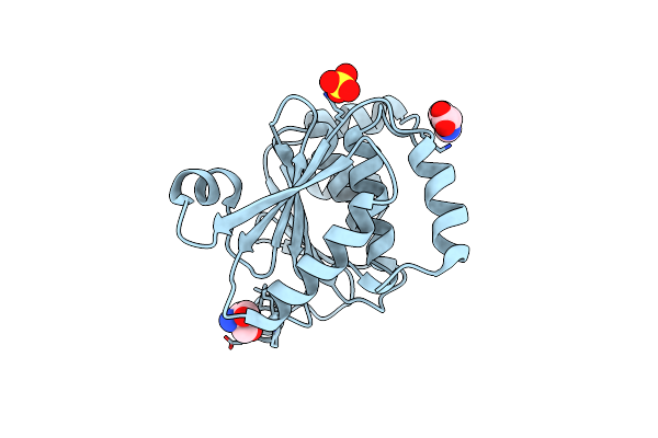 |
Crystal Structure Of The Ntd_N/C Domain Of Alkylhydroperoxide Reductase Ahpf From Enterococcus Faecalis (V583)
Organism: Enterococcus faecalis
Method: X-RAY DIFFRACTION Resolution:2.30 Å Release Date: 2017-11-22 Classification: OXIDOREDUCTASE Ligands: SO4, TRS, PRO |
Organism: Enterococcus faecalis
Method: X-RAY DIFFRACTION
Release Date: 2017-11-22
Ligands: SO4, TRS, PRO
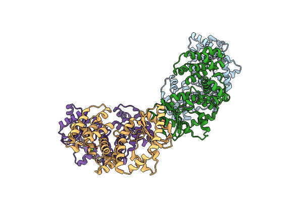 |
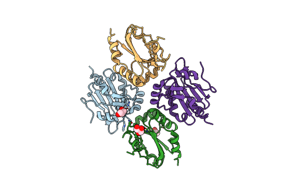
Crystallographic Structure Of The Enzymatically Active N-Terminal Domain Of The Rel Protein From Mycobacterium Tuberculosis
Organism: Mycobacterium tuberculosis (strain atcc 25618 / h37rv)
Method: X-RAY DIFFRACTION Resolution:3.70 Å Release Date: 2017-07-19 Classification: HYDROLASE, TRANSFERASE Ligands: MG |
Organism: Mycobacterium tuberculosis (strain atcc 25618 / h37rv)
Method: X-RAY DIFFRACTION
Release Date: 2017-07-19
Ligands: MG
 |
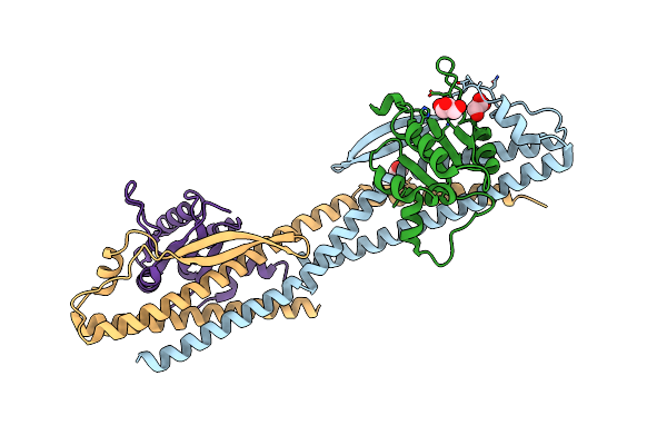
Asymmetry In The Active Site Of Mycobacterium Tuberculosis Ahpe Upon Exposure To Mycothiol
Organism: Mycobacterium tuberculosis h37rv
Method: X-RAY DIFFRACTION Resolution:2.43 Å Release Date: 2016-08-03 Classification: OXIDOREDUCTASE Ligands: GOL, ACT |
Organism: Mycobacterium tuberculosis h37rv
Method: X-RAY DIFFRACTION
Release Date: 2016-08-03
Ligands: GOL, ACT
 |
Crystal Structure Of The Subunit Df-Assembly Of The Eukaryotic V-Atpase.
Organism: Saccharomyces cerevisiae s288c
Method: X-RAY DIFFRACTION Resolution:3.18 Å Release Date: 2014-12-10 Classification: HYDROLASE Ligands: GOL |
Organism: Saccharomyces cerevisiae s288c
Method: X-RAY DIFFRACTION
Release Date: 2014-12-10
Ligands: GOL
 |
Crystal Structure Of The Alkylhydroperoxide Reductase Ahpf From Escherichia Coli
Organism: Escherichia coli
Method: X-RAY DIFFRACTION Resolution:2.00 Å Release Date: 2014-11-05 Classification: OXIDOREDUCTASE Ligands: FAD, SO4, PGE, GOL, CD, PG4, TRS |
Organism: Escherichia coli
Method: X-RAY DIFFRACTION
Release Date: 2014-11-05
Ligands: FAD, SO4, PGE, GOL, CD, PG4, TRS
 |
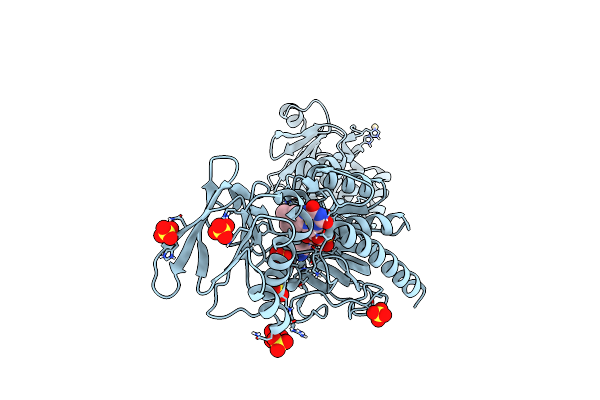
Crystal Structure Of Alkylhydroperoxide Reductase Subunit C From E. Coli
Organism: Escherichia coli
Method: X-RAY DIFFRACTION Resolution:3.33 Å Release Date: 2014-11-05 Classification: OXIDOREDUCTASE |
Organism: Escherichia coli
Method: X-RAY DIFFRACTION
Release Date: 2014-11-05
 |
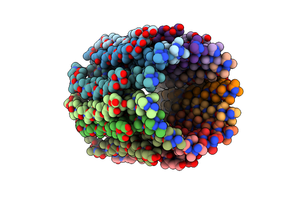
Crystal Structure Of Alkylhydroperoxide Reductase Subunit F From E. Coli At 2.65 Ang Resolution
Organism: Escherichia coli
Method: X-RAY DIFFRACTION Resolution:2.65 Å Release Date: 2014-11-05 Classification: OXIDOREDUCTASE Ligands: SO4, FAD, CD |
Organism: Escherichia coli
Method: X-RAY DIFFRACTION
Release Date: 2014-11-05
Ligands: SO4, FAD, CD
 |
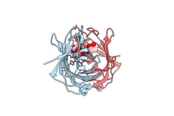
Structure Of The C Ring Of The Cf1Fo Atp Synthases.
Organism: Triticum aestivum
Method: X-RAY DIFFRACTION Resolution:6.00 Å Release Date: 2014-03-19 Classification: HYDROLASE |
Organism: Triticum aestivum
Method: X-RAY DIFFRACTION
Release Date: 2014-03-19
 |
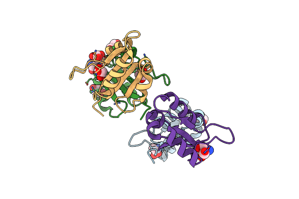
Crystal Structure Of The Fk506 Binding Domain Of Plasmodium Vivax Fkbp35 In Complex With A Tetrapeptide Substrate
Organism: Plasmodium vivax
Method: X-RAY DIFFRACTION Resolution:1.65 Å Release Date: 2013-03-20 Classification: ISOMERASE/SUBSTRATE Ligands: SO4 |
Organism: Plasmodium vivax
Method: X-RAY DIFFRACTION
Release Date: 2013-03-20
Ligands: SO4
 |
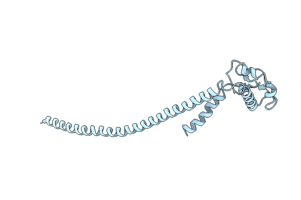
Crystal Structure Of Subunit F Of V-Atpase From S. Cerevisiae
Organism: Saccharomyces cerevisiae
Method: X-RAY DIFFRACTION Resolution:2.33 Å Release Date: 2013-03-20 Classification: HYDROLASE Ligands: TRS, GOL |
Organism: Saccharomyces cerevisiae
Method: X-RAY DIFFRACTION
Release Date: 2013-03-20
Ligands: TRS, GOL
 |
The Structure Of The Peripheral Stalk Subunit E From Pyrococcus Horikoshii
Organism: Pyrococcus horikoshii
Method: X-RAY DIFFRACTION Resolution:3.65 Å Release Date: 2012-05-02 Classification: HYDROLASE |
Organism: Pyrococcus horikoshii
Method: X-RAY DIFFRACTION
Release Date: 2012-05-02