

Search Count: 136
 |
Crystal Structure Of Isolated Death Domain Of Human P53-Induced Protein Pidd (778-873) With Thr788Asp Phosphomimetic Mutation.
Organism: Homo sapiens
Method: X-RAY DIFFRACTION Resolution:1.80 Å Release Date: 2026-07-29 Classification: APOPTOSIS |
Organism: Homo sapiens
Method: X-RAY DIFFRACTION
Release Date: 2026-07-29
 |
D-2-Hydroxyacid Dehydrogenase (D2Hdh) From Haloferax Mediterranei In Complex With Potassium, 2-Ketohexanoic Acid, Nadp+ And Chloride
Organism: Haloferax mediterranei
Method: X-RAY DIFFRACTION Resolution:1.26 Å Release Date: 2025-08-20 Classification: OXIDOREDUCTASE Ligands: NAP, 7N5, K, MG, CL |
Organism: Haloferax mediterranei
Method: X-RAY DIFFRACTION
Release Date: 2025-08-20
Ligands: NAP, 7N5, K, MG, CL
 |
D-2-Hydroxyacid Dehydrogenase (D2-Hdh) From Haloferax Mediterranei Apo-Enzyme (2.25 A Resolution)
Organism: Haloferax mediterranei
Method: X-RAY DIFFRACTION Resolution:2.25 Å Release Date: 2025-08-06 Classification: OXIDOREDUCTASE Ligands: MG, NA |
Organism: Haloferax mediterranei
Method: X-RAY DIFFRACTION
Release Date: 2025-08-06
Ligands: MG, NA
 |
D-2-Hydroxyacid Dehydrogenase (D2Hdh) From Haloferax Mediterranei In Complex With 2-Ketohexanoic Acid, Nad+ And Chloride (1.16 A Resolution)
Organism: Haloferax mediterranei
Method: X-RAY DIFFRACTION Resolution:1.16 Å Release Date: 2025-08-06 Classification: OXIDOREDUCTASE Ligands: NAD, 7N5, ACT, MG, NA, CL |
Organism: Haloferax mediterranei
Method: X-RAY DIFFRACTION
Release Date: 2025-08-06
Ligands: NAD, 7N5, ACT, MG, NA, CL
 |
Substrate-Free D10N,P146A Variant Of Beta-Phosphoglucomutase From Lactococcus Lactis
Organism: Lactococcus lactis subsp. lactis il1403
Method: X-RAY DIFFRACTION Resolution:1.68 Å Release Date: 2024-08-07 Classification: ISOMERASE Ligands: MG, EDO, PO4, TRS |
Organism: Lactococcus lactis subsp. lactis il1403
Method: X-RAY DIFFRACTION
Release Date: 2024-08-07
Ligands: MG, EDO, PO4, TRS
 |
D10N Variant Of Beta-Phosphoglucomutase From Lactococcus Lactis In Complex With Fructose 1,6-Bisphosphate
Organism: Lactococcus lactis subsp. lactis il1403
Method: X-RAY DIFFRACTION Resolution:1.75 Å Release Date: 2024-08-07 Classification: ISOMERASE Ligands: FBP, MG, EDO |
Organism: Lactococcus lactis subsp. lactis il1403
Method: X-RAY DIFFRACTION
Release Date: 2024-08-07
Ligands: FBP, MG, EDO
 |
D10N,P146A Variant Of Beta-Phosphoglucomutase From Lactococcus Lactis In Complex With Fructose 1,6-Bisphosphate
Organism: Lactococcus lactis subsp. lactis il1403
Method: X-RAY DIFFRACTION Resolution:1.23 Å Release Date: 2024-08-07 Classification: ISOMERASE Ligands: FBP, MG, EDO |
Organism: Lactococcus lactis subsp. lactis il1403
Method: X-RAY DIFFRACTION
Release Date: 2024-08-07
Ligands: FBP, MG, EDO
 |
D10N,P146A Variant Of Beta-Phosphoglucomutase From Lactococcus Lactis In Complex With Native Beta-Glucose 1,6-Bisphosphate Intermediate
Organism: Lactococcus lactis subsp. lactis il1403
Method: X-RAY DIFFRACTION Resolution:1.01 Å Release Date: 2024-08-07 Classification: ISOMERASE Ligands: B16, MG, TRS, NA, EDO |
Organism: Lactococcus lactis subsp. lactis il1403
Method: X-RAY DIFFRACTION
Release Date: 2024-08-07
Ligands: B16, MG, TRS, NA, EDO
 |
Crystal Structure Of The Burkholderia Lethal Factor 1 (Blf1) C94S Inactive Mutant In Complex With Human Eif4A - Crystal Form A
Organism: Burkholderia pseudomallei (strain k96243), Homo sapiens
Method: X-RAY DIFFRACTION Resolution:2.52 Å Release Date: 2022-04-13 Classification: TOXIN |
Organism: Burkholderia pseudomallei (strain k96243), Homo sapiens
Method: X-RAY DIFFRACTION
Release Date: 2022-04-13
 |
Crystal Structure Of The Burkholderia Lethal Factor 1 (Blf1) C94S Inactive Mutant In Complex With Human Eif4A - Crystal Form B
Organism: Burkholderia pseudomallei (strain k96243), Homo sapiens
Method: X-RAY DIFFRACTION Resolution:3.00 Å Release Date: 2022-04-13 Classification: TOXIN |
Organism: Burkholderia pseudomallei (strain k96243), Homo sapiens
Method: X-RAY DIFFRACTION
Release Date: 2022-04-13
 |
Structure Of Soluble Smhb Of The Tripartite Alpha-Pore Forming Toxin, Smh, From Serratia Marcescens.
Organism: Serratia marcescens
Method: X-RAY DIFFRACTION Resolution:1.84 Å Release Date: 2021-03-31 Classification: TOXIN Ligands: SO4, EDO |
Organism: Serratia marcescens
Method: X-RAY DIFFRACTION
Release Date: 2021-03-31
Ligands: SO4, EDO
 |
Structure Of Soluble Smhb Crystal Form 2 Of The Tripartite Alpha-Pore Forming Toxin, Smh, From Serratia Marcescens.
Organism: Serratia marcescens
Method: X-RAY DIFFRACTION Resolution:1.86 Å Release Date: 2021-03-31 Classification: TOXIN |
Organism: Serratia marcescens
Method: X-RAY DIFFRACTION
Release Date: 2021-03-31
 |
Structure Of The Smhb Pore Of The Tripartite Alpha-Pore Forming Toxin, Smh, From Serratia Marcescens.
Organism: Serratia marcescens
Method: X-RAY DIFFRACTION Resolution:6.98 Å Release Date: 2021-03-31 Classification: TOXIN |
Organism: Serratia marcescens
Method: X-RAY DIFFRACTION
Release Date: 2021-03-31
 |
Structure Of Soluble Smha Crystal Form 1 Of The Tripartite Alpha-Pore Forming Toxin, Smh, From Serratia Marcescens.
Organism: Serratia marcescens
Method: X-RAY DIFFRACTION Resolution:2.98 Å Release Date: 2021-03-31 Classification: TOXIN Ligands: CA, CL Modified Residue(s): MSE |
Organism: Serratia marcescens
Method: X-RAY DIFFRACTION
Release Date: 2021-03-31
Ligands: CA, CL
 |
Structure Of Soluble Smha Crystal Form 2 Of The Tripartite Alpha-Pore Forming Toxin, Smh, From Serratia Marcescens.
Organism: Serratia marcescens
Method: X-RAY DIFFRACTION Resolution:2.57 Å Release Date: 2021-03-31 Classification: TOXIN |
Organism: Serratia marcescens
Method: X-RAY DIFFRACTION
Release Date: 2021-03-31
 |
Crystal Structure Of The Eimeria Tenella Surface Antigen Protein Sag19
Organism: Eimeria tenella
Method: X-RAY DIFFRACTION Resolution:1.32 Å Release Date: 2021-03-24 Classification: UNKNOWN FUNCTION Ligands: SO4 |
Organism: Eimeria tenella
Method: X-RAY DIFFRACTION
Release Date: 2021-03-24
Ligands: SO4
 |
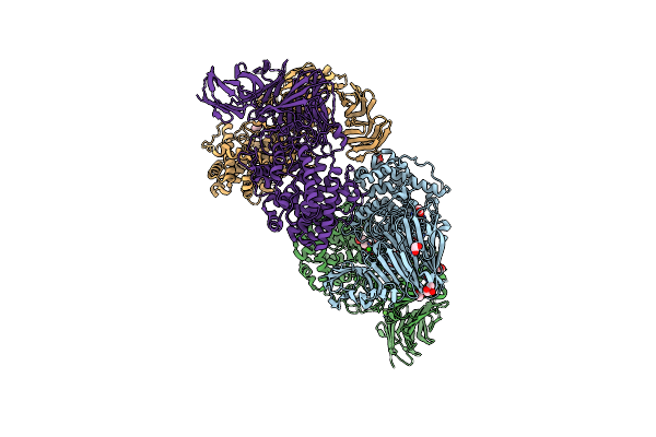
Crystal Structure Of The Burkholderia Lethal Factor 1 (Blf1)
Organism: Burkholderia pseudomallei (strain k96243)
Method: X-RAY DIFFRACTION Resolution:0.99 Å Release Date: 2020-07-15 Classification: TOXIN Ligands: EDO |
Organism: Burkholderia pseudomallei (strain k96243)
Method: X-RAY DIFFRACTION
Release Date: 2020-07-15
Ligands: EDO
 |
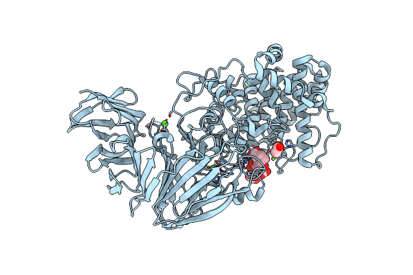
Crystal Structure Of The Catalytic Domain Of A Multi-Domain Alginate Lyase Dp0100 From Thermophilic Bacterium Defluviitalea Phaphyphila
Organism: Defluviitalea phaphyphila
Method: X-RAY DIFFRACTION Resolution:2.07 Å Release Date: 2019-10-30 Classification: LYASE Ligands: MN, MG, CA, ACT, EDO, CAC |
Organism: Defluviitalea phaphyphila
Method: X-RAY DIFFRACTION
Release Date: 2019-10-30
Ligands: MN, MG, CA, ACT, EDO, CAC
 |
Crystal Structure Of The Catalytic Domain Of A Multi-Domain Alginate Lyase Dp0100 From Thermophilic Bacterium Defluviitalea Phaphyphila
Organism: Defluviitalea phaphyphila
Method: X-RAY DIFFRACTION Resolution:2.76 Å Release Date: 2019-10-30 Classification: LYASE Ligands: MN, CA, ACT, MG |
Organism: Defluviitalea phaphyphila
Method: X-RAY DIFFRACTION
Release Date: 2019-10-30
Ligands: MN, CA, ACT, MG
 |
Crystal Structure Of The Catalytic Domain Of A Multi-Domain Alginate Lyase Dp0100 From Thermophilic Bacterium Defluviitalea Phaphyphila
Organism: Defluviitalea phaphyphila
Method: X-RAY DIFFRACTION Resolution:2.85 Å Release Date: 2019-10-30 Classification: LYASE Ligands: MN, CA, MG |
Organism: Defluviitalea phaphyphila
Method: X-RAY DIFFRACTION
Release Date: 2019-10-30
Ligands: MN, CA, MG