

Search Count: 33
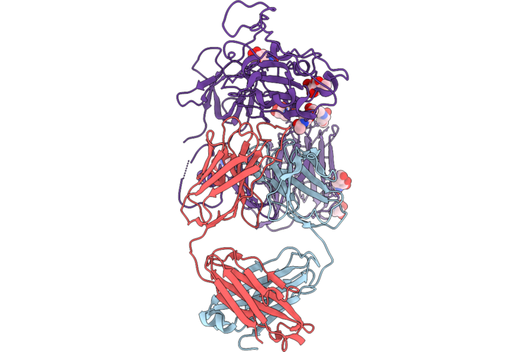 |
Cryoem Structure Of Wiv1 Spike Monomer In Complex With Neutralizing Antibody Va14_26
Organism: Homo sapiens, Bat sars-like coronavirus wiv1
Method: ELECTRON MICROSCOPY Resolution:4.12 Å Release Date: 2026-07-08 Classification: IMMUNE SYSTEM Ligands: NAG |
Organism: Homo sapiens, Bat sars-like coronavirus wiv1
Method: ELECTRON MICROSCOPY
Release Date: 2026-07-08
Ligands: NAG
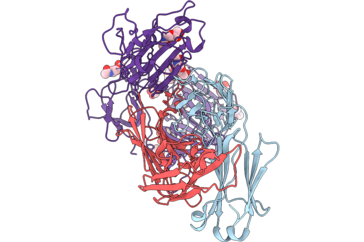 |
Cryoem Structure Of Wiv1 Spike Monomer In Complex With Neutralizing Antibody V1Wt_41
Organism: Homo sapiens, Bat sars-like coronavirus wiv1
Method: ELECTRON MICROSCOPY Resolution:3.57 Å Release Date: 2026-07-08 Classification: IMMUNE SYSTEM Ligands: NAG |
Organism: Homo sapiens, Bat sars-like coronavirus wiv1
Method: ELECTRON MICROSCOPY
Release Date: 2026-07-08
Ligands: NAG
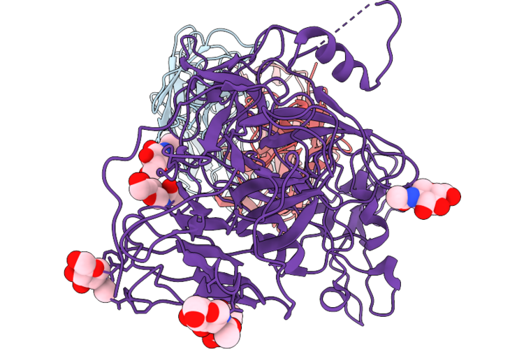 |
Cryoem Structure Of Wiv1 Spike Monomer In Complex With Neutralizing Antibody V1Wt_06
Organism: Homo sapiens, Bat sars-like coronavirus wiv1
Method: ELECTRON MICROSCOPY Resolution:3.98 Å Release Date: 2026-07-08 Classification: IMMUNE SYSTEM Ligands: NAG |
Organism: Homo sapiens, Bat sars-like coronavirus wiv1
Method: ELECTRON MICROSCOPY
Release Date: 2026-07-08
Ligands: NAG
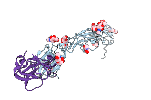 |
Cardioderma Bat Coronavirus Ky43 Receptor Binding Domain In Complex With Human Ceacam6
Organism: Homo sapiens, Cardioderma bat coronavirus
Method: X-RAY DIFFRACTION Resolution:3.01 Å Release Date: 2026-04-08 Classification: VIRAL PROTEIN Ligands: NAG |
Organism: Homo sapiens, Cardioderma bat coronavirus
Method: X-RAY DIFFRACTION
Release Date: 2026-04-08
Ligands: NAG
 |
Cardioderma Bat Coronavirus 2B Receptor Binding Domain In Complex With Human Ceacam6
Organism: Homo sapiens, Cardioderma bat coronavirus
Method: X-RAY DIFFRACTION Resolution:2.99 Å Release Date: 2026-04-08 Classification: VIRAL PROTEIN Ligands: NAG |
Organism: Homo sapiens, Cardioderma bat coronavirus
Method: X-RAY DIFFRACTION
Release Date: 2026-04-08
Ligands: NAG
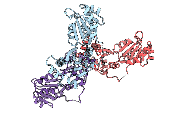 |
Crystal Structure Of The N-Terminal Domain Of The A Subunit Of The Bacillus Cereus Geri Germinant Receptor
Organism: Bacillus cereus atcc 14579
Method: X-RAY DIFFRACTION Resolution:2.80 Å Release Date: 2026-03-25 Classification: PROTEIN TRANSPORT Ligands: ACT |
Organism: Bacillus cereus atcc 14579
Method: X-RAY DIFFRACTION
Release Date: 2026-03-25
Ligands: ACT
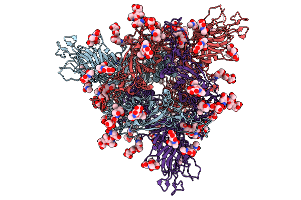 |
Structure Of The Rhgb07 Prefusion, Closed-State Trimeric Spike Protein
Organism: Sarbecovirus sp. rhgb07
Method: ELECTRON MICROSCOPY Release Date: 2025-09-24 Classification: VIRAL PROTEIN Ligands: NAG |
Organism: Sarbecovirus sp. rhgb07
Method: ELECTRON MICROSCOPY
Release Date: 2025-09-24
Ligands: NAG
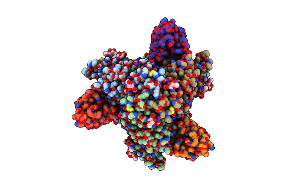 |
Prefusion F Glycoprotein Ectodomain Of Nipah Virus Ectodomain In Complex With Ds90 Nanobody
Organism: Henipavirus nipahense, Vicugna pacos
Method: ELECTRON MICROSCOPY Release Date: 2025-06-04 Classification: VIRAL PROTEIN Ligands: NAG |
Organism: Henipavirus nipahense, Vicugna pacos
Method: ELECTRON MICROSCOPY
Release Date: 2025-06-04
Ligands: NAG
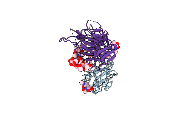 |
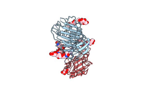
Cryo-Em Structure Of Sars-Cov-2 Spike (Hexapro Variant) In Complex With Nanobody W25 (Map 3, Focus Refinement On Rbd, W25 And Adjacent Ntd)
Organism: Severe acute respiratory syndrome coronavirus 2, Vicugna pacos
Method: ELECTRON MICROSCOPY Release Date: 2023-03-08 Classification: VIRAL PROTEIN Ligands: NAG |
Organism: Severe acute respiratory syndrome coronavirus 2, Vicugna pacos
Method: ELECTRON MICROSCOPY
Release Date: 2023-03-08
Ligands: NAG
 |
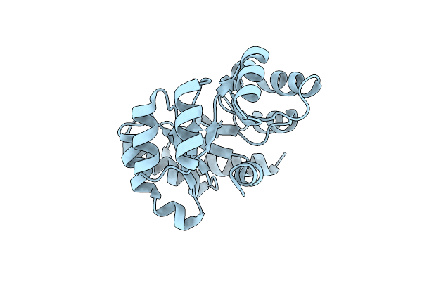
Cryo-Em Structure Of Sars-Cov-2 Spike (Omicron Ba.1 Variant) In Complex With Nanobody W25 (Map 5, Focus Refinement On Rbd, W25 And Adjacent Ntd)
Organism: Severe acute respiratory syndrome coronavirus 2, Vicugna pacos
Method: ELECTRON MICROSCOPY Release Date: 2023-03-08 Classification: VIRAL PROTEIN Ligands: NAG |
Organism: Severe acute respiratory syndrome coronavirus 2, Vicugna pacos
Method: ELECTRON MICROSCOPY
Release Date: 2023-03-08
Ligands: NAG
 |
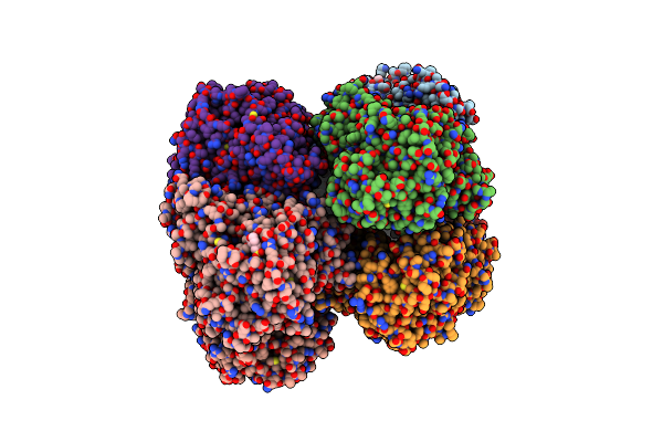
Structure Of A Nanoparticle For A Covid-19 Vaccine Candidate
Organism: Streptococcus pyogenes serotype m49 (strain nz131), Thermotoga maritima (strain atcc 43589 / msb8 / dsm 3109 / jcm 10099)
Method: ELECTRON MICROSCOPY Release Date: 2021-01-13 Classification: VIRUS LIKE PARTICLE |
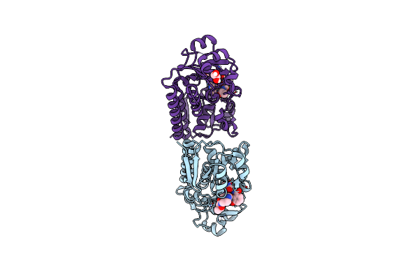 |
Structure Of Siderophore Binding Protein Baub Bound To A Complex Between Two Molecules Of Acinetobactin And Ferric Iron.
Organism: Acinetobacter baumannii
Method: X-RAY DIFFRACTION Resolution:1.90 Å Release Date: 2018-11-21 Classification: METAL TRANSPORT Ligands: FE, OPV, EDO |
Organism: Acinetobacter baumannii
Method: X-RAY DIFFRACTION
Release Date: 2018-11-21
Ligands: FE, OPV, EDO
 |
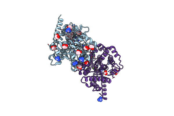
The Structure Of Aerobactin Synthetase Iucc From A Hypervirulent Pathotype Of Klebsiella Pneumoniae
Organism: Klebsiella pneumoniae
Method: X-RAY DIFFRACTION Resolution:2.45 Å Release Date: 2018-04-11 Classification: LIGASE Ligands: MES |
Organism: Klebsiella pneumoniae
Method: X-RAY DIFFRACTION
Release Date: 2018-04-11
Ligands: MES
 |
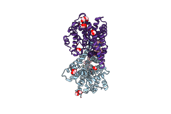
Crystal Structure Of T. Brucei Pde-B1 Catalytic Domain With Inhibitor Npd-038
Organism: Trypanosoma brucei
Method: X-RAY DIFFRACTION Resolution:1.80 Å Release Date: 2018-03-14 Classification: HYDROLASE Ligands: ZN, MG, GAI, EDO, FMT, 8Z4 |
Organism: Trypanosoma brucei
Method: X-RAY DIFFRACTION
Release Date: 2018-03-14
Ligands: ZN, MG, GAI, EDO, FMT, 8Z4
 |
Crystal Structure Of T. Brucei Pde-B1 Catalytic Domain With Inhibitor Npd-039
Organism: Trypanosoma brucei
Method: X-RAY DIFFRACTION Resolution:2.01 Å Release Date: 2018-03-14 Classification: HYDROLASE Ligands: ZN, MG, FMT, GAI, 908, GOL, PEG |
Organism: Trypanosoma brucei
Method: X-RAY DIFFRACTION
Release Date: 2018-03-14
Ligands: ZN, MG, FMT, GAI, 908, GOL, PEG
 |
Crystal Structure Of T. Brucei Pde-B1 Catalytic Domain With Inhibitor Npd-937
Organism: Trypanosoma brucei
Method: X-RAY DIFFRACTION Resolution:2.14 Å Release Date: 2018-03-14 Classification: HYDROLASE Ligands: ZN, MG, FMT, GAI, GOL, CVA |
Organism: Trypanosoma brucei
Method: X-RAY DIFFRACTION
Release Date: 2018-03-14
Ligands: ZN, MG, FMT, GAI, GOL, CVA
 |
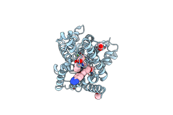
Crystal Structure Of T. Brucei Pde-B1 Catalytic Domain With Inhibitor Npd-340
Organism: Trypanosoma brucei
Method: X-RAY DIFFRACTION Resolution:2.25 Å Release Date: 2018-03-14 Classification: HYDROLASE Ligands: ZN, MG, GOL, FMT, CVL |
Organism: Trypanosoma brucei
Method: X-RAY DIFFRACTION
Release Date: 2018-03-14
Ligands: ZN, MG, GOL, FMT, CVL
 |
Crystal Structure Of Human Phosphodiesterase 4B Catalytic Domain With Inhibitor Npd-001
Organism: Homo sapiens
Method: X-RAY DIFFRACTION Resolution:2.40 Å Release Date: 2018-03-14 Classification: HYDROLASE Ligands: ZN, MG, ACT, 6M5 |
Organism: Homo sapiens
Method: X-RAY DIFFRACTION
Release Date: 2018-03-14
Ligands: ZN, MG, ACT, 6M5
 |
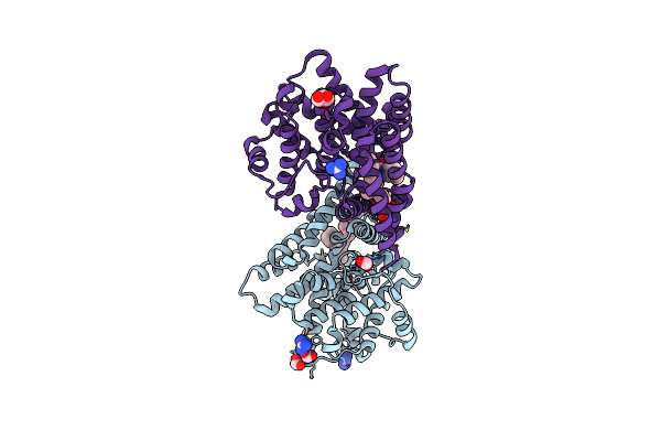
Crystal Structure Of Human Phosphodiesterase 4D2 Catalytic Domain With Inhibitor Npd-001
Organism: Homo sapiens
Method: X-RAY DIFFRACTION Resolution:2.25 Å Release Date: 2018-03-14 Classification: HYDROLASE Ligands: ZN, MG, EDO, DTT, 6M5, EPE, PEG |
Organism: Homo sapiens
Method: X-RAY DIFFRACTION
Release Date: 2018-03-14
Ligands: ZN, MG, EDO, DTT, 6M5, EPE, PEG
 |
Crystal Structure Of T. Brucei Pde-B1 Catalytic Domain With Inhibitor Npd-008
Organism: Trypanosoma brucei
Method: X-RAY DIFFRACTION Resolution:1.83 Å Release Date: 2017-11-29 Classification: HYDROLASE Ligands: ZN, MG, LQY, FMT, GAI, GOL |
Organism: Trypanosoma brucei
Method: X-RAY DIFFRACTION
Release Date: 2017-11-29
Ligands: ZN, MG, LQY, FMT, GAI, GOL