

Search Count: 42
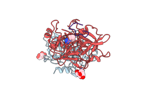 |
Crystal Structures Of The Serine Protease Domain Of Murine Plasma Kallikrein With Peptide Inhibitor Mupain-1-16
Organism: Mus musculus, Synthemiopsis
Method: X-RAY DIFFRACTION Resolution:2.77 Å Release Date: 2019-07-10 Classification: HYDROLASE Ligands: MAN, MRZ |
Organism: Mus musculus, Synthemiopsis
Method: X-RAY DIFFRACTION
Release Date: 2019-07-10
Ligands: MAN, MRZ
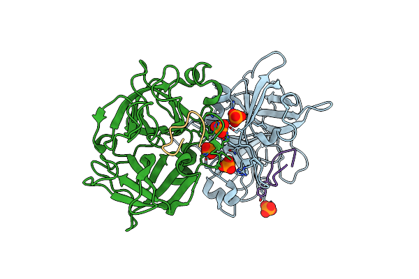 |
The Crystal Structure Of Mupain-1-Ig In Complex With Mupa-Spd At Ph8.5
Organism: Mus musculus, Phage display vector ptdisp
Method: X-RAY DIFFRACTION Resolution:2.53 Å Release Date: 2019-02-20 Classification: HYDROLASE INHIBITOR/HYDROLASE Ligands: PO4 |
Organism: Mus musculus, Phage display vector ptdisp
Method: X-RAY DIFFRACTION
Release Date: 2019-02-20
Ligands: PO4
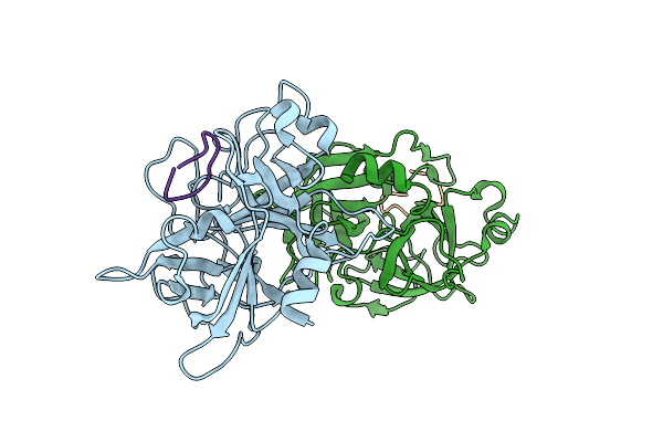 |
The Crystal Structure Of Mupain-1-Ig-2 In Complex With Mupa-Spd At Ph8.5
Organism: Mus musculus, Phage display vector ptdisp
Method: X-RAY DIFFRACTION Resolution:2.49 Å Release Date: 2019-02-20 Classification: HYDROLASE/HYDROLASE INHIBITOR |
Organism: Mus musculus, Phage display vector ptdisp
Method: X-RAY DIFFRACTION
Release Date: 2019-02-20
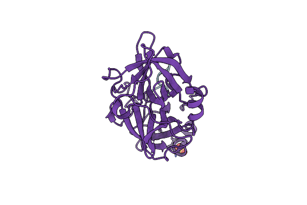 |
Crystal Structure Of Upa In Complex With Upain-2-2
Organism: Homo sapiens, Phage display vector ptdisp
Method: X-RAY DIFFRACTION Resolution:1.46 Å Release Date: 2018-07-11 Classification: HYDROLASE/HYDROLASE INHIBITOR Ligands: SO4 |
Organism: Homo sapiens, Phage display vector ptdisp
Method: X-RAY DIFFRACTION
Release Date: 2018-07-11
Ligands: SO4
 |
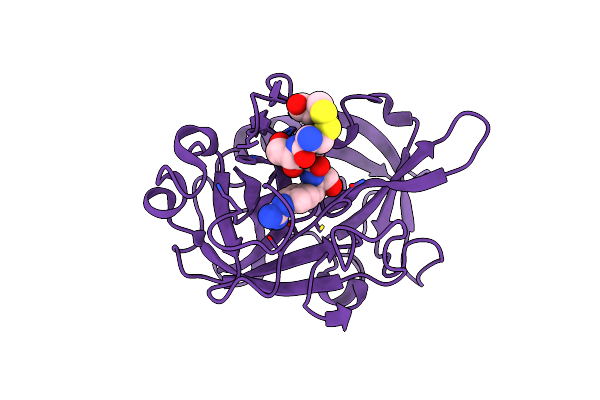
Crystal Structure Of Upa In Complex With Upain-2-2-W3A
Organism: Homo sapiens, Phage display vector ptdisp
Method: X-RAY DIFFRACTION Resolution:1.64 Å Release Date: 2018-07-11 Classification: HYDROLASE/HYDROLASE INHIBITOR |
Organism: Homo sapiens, Phage display vector ptdisp
Method: X-RAY DIFFRACTION
Release Date: 2018-07-11
 |
Crystal Structure Of Upa In Complex With Upain-2-3-W3A
Organism: Homo sapiens, Phage display vector ptdisp
Method: X-RAY DIFFRACTION Resolution:1.75 Å Release Date: 2018-07-11 Classification: HYDROLASE/HYDROLASE INHIBITOR Ligands: ALA, CYS |
Organism: Homo sapiens, Phage display vector ptdisp
Method: X-RAY DIFFRACTION
Release Date: 2018-07-11
Ligands: ALA, CYS
 |
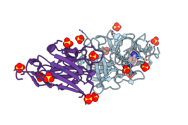
The Catalytic Domain Of Murine Urokinase-Type Plasminogen Activator In Complex With The Allosteric Inhibitory Nanobody Nb7
Organism: Mus musculus, Vicugna pacos
Method: X-RAY DIFFRACTION Resolution:2.55 Å Release Date: 2017-06-28 Classification: Hydrolase/Antibody Ligands: SO4, EDO |
Organism: Mus musculus, Vicugna pacos
Method: X-RAY DIFFRACTION
Release Date: 2017-06-28
Ligands: SO4, EDO
 |
The P-Aminobenzamidine Active Site Inhibited Catalytic Domain Of Murine Urokinase-Type Plasminogen Activator In Complex With The Allosteric Inhibitory Nanobody Nb7
Organism: Mus musculus, Vicugna pacos
Method: X-RAY DIFFRACTION Resolution:2.63 Å Release Date: 2017-06-28 Classification: HYDROLASE Ligands: SO4, EDO, PBZ |
Organism: Mus musculus, Vicugna pacos
Method: X-RAY DIFFRACTION
Release Date: 2017-06-28
Ligands: SO4, EDO, PBZ
 |
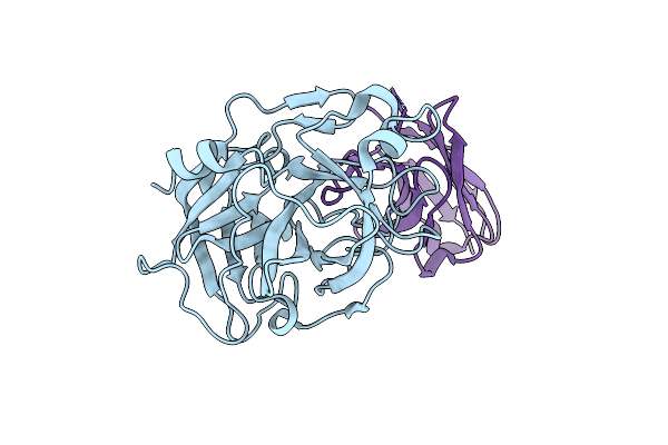
The Egr-Cmk Active Site Inhibited Catalytic Domain Of Murine Urokinase-Type Plasminogen Activator In Complex With The Allosteric Inhibitory Nanobody Nb7
Organism: Mus musculus, Vicugna pacos
Method: X-RAY DIFFRACTION Resolution:2.60 Å Release Date: 2017-06-28 Classification: Hydrolase/Antibody Ligands: 0GJ, SO4, EDO |
Organism: Mus musculus, Vicugna pacos
Method: X-RAY DIFFRACTION
Release Date: 2017-06-28
Ligands: 0GJ, SO4, EDO
 |
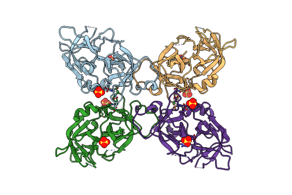
The Catalytic Domain Of Murine Urokinase-Type Plasminogen Activator In Complex With The Active Site Binding Inhibitory Nanobody Nb22
Organism: Mus musculus, Vicugna pacos
Method: X-RAY DIFFRACTION Resolution:2.30 Å Release Date: 2017-06-28 Classification: HYDROLASE |
Organism: Mus musculus, Vicugna pacos
Method: X-RAY DIFFRACTION
Release Date: 2017-06-28
 |
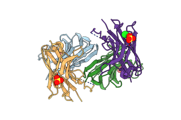
The Ligand Free Catalytic Domain Of Murine Urokinase-Type Plasminogen Activator
Organism: Mus musculus
Method: X-RAY DIFFRACTION Resolution:3.05 Å Release Date: 2017-06-28 Classification: HYDROLASE Ligands: SO4, NI |
Organism: Mus musculus
Method: X-RAY DIFFRACTION
Release Date: 2017-06-28
Ligands: SO4, NI
 |
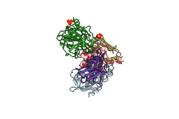
Crystal Structure Of A Nanobody Raised Against Urokinase-Type Plasminogen Activator
Organism: Camelus dromedarius
Method: X-RAY DIFFRACTION Resolution:2.16 Å Release Date: 2016-06-01 Classification: IMMUNE SYSTEM Ligands: SO4, CL |
Organism: Camelus dromedarius
Method: X-RAY DIFFRACTION
Release Date: 2016-06-01
Ligands: SO4, CL
 |
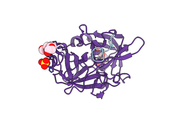
Crystal Structure Of Upa In Complex With A Camelid-Derived Antibody Fragment
Organism: Homo sapiens, Vicugna pacos
Method: X-RAY DIFFRACTION Resolution:1.97 Å Release Date: 2016-06-01 Classification: HYDROLASE/INHIBITOR Ligands: GOL, SO4, TWN, MES |
Organism: Homo sapiens, Vicugna pacos
Method: X-RAY DIFFRACTION
Release Date: 2016-06-01
Ligands: GOL, SO4, TWN, MES
 |
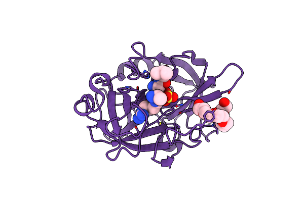
The Crystal Structure Of Upain-1-W3A In Complex With Upa At Ph5.5
Organism: Homo sapiens, Synthetic construct
Method: X-RAY DIFFRACTION Resolution:1.36 Å Release Date: 2016-05-18 Classification: HYDROLASE/HYDROLASE INHIBITOR Ligands: SO4, PG4 |
Organism: Homo sapiens, Synthetic construct
Method: X-RAY DIFFRACTION
Release Date: 2016-05-18
Ligands: SO4, PG4
 |
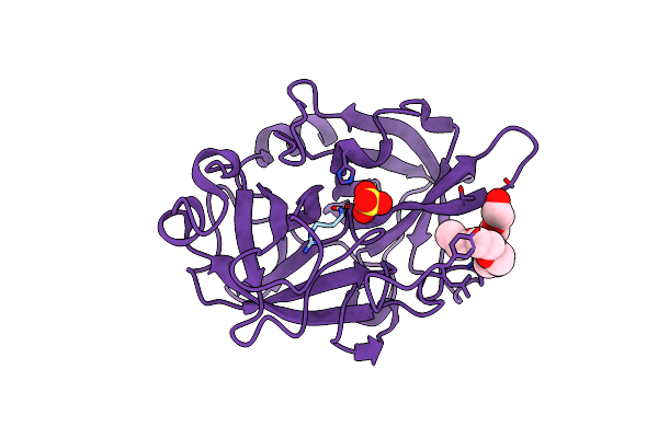
The Crystal Structure Of Upain-1-W3A In Complex With Upa At Ph7.4
Organism: Homo sapiens, Synthetic construct
Method: X-RAY DIFFRACTION Resolution:1.29 Å Release Date: 2016-05-18 Classification: HYDROLASE/HYDROLASE INHIBITOR Ligands: SO4, P6G |
Organism: Homo sapiens, Synthetic construct
Method: X-RAY DIFFRACTION
Release Date: 2016-05-18
Ligands: SO4, P6G
 |
The Crystal Structure Of Upain-1-W3A In Complex With Upa At Ph9.0
Organism: Homo sapiens, Synthetic construct
Method: X-RAY DIFFRACTION Resolution:1.36 Å Release Date: 2016-05-18 Classification: HYDROLASE/HYDROLASE INHIBITOR Ligands: SO4, P6G |
Organism: Homo sapiens, Synthetic construct
Method: X-RAY DIFFRACTION
Release Date: 2016-05-18
Ligands: SO4, P6G
 |
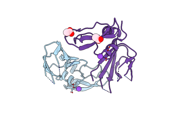
The Crystal Structure Of Upain-1-W3A In Complex With Inactive Upa (Upa-S195A) At Ph7.4
Organism: Homo sapiens, Synthetic construct
Method: X-RAY DIFFRACTION Resolution:1.85 Å Release Date: 2016-05-18 Classification: HYDROLASE/HYDROLASE INHIBITOR |
Organism: Homo sapiens, Synthetic construct
Method: X-RAY DIFFRACTION
Release Date: 2016-05-18
 |
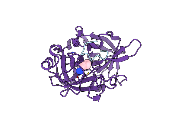
Crystal Structure Of A Hepatocyte Growth Factor Activator Inhibitor-1 (Hai-1) Fragment Covering The Pkd-Like 'Internal' Domain And Kunitz Domain 1
Organism: Homo sapiens
Method: X-RAY DIFFRACTION Resolution:2.10 Å Release Date: 2016-05-18 Classification: HYDROLASE Ligands: K, ACT |
Organism: Homo sapiens
Method: X-RAY DIFFRACTION
Release Date: 2016-05-18
Ligands: K, ACT
 |
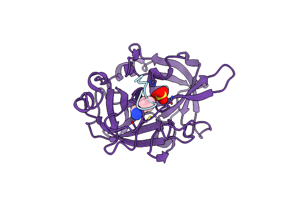
The Crystal Structure Of Mupain-1-16-D9A In Complex With Murinised Human Upa At Ph7.4
Organism: Homo sapiens, Synthetic construct
Method: X-RAY DIFFRACTION Resolution:1.90 Å Release Date: 2015-10-28 Classification: HYDROLASE INHIBITOR/HYDROLASE Ligands: MRZ |
Organism: Homo sapiens, Synthetic construct
Method: X-RAY DIFFRACTION
Release Date: 2015-10-28
Ligands: MRZ
 |
The Crystal Structure Of Mupain-1-17 In Complex With Murinised Human Upa
Organism: Homo sapiens, Synthetic construct
Method: X-RAY DIFFRACTION Resolution:2.10 Å Release Date: 2015-10-21 Classification: HYDROLASE INHIBITOR/HYDROLASE Ligands: MRZ, SO4 |
Organism: Homo sapiens, Synthetic construct
Method: X-RAY DIFFRACTION
Release Date: 2015-10-21
Ligands: MRZ, SO4