

Search Count: 37
 |
Crystal Structure Of Protein-Asparaginase From Amycolatopsis Deserti
Organism: Amycolatopsis deserti
Method: X-RAY DIFFRACTION Resolution:2.08 Å Release Date: 2026-04-15 Classification: HYDROLASE |
Organism: Amycolatopsis deserti
Method: X-RAY DIFFRACTION
Release Date: 2026-04-15
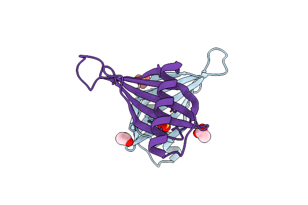 |
X-Ray Structure Of Mnei Mutant Mut9 (E23A, C41A, Y65R, S76Y)
Organism: Dioscoreophyllum cumminsii
Method: X-RAY DIFFRACTION Resolution:1.55 Å Release Date: 2024-02-07 Classification: PLANT PROTEIN Ligands: ACT, SO4 |
Organism: Dioscoreophyllum cumminsii
Method: X-RAY DIFFRACTION
Release Date: 2024-02-07
Ligands: ACT, SO4
 |
X-Ray Structure Of The Single Chain Monellin Derivative Mnei
Organism: Dioscoreophyllum cumminsii
Method: X-RAY DIFFRACTION Resolution:1.19 Å Release Date: 2024-02-07 Classification: PLANT PROTEIN Ligands: ACT, GOL, NA |
Organism: Dioscoreophyllum cumminsii
Method: X-RAY DIFFRACTION
Release Date: 2024-02-07
Ligands: ACT, GOL, NA
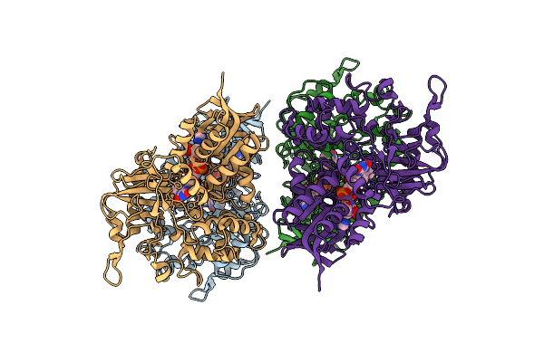 |
Hyper-Thermostable Ancestral L-Amino Acid Oxidase 2 (Htanclaao2)
Organism: Synthetic construct
Method: X-RAY DIFFRACTION Resolution:2.20 Å Release Date: 2023-10-04 Classification: OXIDOREDUCTASE Ligands: FAD |
Organism: Synthetic construct
Method: X-RAY DIFFRACTION
Release Date: 2023-10-04
Ligands: FAD
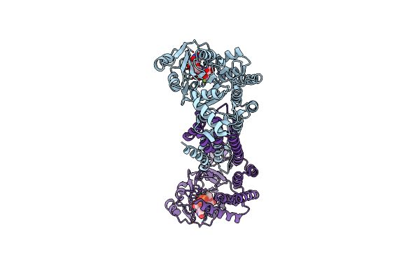 |
Thus Glycosin S-Glycosyltransferase
Organism: Bacillus thuringiensis serovar andalousiensis bgsc 4aw1
Method: X-RAY DIFFRACTION Resolution:2.06 Å Release Date: 2022-04-13 Classification: BIOSYNTHETIC PROTEIN Ligands: MG, U2F |
Organism: Bacillus thuringiensis serovar andalousiensis bgsc 4aw1
Method: X-RAY DIFFRACTION
Release Date: 2022-04-13
Ligands: MG, U2F
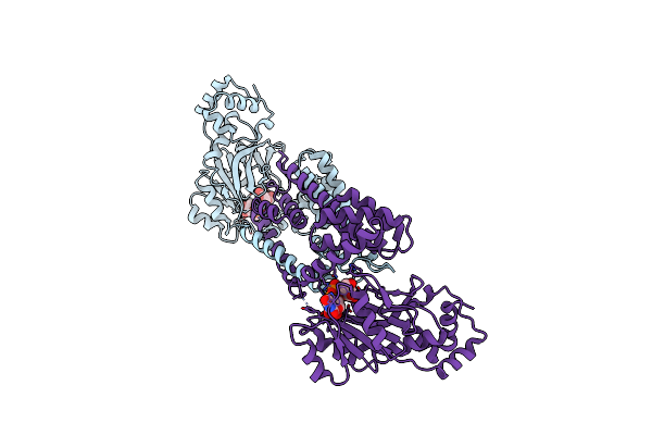 |
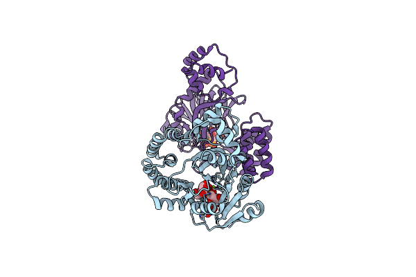
Suns Glycosin S-Glycosyltransferase
Organism: Bacillus subtilis (strain 168)
Method: X-RAY DIFFRACTION Resolution:3.00 Å Release Date: 2022-04-13 Classification: TRANSFERASE Ligands: UPG |
Organism: Bacillus subtilis (strain 168)
Method: X-RAY DIFFRACTION
Release Date: 2022-04-13
Ligands: UPG
 |
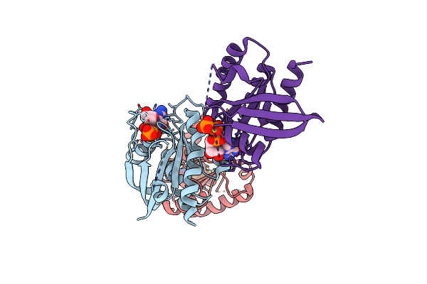
Suns Glycosin S-Glycosyltransferase
Organism: Bacillus subtilis (strain 168)
Method: X-RAY DIFFRACTION Resolution:2.10 Å Release Date: 2022-04-13 Classification: BIOSYNTHETIC PROTEIN Ligands: UPG, MG |
Organism: Bacillus subtilis (strain 168)
Method: X-RAY DIFFRACTION
Release Date: 2022-04-13
Ligands: UPG, MG
 |
Crystal Structure Of Wild-Type Kras4B(1-169) In Complex With Gmppnp And Mg Ion
Organism: Homo sapiens
Method: X-RAY DIFFRACTION Resolution:2.50 Å Release Date: 2021-02-10 Classification: ONCOPROTEIN Ligands: GNP, MG |
Organism: Homo sapiens
Method: X-RAY DIFFRACTION
Release Date: 2021-02-10
Ligands: GNP, MG
 |
Nukacin Isk-1 In Active State
Organism: Staphylococcus warneri
Method: SOLUTION NMR Release Date: 2018-11-28 Classification: ANTIBIOTIC |
Organism: Staphylococcus warneri
Method: SOLUTION NMR
Release Date: 2018-11-28
 |
Nukacin Isk-1 In Inactive State
Organism: Staphylococcus warneri
Method: SOLUTION NMR Release Date: 2018-11-28 Classification: ANTIBIOTIC |
Organism: Staphylococcus warneri
Method: SOLUTION NMR
Release Date: 2018-11-28
 |
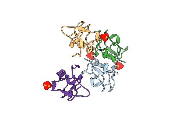
Crystal Structure Of Notched-Fin Eelpout Type Iii Antifreeze Protein (Nfe6, Afp), C2221 Form.
Organism: Zoarces elongatus
Method: X-RAY DIFFRACTION Resolution:1.19 Å Release Date: 2018-05-16 Classification: ANTIFREEZE PROTEIN Ligands: SO4 |
Organism: Zoarces elongatus
Method: X-RAY DIFFRACTION
Release Date: 2018-05-16
Ligands: SO4
 |
Crystal Structure Of Notched-Fin Eelpout Type Iii Antifreeze Protein (Nfe6, Afp), P212121 Form
Organism: Zoarces elongatus
Method: X-RAY DIFFRACTION Resolution:1.00 Å Release Date: 2018-05-16 Classification: ANTIFREEZE PROTEIN Ligands: SO4 |
Organism: Zoarces elongatus
Method: X-RAY DIFFRACTION
Release Date: 2018-05-16
Ligands: SO4
 |
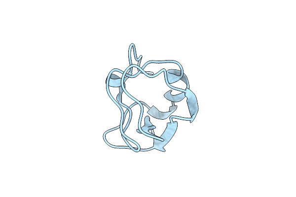
Crystal Structure Of Notched-Fin Eelpout Type Iii Antifreeze Protein A20V Mutant (Nfe6, Afp), C2221 Form
Organism: Zoarces elongatus
Method: X-RAY DIFFRACTION Resolution:1.30 Å Release Date: 2018-05-16 Classification: ANTIFREEZE PROTEIN Ligands: ACT |
Organism: Zoarces elongatus
Method: X-RAY DIFFRACTION
Release Date: 2018-05-16
Ligands: ACT
 |
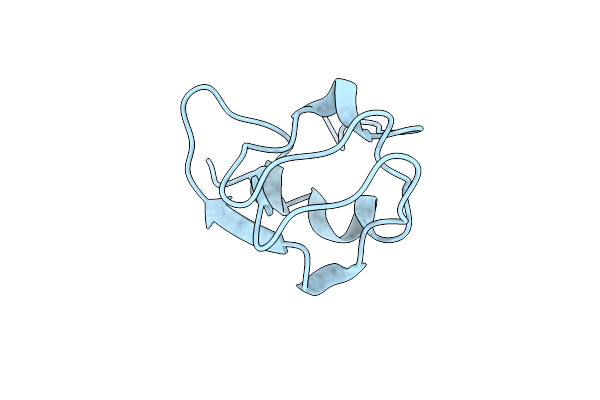
Crystal Structure Of Notched-Fin Eelpout Type Iii Antifreeze Protein A20I Mutant (Nfe6, Afp), P212121 Form
Organism: Zoarces elongatus
Method: X-RAY DIFFRACTION Resolution:1.00 Å Release Date: 2018-05-16 Classification: ANTIFREEZE PROTEIN |
Organism: Zoarces elongatus
Method: X-RAY DIFFRACTION
Release Date: 2018-05-16
 |
Crystal Structure Of Notched-Fin Eelpout Type Iii Antifreeze Protein A20L Mutant (Nfe6, Afp), P21 Form
Organism: Zoarces elongatus
Method: X-RAY DIFFRACTION Resolution:0.97 Å Release Date: 2018-05-16 Classification: ANTIFREEZE PROTEIN |
Organism: Zoarces elongatus
Method: X-RAY DIFFRACTION
Release Date: 2018-05-16
 |
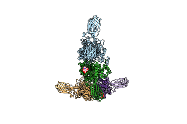
Crystal Structure Of Notched-Fin Eelpout Type Iii Antifreeze Protein A20T Mutant (Nfe6, Afp), P21 Form
Organism: Zoarces elongatus
Method: X-RAY DIFFRACTION Resolution:0.98 Å Release Date: 2018-05-16 Classification: ANTIFREEZE PROTEIN |
Organism: Zoarces elongatus
Method: X-RAY DIFFRACTION
Release Date: 2018-05-16
 |
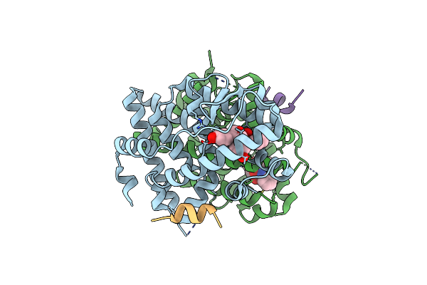
Crystal Structure Of Rhodopsin Bound To Visual Arrestin Determined By X-Ray Free Electron Laser
Organism: Enterobacteria phage rb55, Homo sapiens, Mus musculus
Method: X-RAY DIFFRACTION Resolution:3.01 Å Release Date: 2017-08-09 Classification: SIGNALING PROTEIN |
Organism: Enterobacteria phage rb55, Homo sapiens, Mus musculus
Method: X-RAY DIFFRACTION
Release Date: 2017-08-09
 |
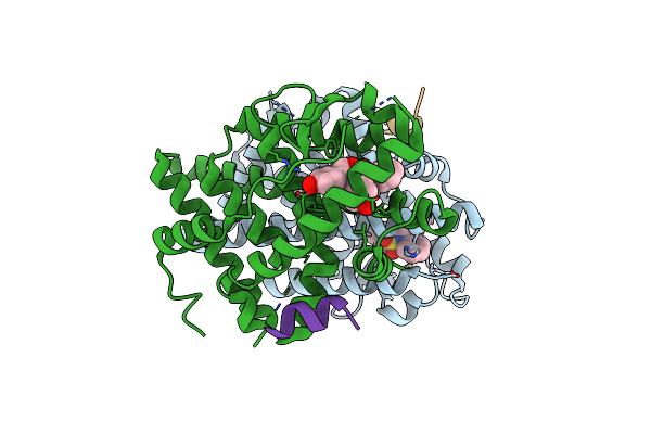
Crystal Structure Of The Er-Alpha Ligand-Binding Domain (Y537S) In Complex With Oxabicyclic Heptene Sulfonamide (Obhs-N)
Organism: Homo sapiens
Method: X-RAY DIFFRACTION Resolution:2.39 Å Release Date: 2016-11-16 Classification: TRANSCRIPTION Ligands: OB1 |
Organism: Homo sapiens
Method: X-RAY DIFFRACTION
Release Date: 2016-11-16
Ligands: OB1
 |
Crystal Structure Of The Er-Alpha Ligand-Binding Domain (Y537S) In Complex With An N-Methyl Substituted Obhs-N Derivative
Organism: Homo sapiens
Method: X-RAY DIFFRACTION Resolution:1.82 Å Release Date: 2016-11-16 Classification: TRANSCRIPTION Ligands: OB2 |
Organism: Homo sapiens
Method: X-RAY DIFFRACTION
Release Date: 2016-11-16
Ligands: OB2
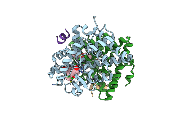 |
Crystal Structure Of The Er-Alpha Ligand-Binding Domain (Y537S) In Complex With An N-Methyl, 2-Chlorobenzyl Obhs-N Derivative
Organism: Homo sapiens
Method: X-RAY DIFFRACTION Resolution:1.85 Å Release Date: 2016-11-16 Classification: TRANSCRIPTION Ligands: OB3 |
Organism: Homo sapiens
Method: X-RAY DIFFRACTION
Release Date: 2016-11-16
Ligands: OB3