

Search Count: 69
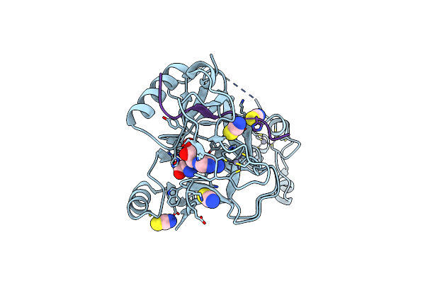 |
Crystal Structure Of Sestd2 In Complex With H3.3S31Phk36M Peptide
Organism: Homo sapiens
Method: X-RAY DIFFRACTION Resolution:1.78 Å Release Date: 2020-02-26 Classification: TRANSFERASE Ligands: ZN, SAH, SCN |
Organism: Homo sapiens
Method: X-RAY DIFFRACTION
Release Date: 2020-02-26
Ligands: ZN, SAH, SCN
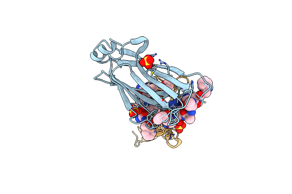 |
Crystal Structure Of Af9 Yeats Domain In Complex With A Peptide Inhibitor "Phq-H3(Q5-K9)" Modified At K9 With 2-Furancarboyl Group
Organism: Homo sapiens
Method: X-RAY DIFFRACTION Resolution:1.90 Å Release Date: 2018-11-07 Classification: TRANSCRIPTION Ligands: SO4, FOA |
Organism: Homo sapiens
Method: X-RAY DIFFRACTION
Release Date: 2018-11-07
Ligands: SO4, FOA
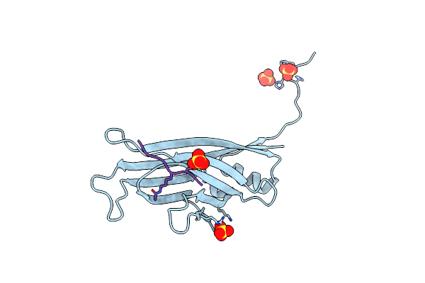 |
Enl Yeats In Complex With Histone H3 Acetylation At K27
Organism: Homo sapiens
Method: X-RAY DIFFRACTION Resolution:2.70 Å Release Date: 2017-03-08 Classification: TRANSCRIPTION Ligands: SO4 |
Organism: Homo sapiens
Method: X-RAY DIFFRACTION
Release Date: 2017-03-08
Ligands: SO4
 |
Crystal Structure Of Setd2 Bound To Histone H3.3 K36M Peptide
Organism: Homo sapiens
Method: X-RAY DIFFRACTION Resolution:2.05 Å Release Date: 2016-11-02 Classification: TRANSFERASE Ligands: ZN, SAH, SCN |
Organism: Homo sapiens
Method: X-RAY DIFFRACTION
Release Date: 2016-11-02
Ligands: ZN, SAH, SCN
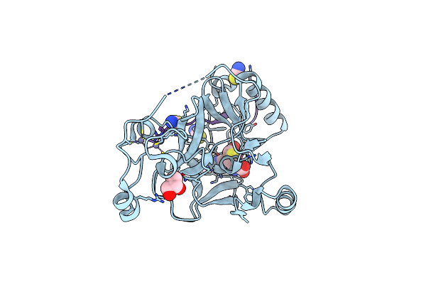 |
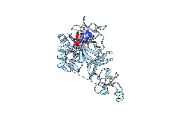
Crystal Structure Of Setd2 Bound To Histone H3.3 K36I Peptide
Organism: Homo sapiens
Method: X-RAY DIFFRACTION Resolution:1.50 Å Release Date: 2016-11-02 Classification: TRANSFERASE Ligands: SAH, SCN, GOL, ZN |
Organism: Homo sapiens
Method: X-RAY DIFFRACTION
Release Date: 2016-11-02
Ligands: SAH, SCN, GOL, ZN
 |
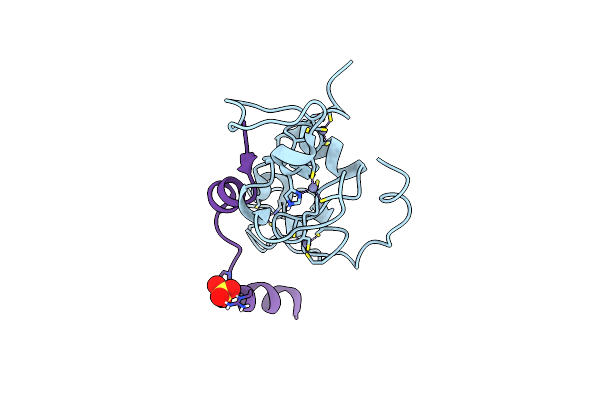
Crystal Structure Of Setd2 Bound To Sah
Organism: Homo sapiens
Method: X-RAY DIFFRACTION Resolution:2.40 Å Release Date: 2016-11-02 Classification: TRANSFERASE Ligands: ZN, SAH |
Organism: Homo sapiens
Method: X-RAY DIFFRACTION
Release Date: 2016-11-02
Ligands: ZN, SAH
 |
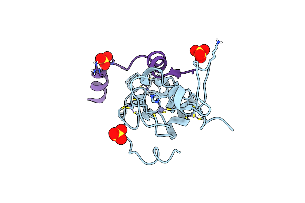
Crystal Structure Of Moz Double Phd Finger In Complex With Histone H3 Butyrylation At K14
Organism: Homo sapiens
Method: X-RAY DIFFRACTION Resolution:1.70 Å Release Date: 2016-10-26 Classification: TRANSFERASE Ligands: ZN, SO4 |
Organism: Homo sapiens
Method: X-RAY DIFFRACTION
Release Date: 2016-10-26
Ligands: ZN, SO4
 |
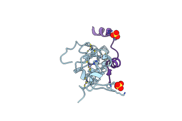
Crystal Structure Of Moz Double Phd Finger Domain In Complex With Histone H3 Crotonylation At K14
Organism: Homo sapiens
Method: X-RAY DIFFRACTION Resolution:1.65 Å Release Date: 2016-10-26 Classification: TRANSFERASE Ligands: ZN, SO4 |
Organism: Homo sapiens
Method: X-RAY DIFFRACTION
Release Date: 2016-10-26
Ligands: ZN, SO4
 |
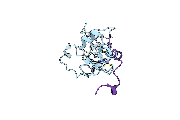
Crystal Structrue Of Moz Double Phd Finger In Complex With Histone H3 Propionylation At K14
Organism: Homo sapiens
Method: X-RAY DIFFRACTION Resolution:1.55 Å Release Date: 2016-10-26 Classification: TRANSFERASE Ligands: ZN, SO4 |
Organism: Homo sapiens
Method: X-RAY DIFFRACTION
Release Date: 2016-10-26
Ligands: ZN, SO4
 |
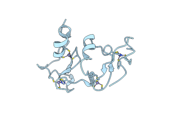
Crystal Structure Of Moz Double Phd Finger Mutant-S210D/N235R In Complex With Histone H3 Crotonylation At K14
Organism: Homo sapiens
Method: X-RAY DIFFRACTION Resolution:1.40 Å Release Date: 2016-10-26 Classification: TRANSFERASE Ligands: ZN |
Organism: Homo sapiens
Method: X-RAY DIFFRACTION
Release Date: 2016-10-26
Ligands: ZN
 |
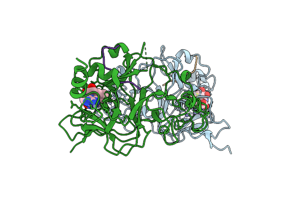
Crystal Structure Of Dpf2 Double Phd Finger
Organism: Homo sapiens
Method: X-RAY DIFFRACTION Resolution:2.60 Å Release Date: 2016-10-26 Classification: METAL BINDING PROTEIN Ligands: ZN |
Organism: Homo sapiens
Method: X-RAY DIFFRACTION
Release Date: 2016-10-26
Ligands: ZN
 |
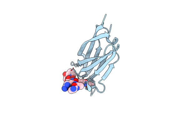
Structure Of G9A Set-Domain With Histone H3K9Norleucine Mutant Peptide And Bound S-Adenosylmethionine
Organism: Homo sapiens
Method: X-RAY DIFFRACTION Resolution:1.48 Å Release Date: 2016-09-14 Classification: TRANSFERASE Ligands: ZN, SAM |
Organism: Homo sapiens
Method: X-RAY DIFFRACTION
Release Date: 2016-09-14
Ligands: ZN, SAM
 |
Crystal Structure Of Yeats2 Yeats Bound To H3K27Cr Peptide
Organism: Homo sapiens
Method: X-RAY DIFFRACTION Resolution:2.10 Å Release Date: 2016-07-20 Classification: PROTEIN BINDING |
Organism: Homo sapiens
Method: X-RAY DIFFRACTION
Release Date: 2016-07-20
 |
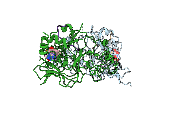
Structure Of G9A Set-Domain With Histone H3K9Ala Mutant Peptide And Bound S-Adenosylmethionine
Organism: Homo sapiens
Method: X-RAY DIFFRACTION Resolution:1.67 Å Release Date: 2016-07-06 Classification: TRANSFERASE Ligands: ZN, SAM |
Organism: Homo sapiens
Method: X-RAY DIFFRACTION
Release Date: 2016-07-06
Ligands: ZN, SAM
 |
Structure Of G9A Set-Domain With Histone H3K9M Mutant Peptide And Bound S-Adenosylmethionine
Organism: Homo sapiens
Method: X-RAY DIFFRACTION Resolution:1.85 Å Release Date: 2016-07-06 Classification: TRANSFERASE Ligands: ZN, SAM |
Organism: Homo sapiens
Method: X-RAY DIFFRACTION
Release Date: 2016-07-06
Ligands: ZN, SAM
 |
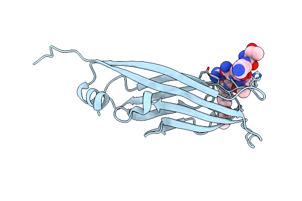
Structure Of G9A Set-Domain With Histone H3K9M Peptide And Excess Sah
Organism: Homo sapiens
Method: X-RAY DIFFRACTION Resolution:1.72 Å Release Date: 2016-07-06 Classification: TRANSFERASE Ligands: ZN, SAM |
Organism: Homo sapiens
Method: X-RAY DIFFRACTION
Release Date: 2016-07-06
Ligands: ZN, SAM
 |
Af9 Yeats In Complex With Histone H3 Crotonylation At K9
Organism: Homo sapiens
Method: X-RAY DIFFRACTION Resolution:2.70 Å Release Date: 2016-04-20 Classification: TRANSCRIPTION/PEPTIDE |
Organism: Homo sapiens
Method: X-RAY DIFFRACTION
Release Date: 2016-04-20
 |
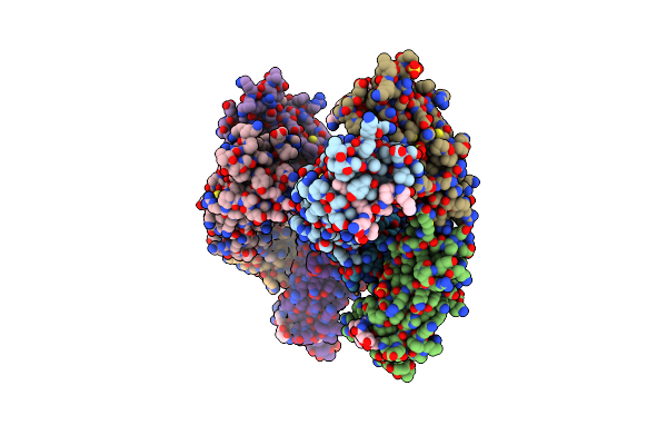
Brd3 Second Bromodomain In Complex With Histone H3 Acetylation At K18
Organism: Homo sapiens
Method: X-RAY DIFFRACTION Resolution:2.60 Å Release Date: 2016-04-20 Classification: SIGNALING PROTEIN/PEPTIDE Ligands: EDO, CL |
Organism: Homo sapiens
Method: X-RAY DIFFRACTION
Release Date: 2016-04-20
Ligands: EDO, CL
 |
Af9 Yeats In Complex With Histone H3 Crotonylation At K18
Organism: Homo sapiens
Method: X-RAY DIFFRACTION Resolution:2.81 Å Release Date: 2016-04-20 Classification: TRANSCRIPTION/PEPTIDE Ligands: CU, SO4 |
Organism: Homo sapiens
Method: X-RAY DIFFRACTION
Release Date: 2016-04-20
Ligands: CU, SO4
 |
Crystal Structure Of Dnmt3A Add Domain Bound To H3 Peptide
Organism: Homo sapiens
Method: X-RAY DIFFRACTION Resolution:2.41 Å Release Date: 2015-05-13 Classification: TRANSFERASE Ligands: ZN |
Organism: Homo sapiens
Method: X-RAY DIFFRACTION
Release Date: 2015-05-13
Ligands: ZN