

Search Count: 45
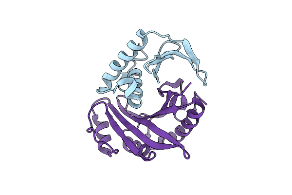 |
1.75 Angstrom Resolution Crystal Structure Of The Toxic C-Terminal Tip Of Cdia From Pseudomonas Aeruginosa In Complex With Immune Protein
Organism: Pseudomonas aeruginosa, Enterobacter cloacae
Method: X-RAY DIFFRACTION Resolution:1.75 Å Release Date: 2019-05-01 Classification: TOXIN |
Organism: Pseudomonas aeruginosa, Enterobacter cloacae
Method: X-RAY DIFFRACTION
Release Date: 2019-05-01
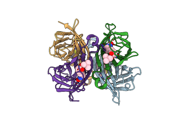 |
A Hyrdrogen Producing Hybrid Streptavidin-Diiron Catalyst
Organism: Streptomyces avidinii
Method: X-RAY DIFFRACTION Resolution:2.05 Å Release Date: 2018-04-04 Classification: Biotin Binding Protein Ligands: BN7 |
Organism: Streptomyces avidinii
Method: X-RAY DIFFRACTION
Release Date: 2018-04-04
Ligands: BN7
 |
Crystal Structure Of A Designed Mn Binding Peptide
Organism: Synthetic construct
Method: X-RAY DIFFRACTION Resolution:1.75 Å Release Date: 2015-10-21 Classification: DE NOVO PROTEIN Ligands: MN |
Organism: Synthetic construct
Method: X-RAY DIFFRACTION
Release Date: 2015-10-21
Ligands: MN
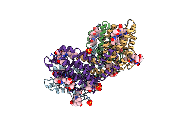 |
Crystal Structure Of The B-Phycoerythrin From The Red Algae Porphyridium Cruentum At Ph8
Organism: Porphyridium purpureum
Method: X-RAY DIFFRACTION Resolution:1.70 Å Release Date: 2012-10-03 Classification: PHOTOSYNTHESIS Ligands: PEB, SO4 |
Organism: Porphyridium purpureum
Method: X-RAY DIFFRACTION
Release Date: 2012-10-03
Ligands: PEB, SO4
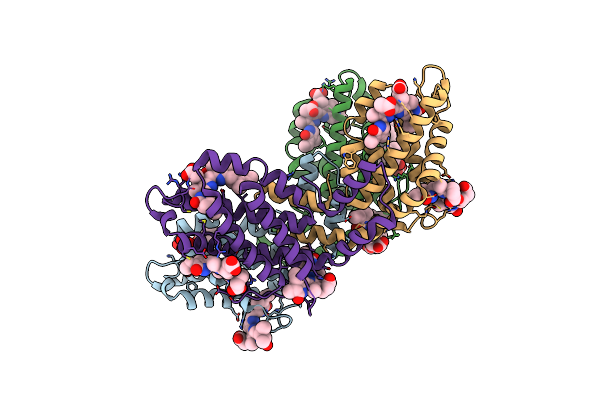 |
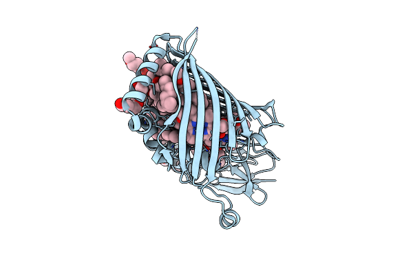
Crystal Structure Of The B-Phycoerythrin From The Red Algae Porphyridium Cruentum At Ph5
Organism: Porphyridium purpureum
Method: X-RAY DIFFRACTION Resolution:1.85 Å Release Date: 2012-10-03 Classification: PHOTOSYNTHESIS Ligands: PEB, SO4 |
Organism: Porphyridium purpureum
Method: X-RAY DIFFRACTION
Release Date: 2012-10-03
Ligands: PEB, SO4
 |
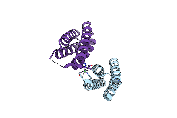
Structure Of The Fmo Protein From Pelodictyon Phaeum
Organism: Pelodictyon phaeum
Method: X-RAY DIFFRACTION Resolution:1.99 Å Release Date: 2012-01-25 Classification: PHOTOSYNTHESIS Ligands: BCL, PG4 |
Organism: Pelodictyon phaeum
Method: X-RAY DIFFRACTION
Release Date: 2012-01-25
Ligands: BCL, PG4
 |
Three Dimensional Structure Of The Small Metal Binding Protein, Smbp
Organism: Nitrosomonas europaea
Method: X-RAY DIFFRACTION Resolution:1.90 Å Release Date: 2011-11-09 Classification: METAL BINDING PROTEIN Ligands: NI |
Organism: Nitrosomonas europaea
Method: X-RAY DIFFRACTION
Release Date: 2011-11-09
Ligands: NI
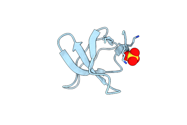 |
Crystal Structure Of The R21D Mutant Of Alpha-Spectrin Sh3 Domain. Crystal Obtained In Ammonium Sulphate At Ph 4.
Organism: Gallus gallus
Method: X-RAY DIFFRACTION Resolution:2.60 Å Release Date: 2011-01-19 Classification: SIGNALING PROTEIN Ligands: SO4 |
Organism: Gallus gallus
Method: X-RAY DIFFRACTION
Release Date: 2011-01-19
Ligands: SO4
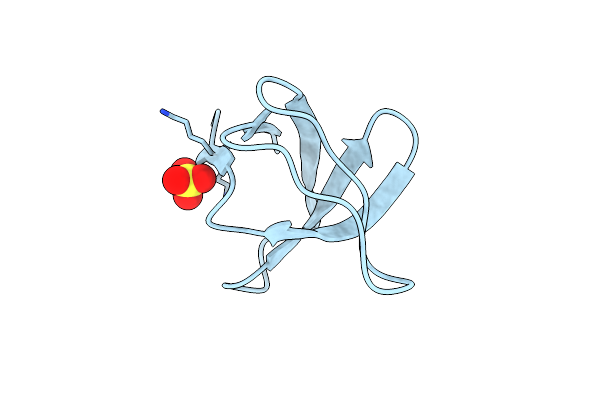 |
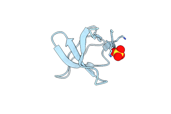
Crystal Structure Of The R21D Mutant Of Alpha-Spectrin Sh3 Domain. Crystal Obtained In Ammonium Sulphate At Ph 5.
Organism: Gallus gallus
Method: X-RAY DIFFRACTION Resolution:1.75 Å Release Date: 2011-01-19 Classification: SIGNALING PROTEIN Ligands: SO4 |
Organism: Gallus gallus
Method: X-RAY DIFFRACTION
Release Date: 2011-01-19
Ligands: SO4
 |
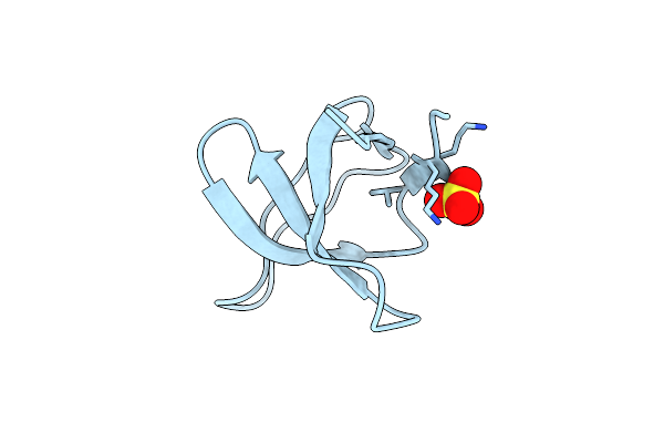
Crystal Structure Of The R21D Mutant Of Alpha-Spectrin Sh3 Domain. Crystal Obtained In Ammonium Sulphate At Ph 6.
Organism: Gallus gallus
Method: X-RAY DIFFRACTION Resolution:1.10 Å Release Date: 2011-01-19 Classification: SIGNALING PROTEIN Ligands: SO4 |
Organism: Gallus gallus
Method: X-RAY DIFFRACTION
Release Date: 2011-01-19
Ligands: SO4
 |
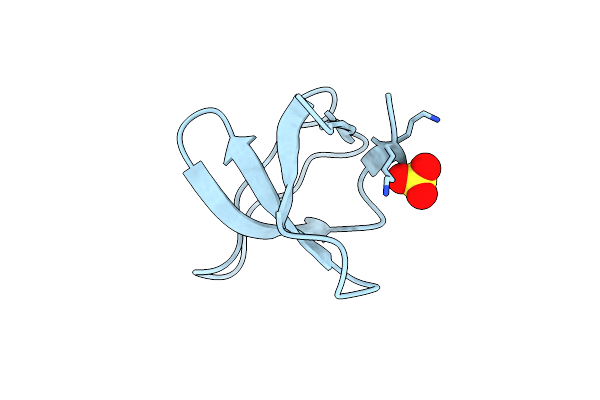
Crystal Structure Of The R21D Mutant Of Alpha-Spectrin Sh3 Domain. Crystal Obtained In Ammonium Sulphate At Ph 7
Organism: Gallus gallus
Method: X-RAY DIFFRACTION Resolution:1.60 Å Release Date: 2011-01-19 Classification: SIGNALING PROTEIN Ligands: SO4 |
Organism: Gallus gallus
Method: X-RAY DIFFRACTION
Release Date: 2011-01-19
Ligands: SO4
 |
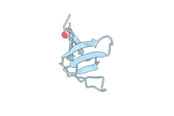
Crystal Structure Of The R21D Mutant Of Alpha-Spectrin Sh3 Domain. Crystal Obtained In Ammonium Sulphate At Ph 9.
Organism: Gallus gallus
Method: X-RAY DIFFRACTION Resolution:1.70 Å Release Date: 2011-01-19 Classification: SIGNALING PROTEIN Ligands: SO4 |
Organism: Gallus gallus
Method: X-RAY DIFFRACTION
Release Date: 2011-01-19
Ligands: SO4
 |
Crystal Structure Of The R21D Mutant Of Alpha-Spectrin Sh3 Domain. Hexagonal Crystal Obtained In Sodium Formate At Ph 6.5.
Organism: Gallus gallus
Method: X-RAY DIFFRACTION Resolution:1.10 Å Release Date: 2011-01-19 Classification: SIGNALING PROTEIN Ligands: FMT |
Organism: Gallus gallus
Method: X-RAY DIFFRACTION
Release Date: 2011-01-19
Ligands: FMT
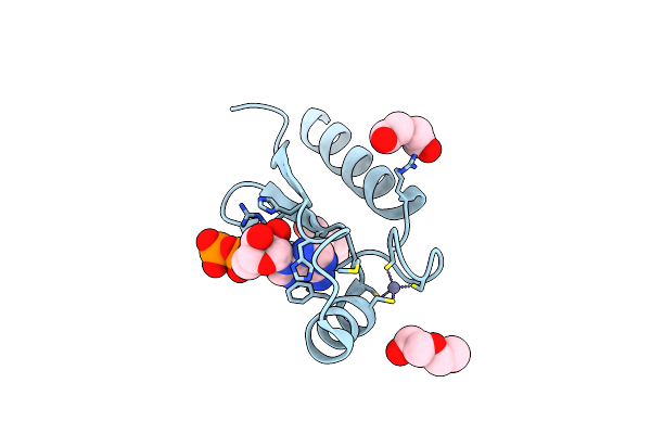 |
A Non-Biological Atp Binding Protein With A Single Point Mutation (D65V), That Contributes To Optimized Folding And Ligand Binding, Crystallized In The Presence Of 100 Mm Atp.
Organism: Synthetic construct
Method: X-RAY DIFFRACTION Resolution:2.55 Å Release Date: 2010-09-22 Classification: DE NOVO PROTEIN Ligands: ZN, ADP, PEG, CL |
Organism: Synthetic construct
Method: X-RAY DIFFRACTION
Release Date: 2010-09-22
Ligands: ZN, ADP, PEG, CL
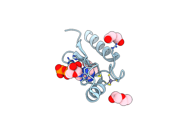 |
A Non-Biological Atp Binding Protein With A Single Point Mutation (D65V), That Contributes To Optimized Folding And Ligand Binding
Organism: Synthetic construct
Method: X-RAY DIFFRACTION Resolution:2.55 Å Release Date: 2010-09-22 Classification: DE NOVO PROTEIN Ligands: ZN, ADP, PEG, CL |
Organism: Synthetic construct
Method: X-RAY DIFFRACTION
Release Date: 2010-09-22
Ligands: ZN, ADP, PEG, CL
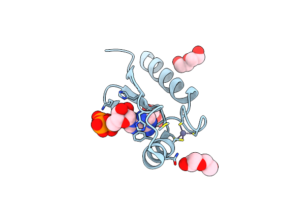 |
Crystal Structure Of A Non-Biological Atp Binding Protein With A Tyr-Phe Mutation Within The Ligand Binding Domain
Organism: Synthetic construct
Method: X-RAY DIFFRACTION Resolution:2.70 Å Release Date: 2010-09-22 Classification: DE NOVO PROTEIN Ligands: ZN, ATP, PEG, CL |
Organism: Synthetic construct
Method: X-RAY DIFFRACTION
Release Date: 2010-09-22
Ligands: ZN, ATP, PEG, CL
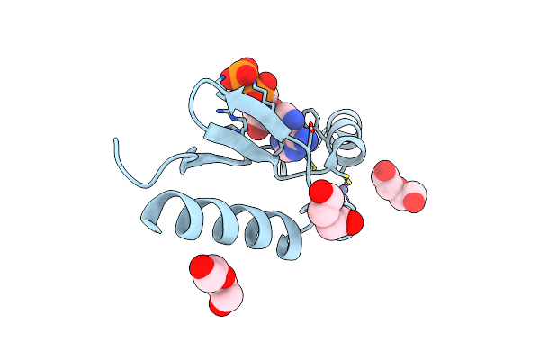 |
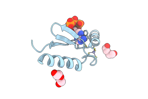
X-Ray Structure Of A Non-Biological Atp Binding Protein Determined In The Presence Of 10 Mm Atp At 2.6 A After 3 Weeks Of Incubation
Organism: Synthetic construct
Method: X-RAY DIFFRACTION Resolution:2.60 Å Release Date: 2010-09-22 Classification: DE NOVO PROTEIN Ligands: ZN, ADP, PEG, CL |
Organism: Synthetic construct
Method: X-RAY DIFFRACTION
Release Date: 2010-09-22
Ligands: ZN, ADP, PEG, CL
 |
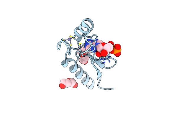
X-Ray Structure Of A Non-Biological Atp Binding Protein Determined In The Presence Of 10 Mm Atp At 2.0 A By Multi-Wavelength Anomalous Dispersion
Organism: Synthetic construct
Method: X-RAY DIFFRACTION Resolution:2.00 Å Release Date: 2010-09-22 Classification: DE NOVO PROTEIN Ligands: ZN, ATP, PEG, CL |
Organism: Synthetic construct
Method: X-RAY DIFFRACTION
Release Date: 2010-09-22
Ligands: ZN, ATP, PEG, CL
 |
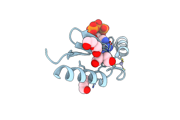
X-Ray Structure Of A Non-Biological Atp Binding Protein Determined At 2.8 A By Multi-Wavelength Anomalous Dispersion
Organism: Synthetic construct
Method: X-RAY DIFFRACTION Resolution:2.80 Å Release Date: 2010-09-22 Classification: DE NOVO PROTEIN Ligands: PEG, ZN, ADP, CL |
Organism: Synthetic construct
Method: X-RAY DIFFRACTION
Release Date: 2010-09-22
Ligands: PEG, ZN, ADP, CL
 |
1.8 A Crystal Structure Of A Non-Biological Protein With Bound Atp In A Novel Bent Conformation
Organism: Unidentified
Method: X-RAY DIFFRACTION Resolution:1.80 Å Release Date: 2009-06-30 Classification: DE NOVO PROTEIN Ligands: ZN, ATP, PEG |
Organism: Unidentified
Method: X-RAY DIFFRACTION
Release Date: 2009-06-30
Ligands: ZN, ATP, PEG