
Search Count: 2,209
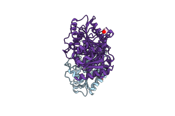 |
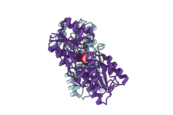
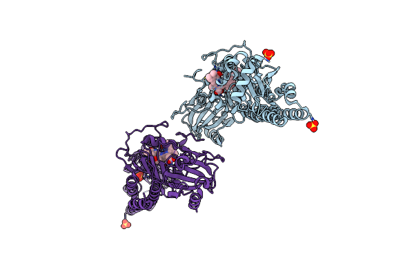
Organism: Agrobacterium tumefaciens
Method: X-RAY DIFFRACTION Resolution:3.13 Å Release Date: 2026-04-15 Classification: HYDROLASE Ligands: EDO |
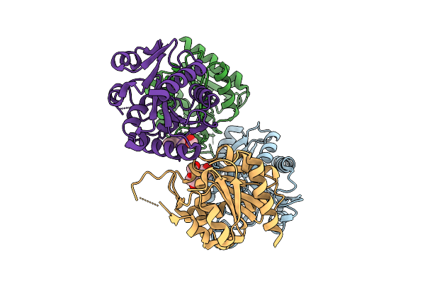 |
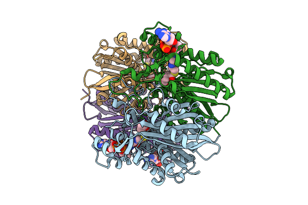
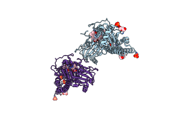
Organism: Agrobacterium tumefaciens
Method: X-RAY DIFFRACTION Resolution:1.83 Å Release Date: 2026-04-15 Classification: HYDROLASE Ligands: PEG |
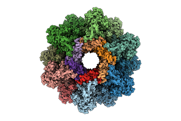 |
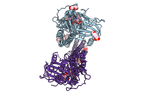
Organism: Agrobacterium phage 7-7-1
Method: ELECTRON MICROSCOPY Release Date: 2025-12-10 Classification: VIRUS |
 |
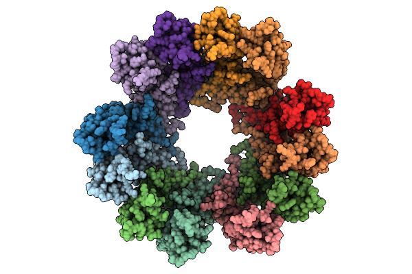
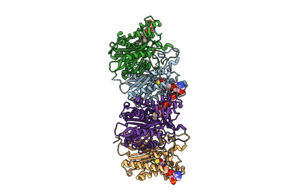
Organism: Agrobacterium phage 7-7-1
Method: ELECTRON MICROSCOPY Release Date: 2025-12-10 Classification: VIRAL PROTEIN |
 |
Organism: Agrobacterium phage 7-7-1
Method: ELECTRON MICROSCOPY Release Date: 2025-12-10 Classification: VIRUS |
 |
Organism: Agrobacterium phage 7-7-1
Method: ELECTRON MICROSCOPY Release Date: 2025-12-10 Classification: VIRUS |
 |
Organism: Agrobacterium phage 7-7-1
Method: ELECTRON MICROSCOPY Release Date: 2025-12-10 Classification: VIRUS |
 |
Organism: Agrobacterium phage 7-7-1
Method: ELECTRON MICROSCOPY Release Date: 2025-12-10 Classification: VIRUS |
 |
Organism: Agrobacterium phage 7-7-1
Method: ELECTRON MICROSCOPY Release Date: 2025-12-10 Classification: VIRUS |
 |
Organism: Agrobacterium phage 7-7-1
Method: ELECTRON MICROSCOPY Release Date: 2025-12-10 Classification: VIRUS |
 |
Organism: Agrobacterium fabrum str. c58
Method: X-RAY DIFFRACTION Resolution:2.53 Å Release Date: 2025-11-26 Classification: TRANSCRIPTION Ligands: A1EGE |
 |
Organism: Agrobacterium radiobacter
Method: ELECTRON MICROSCOPY Release Date: 2025-11-12 Classification: OXIDOREDUCTASE Ligands: HEM, NDP |
 |
Organism: Agrobacterium fabrum str. c58
Method: X-RAY DIFFRACTION Resolution:2.50 Å Release Date: 2025-10-29 Classification: BIOSYNTHETIC PROTEIN Ligands: GRA |
 |
Organism: Agrobacterium fabrum str. c58
Method: X-RAY DIFFRACTION Resolution:1.79 Å Release Date: 2025-10-08 Classification: SIGNALING PROTEIN Ligands: EL5, MPD, PEG, MES, CL |
 |
Organism: Agrobacterium fabrum (strain c58 / atcc 33970)
Method: X-RAY DIFFRACTION Resolution:2.15 Å Release Date: 2025-08-20 Classification: BIOSYNTHETIC PROTEIN Ligands: COA |
 |
Organism: Agrobacterium fabrum str. c58
Method: X-RAY DIFFRACTION Resolution:2.15 Å Release Date: 2025-05-14 Classification: SIGNALING PROTEIN Ligands: EL5, SO4, CL, EDO |
 |
Organism: Agrobacterium fabrum str. c58
Method: X-RAY DIFFRACTION Resolution:2.20 Å Release Date: 2025-05-14 Classification: SIGNALING PROTEIN Ligands: EL5, SO4 |
 |
Organism: Agrobacterium fabrum str. c58
Method: X-RAY DIFFRACTION Resolution:2.54 Å Release Date: 2025-05-14 Classification: SIGNALING PROTEIN Ligands: EL5, SO4 |
 |
Organism: Agrobacterium fabrum str. c58
Method: X-RAY DIFFRACTION Resolution:2.43 Å Release Date: 2025-05-14 Classification: SIGNALING PROTEIN Ligands: EL5, SO4, PGE, PEG, CL |
 |
Organism: Agrobacterium fabrum str. c58
Method: X-RAY DIFFRACTION Resolution:2.40 Å Release Date: 2025-05-14 Classification: SIGNALING PROTEIN Ligands: EL5, SO4, GOL, PEG |