

Search Count: 66
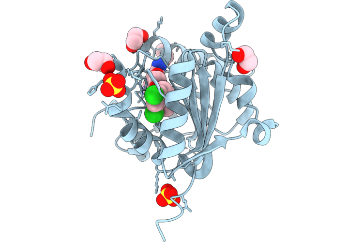 |
Mettl1 Bound To The Sam Competitive Small Molecule Inhibitor Stm9005
Organism: Homo sapiens
Method: X-RAY DIFFRACTION Resolution:1.44 Å Release Date: 2026-08-26 Classification: TRANSFERASE Ligands: A1JKJ, SO4, EDO, CL |
Organism: Homo sapiens
Method: X-RAY DIFFRACTION
Release Date: 2026-08-26
Ligands: A1JKJ, SO4, EDO, CL
 |
Solution Structure Of Murinotoxin-1
|
Organism: Pterinochilus murinus
Method: SOLUTION NMR
Release Date: 2025-09-10
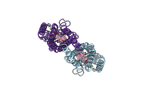 |
Human Ebp Complexed With Compound 1
Organism: Homo sapiens
Method: ELECTRON MICROSCOPY Release Date: 2024-06-19 Classification: ISOMERASE/INHIBITOR Ligands: A1AEU |
Organism: Homo sapiens
Method: ELECTRON MICROSCOPY
Release Date: 2024-06-19
Ligands: A1AEU
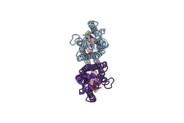 |
Human Ebp Complexed With Compound 3A
Organism: Homo sapiens
Method: ELECTRON MICROSCOPY Release Date: 2024-06-19 Classification: ISOMERASE/INHIBITOR Ligands: A1AEV |
Organism: Homo sapiens
Method: ELECTRON MICROSCOPY
Release Date: 2024-06-19
Ligands: A1AEV
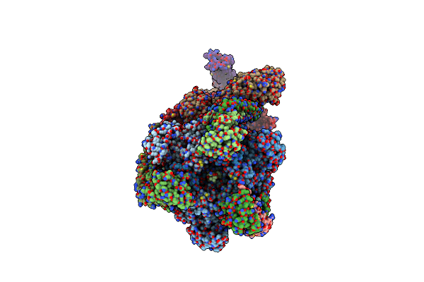 |
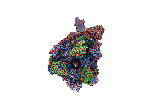
Intact V-Atpase State 2 And Synaptophysin Complex In Mouse Brain Isolated Synaptic Vesicles
Organism: Mus musculus
Method: ELECTRON MICROSCOPY Release Date: 2024-06-19 Classification: MEMBRANE PROTEIN |
Organism: Mus musculus
Method: ELECTRON MICROSCOPY
Release Date: 2024-06-19
 |
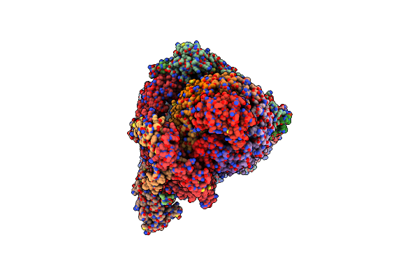
Intact V-Atpase State 3 And Synaptophysin Complex In Mouse Brain Isolated Synaptic Vesicles
Organism: Mus musculus
Method: ELECTRON MICROSCOPY Release Date: 2024-06-19 Classification: MEMBRANE PROTEIN |
Organism: Mus musculus
Method: ELECTRON MICROSCOPY
Release Date: 2024-06-19
 |
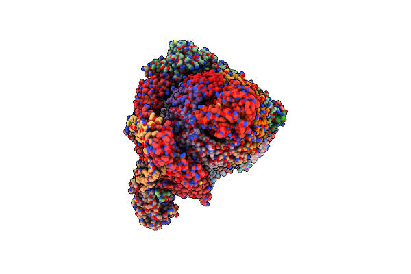
Intact V-Atpase State 3 In Synaptophysin Knock-Out Isolated Synaptic Vesicles
Organism: Mus musculus
Method: ELECTRON MICROSCOPY Release Date: 2024-06-19 Classification: MEMBRANE PROTEIN |
Organism: Mus musculus
Method: ELECTRON MICROSCOPY
Release Date: 2024-06-19
 |
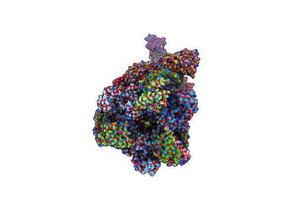
Intact V-Atpase State 2 In Synaptophysin Knock-Out Isolated Synaptic Vesicles
Organism: Mus musculus
Method: ELECTRON MICROSCOPY Release Date: 2024-06-19 Classification: MEMBRANE PROTEIN |
Organism: Mus musculus
Method: ELECTRON MICROSCOPY
Release Date: 2024-06-19
 |
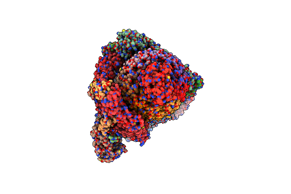
Intact V-Atpase State 1 And Synaptophysin Complex In Mouse Brain Isolated Synaptic Vesicles
Organism: Mus musculus
Method: ELECTRON MICROSCOPY Release Date: 2024-06-19 Classification: MEMBRANE PROTEIN |
Organism: Mus musculus
Method: ELECTRON MICROSCOPY
Release Date: 2024-06-19
 |
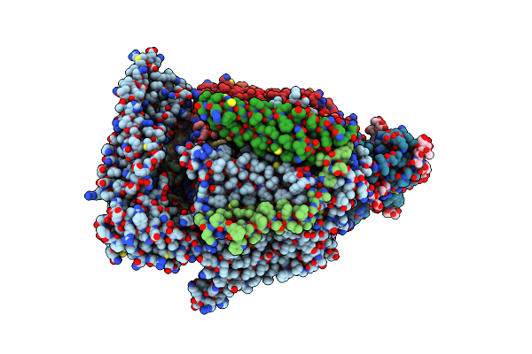
Intact V-Atpase State 1 In Synaptophysin Knock-Out Isolated Synaptic Vesicles
Organism: Mus musculus
Method: ELECTRON MICROSCOPY Release Date: 2024-06-19 Classification: MEMBRANE PROTEIN |
Organism: Mus musculus
Method: ELECTRON MICROSCOPY
Release Date: 2024-06-19
 |
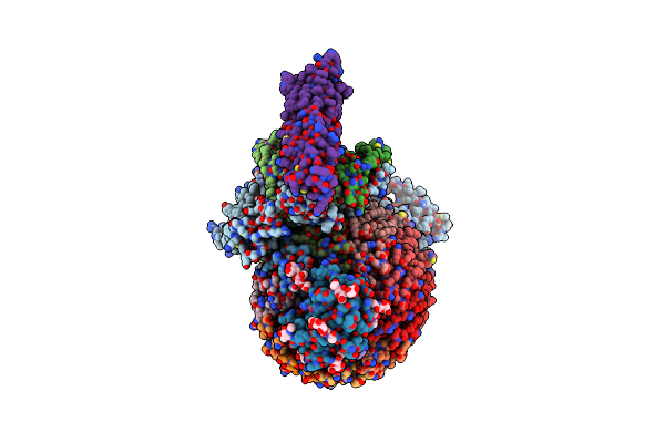
V0-Only V-Atpase In Synaptophysin Gene Knock-Out Mouse Brain Isolated Synaptic Vesicles
Organism: Mus musculus
Method: ELECTRON MICROSCOPY Release Date: 2024-06-19 Classification: MEMBRANE PROTEIN Ligands: NAG, BMA |
Organism: Mus musculus
Method: ELECTRON MICROSCOPY
Release Date: 2024-06-19
Ligands: NAG, BMA
 |
V0-Only V-Atpase And Synaptophysin Complex In Mouse Brain Isolated Synaptic Vesicles
Organism: Mus musculus
Method: ELECTRON MICROSCOPY Release Date: 2024-06-19 Classification: MEMBRANE PROTEIN Ligands: NAG |
Organism: Mus musculus
Method: ELECTRON MICROSCOPY
Release Date: 2024-06-19
Ligands: NAG
 |
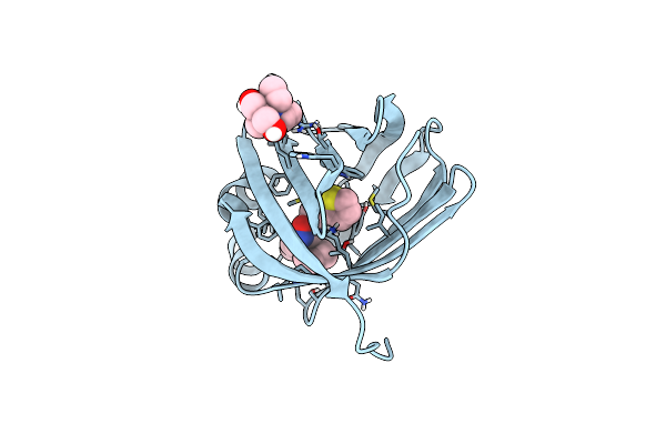
Crystal Structure Of Human Cellular Retinol Binding Protein 1 In Complex With Methyl({3-[1-(4-Methylphenyl)Cyclopentyl]-1,2,4-Oxadiazol-5-Yl}Methyl)[(1-Methylpyrazol-4-Yl)Methyl]Amine
Organism: Homo sapiens
Method: X-RAY DIFFRACTION Resolution:1.47 Å Release Date: 2023-10-11 Classification: Retinol-binding protein Ligands: ZCF, BTB |
Organism: Homo sapiens
Method: X-RAY DIFFRACTION
Release Date: 2023-10-11
Ligands: ZCF, BTB
 |
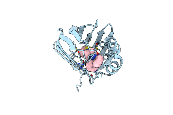
Crystal Structure Of Human Cellular Retinol Binding Protein 1 In Complex With N-Methyl-1-{3-[1-(4-Methylphenyl)Cyclopentyl]-1,2,4-Oxadiazol-5-Yl}-N-(2-Thienylmethyl)Methanamine
Organism: Homo sapiens
Method: X-RAY DIFFRACTION Resolution:1.13 Å Release Date: 2023-10-04 Classification: Retinol-binding protein Ligands: Z5H, BTB |
Organism: Homo sapiens
Method: X-RAY DIFFRACTION
Release Date: 2023-10-04
Ligands: Z5H, BTB
 |
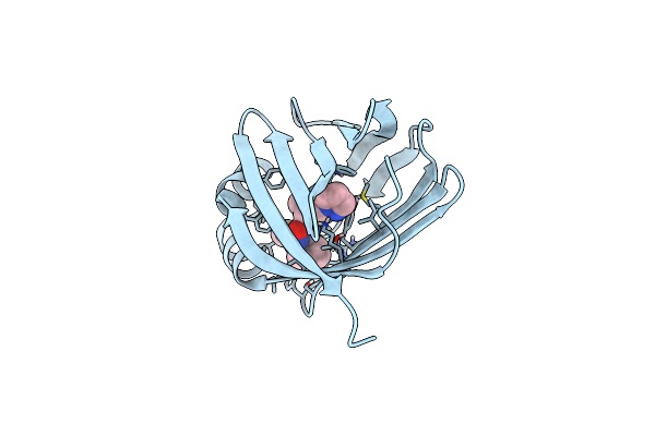
Crystal Structure Of Human Cellular Retinol Binding Protein 1 In Complex With {[3-(Diphenylmethyl)-1,2,4-Oxadiazol-5-Yl]Methyl}(Methyl)[1-(Thiophen-2-Yl)Ethyl]Amine
Organism: Homo sapiens
Method: X-RAY DIFFRACTION Resolution:1.80 Å Release Date: 2023-10-04 Classification: Retinol-binding protein Ligands: ZA6 |
Organism: Homo sapiens
Method: X-RAY DIFFRACTION
Release Date: 2023-10-04
Ligands: ZA6
 |
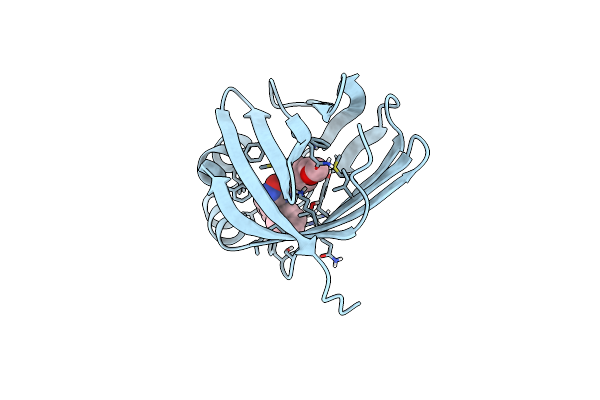
Crystal Structure Of Human Cellular Retinol Binding Protein 1 In Complex With N-Ethyl-N-({3-[1-(4-Methylphenyl)Cyclopentyl]-1,2,4-Oxadiazol-5-Yl}Methyl)-2-(1H-Pyrazol-1-Yl)Ethanamine
Organism: Homo sapiens
Method: X-RAY DIFFRACTION Resolution:1.55 Å Release Date: 2023-10-04 Classification: retinol-binding protein Ligands: ZDF |
Organism: Homo sapiens
Method: X-RAY DIFFRACTION
Release Date: 2023-10-04
Ligands: ZDF
 |
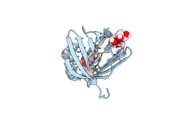
Crystal Structure Of Human Cellular Retinol Binding Protein 1 In Complex With 1-{[3-(Diphenylmethyl)-1,2,4-Oxadiazol-5-Yl]Methyl}-4-(Methoxymethyl)Piperidine
Organism: Homo sapiens
Method: X-RAY DIFFRACTION Resolution:1.85 Å Release Date: 2023-10-04 Classification: Retinol-binding protein Ligands: ZDK |
Organism: Homo sapiens
Method: X-RAY DIFFRACTION
Release Date: 2023-10-04
Ligands: ZDK
 |
Crystal Structure Of Human Cellular Retinol Binding Protein 1 In Complex With 4-(Hydroxymethyl)-1-[(4-Methoxy-5,6,7,8-Tetrahydronaphthalen-1-Yl)Sulfonyl]Piperidin-4-Ol
Organism: Homo sapiens
Method: X-RAY DIFFRACTION Resolution:1.30 Å Release Date: 2023-10-04 Classification: Retinol-binding protein Ligands: ZE2, BTB |
Organism: Homo sapiens
Method: X-RAY DIFFRACTION
Release Date: 2023-10-04
Ligands: ZE2, BTB
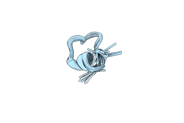 |
Structure Of Elevenin-Vc1 From Venom Of The Australian Cone Snail Conus Victoriae
|
Organism: Conus victoriae
Method: SOLUTION NMR
Release Date: 2023-09-13
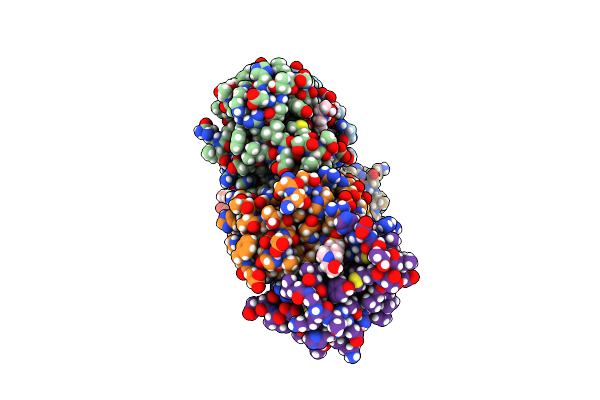 |
Conotoxin Mu8.1 From Conus Mucronatus
Organism: Conus mucronatus
Method: X-RAY DIFFRACTION Resolution:2.12 Å Release Date: 2022-11-16 Classification: TOXIN Ligands: EPE |
Organism: Conus mucronatus
Method: X-RAY DIFFRACTION
Release Date: 2022-11-16
Ligands: EPE