

Search Count: 5
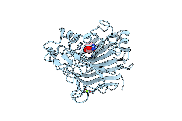 |
X-Ray Structure Analysis Of Beita-1,3-Glucanase
Organism: Actinobacteria bacterium
Method: X-RAY DIFFRACTION Resolution:1.14 Å Release Date: 2022-03-02 Classification: HYDROLASE Ligands: TRS, MG |
Organism: Actinobacteria bacterium
Method: X-RAY DIFFRACTION
Release Date: 2022-03-02
Ligands: TRS, MG
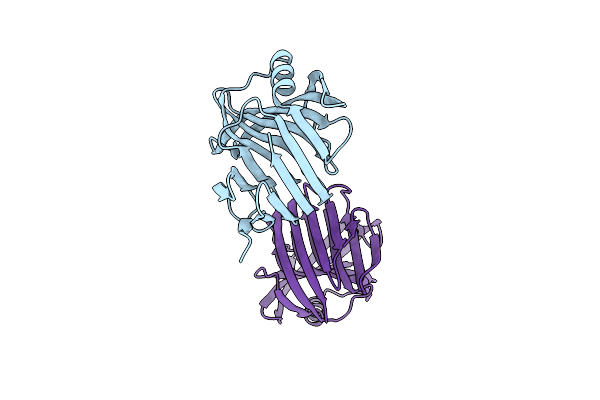 |
X-Ray Structure Analysis Of Xylanase
Organism: Actinobacteria bacterium
Method: X-RAY DIFFRACTION Resolution:1.90 Å Release Date: 2021-10-13 Classification: HYDROLASE Ligands: IOD |
Organism: Actinobacteria bacterium
Method: X-RAY DIFFRACTION
Release Date: 2021-10-13
Ligands: IOD
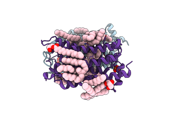 |
Crystal Structure Of The 48C12 Heliorhodopsin In The Violet Form At Ph 8.8
Organism: Actinobacteria bacterium
Method: X-RAY DIFFRACTION Resolution:1.50 Å Release Date: 2019-12-11 Classification: MEMBRANE PROTEIN Ligands: LFA, OLC, SO4, RET, GOL, OLA |
Organism: Actinobacteria bacterium
Method: X-RAY DIFFRACTION
Release Date: 2019-12-11
Ligands: LFA, OLC, SO4, RET, GOL, OLA
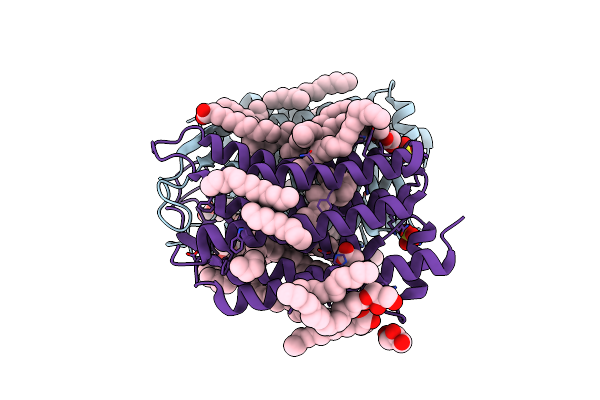 |
Crystal Structure Of The 48C12 Heliorhodopsin In The Blue Form At Ph 4.3
Organism: Actinobacteria bacterium
Method: X-RAY DIFFRACTION Resolution:1.50 Å Release Date: 2019-12-11 Classification: MEMBRANE PROTEIN Ligands: ACT, OLA, GOL, OLC, LFA, SO4, RET |
Organism: Actinobacteria bacterium
Method: X-RAY DIFFRACTION
Release Date: 2019-12-11
Ligands: ACT, OLA, GOL, OLC, LFA, SO4, RET
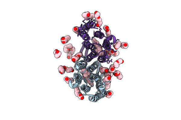 |
Crystal Structure Of Bacterial Heliorhodopsin 48C12
Organism: Actinobacteria bacterium
Method: X-RAY DIFFRACTION Resolution:2.70 Å Release Date: 2019-12-04 Classification: SIGNALING PROTEIN Ligands: RET, PGE, PLM, PEG |
Organism: Actinobacteria bacterium
Method: X-RAY DIFFRACTION
Release Date: 2019-12-04
Ligands: RET, PGE, PLM, PEG