
Search Count: 550
All
Selected
 |
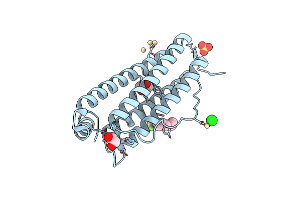
Crystal Structure Of L-Threonate 3-Dehydrogenase From Paracoccus Litorisediminis (Ligand-Free Form)
Organism: Paracoccus litorisediminis
Method: X-RAY DIFFRACTION Resolution:1.90 Å Release Date: 2026-03-18 Classification: OXIDOREDUCTASE Ligands: SO4, GOL, PEG |
 |
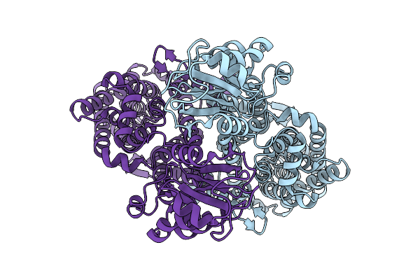
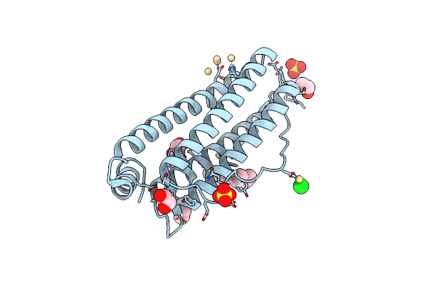
Crystal Structure Of L-Threonate 3-Dehydrogenase From Paracoccus Litorisediminis (Nadp+ And Tartronate Bound Form)
Organism: Paracoccus litorisediminis
Method: X-RAY DIFFRACTION Resolution:2.08 Å Release Date: 2026-03-18 Classification: OXIDOREDUCTASE Ligands: NAP, TTN |
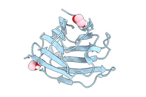 |
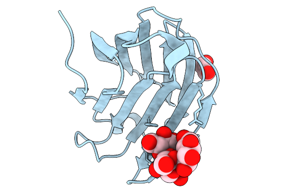
Crystal Structure Of Human Galectin-10 Produced By Cell-Free Protein Synthesis
Organism: Homo sapiens
Method: X-RAY DIFFRACTION Resolution:1.60 Å Release Date: 2026-02-18 Classification: SUGAR BINDING PROTEIN Ligands: EDO |
 |
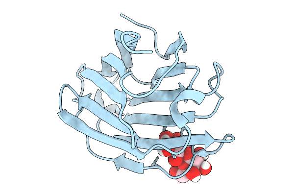
Crystal Structure Of E33A Mutant Of Human Galectin-10 Produced By Cell-Free Protein Synthesis In Complex With Raffinose
Organism: Homo sapiens
Method: X-RAY DIFFRACTION Resolution:1.57 Å Release Date: 2026-02-18 Classification: SUGAR BINDING PROTEIN Ligands: EDO |
 |
Crystal Structure Of Human Galectin-10 Produced By Cell-Free Protein Synthesis In Complex With Sucrose
Organism: Homo sapiens
Method: X-RAY DIFFRACTION Resolution:1.64 Å Release Date: 2026-02-18 Classification: SUGAR BINDING PROTEIN |
 |
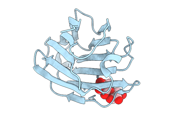
Crystal Structure Of Human Galectin-10 Produced By Cell-Free Protein Synthesis In Complex With Lactose
Organism: Homo sapiens
Method: X-RAY DIFFRACTION Resolution:1.57 Å Release Date: 2026-02-18 Classification: SUGAR BINDING PROTEIN |
 |
Crystal Structure Of Human Galectin-10 Produced By Cell-Free Protein Synthesis In Complex With Trehalose
Organism: Homo sapiens
Method: X-RAY DIFFRACTION Resolution:1.78 Å Release Date: 2026-02-18 Classification: SUGAR BINDING PROTEIN Ligands: EDO |
 |
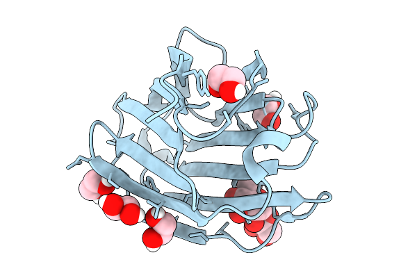
Crystal Structure Of Human Galectin-10 Produced By Cell-Free Protein Synthesis In Complex With Maltose
Organism: Homo sapiens
Method: X-RAY DIFFRACTION Resolution:2.31 Å Release Date: 2026-02-18 Classification: SUGAR BINDING PROTEIN |
 |
Crystal Structure Of Human Galectin-10 Produced By Cell-Free Protein Synthesis In Complex With Melezitose
Organism: Homo sapiens
Method: X-RAY DIFFRACTION Resolution:1.99 Å Release Date: 2026-02-18 Classification: SUGAR BINDING PROTEIN Ligands: EDO |
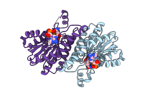 |
Crystal Structure Of L-2-Keto-3-Deoxypentonate 4-Dehydrogenase Bound To Nad(H)
Organism: Herbaspirillum huttiense
Method: X-RAY DIFFRACTION Resolution:2.48 Å Release Date: 2025-11-26 Classification: OXIDOREDUCTASE Ligands: NAD |
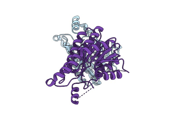 |
Crysral Structure Of 2-Keto-3-Deoxypentonate 4-Dehydrogenase From Herbaspirillum Huttiense (Apo Form)
Organism: Herbaspirillum huttiense
Method: X-RAY DIFFRACTION Resolution:2.27 Å Release Date: 2025-10-15 Classification: OXIDOREDUCTASE |
 |
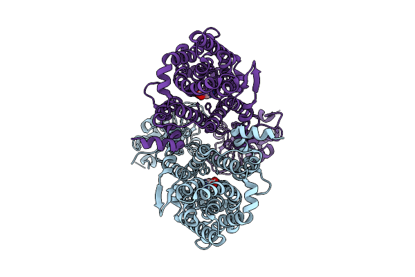
X-Ray Crystal Structure Of Asp/Ala Exchanger Aspt At Outward-Facing Conformation
Organism: Tetragenococcus halophilus
Method: X-RAY DIFFRACTION Resolution:3.45 Å Release Date: 2025-10-08 Classification: MEMBRANE PROTEIN |
 |
Organism: Tetragenococcus halophilus
Method: ELECTRON MICROSCOPY Release Date: 2025-08-06 Classification: TRANSPORT PROTEIN Ligands: ASP |
 |
Organism: Tetragenococcus halophilus
Method: ELECTRON MICROSCOPY Resolution:3.56 Å Release Date: 2025-08-06 Classification: TRANSPORT PROTEIN |
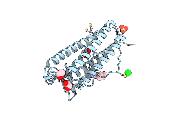 |
Crystal Structure Of Horse Spleen L-Ferritin Mutant (E53F/E56F/E57F/R59F/E60F/E63F) With Nile Red
Organism: Equus caballus
Method: X-RAY DIFFRACTION Resolution:1.60 Å Release Date: 2025-03-05 Classification: METAL BINDING PROTEIN Ligands: CD, SO4, CL, EDO, A1L52 |
 |
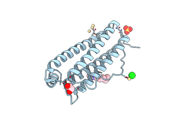
Crystal Structure Of Horse Spleen L-Ferritin Mutant (E53F/E56F/E57F/R59F/E60F/E63F)
Organism: Equus caballus
Method: X-RAY DIFFRACTION Resolution:1.50 Å Release Date: 2025-03-05 Classification: METAL BINDING PROTEIN Ligands: CD, SO4, CL, EDO |
 |
Crystal Structure Of Horse Spleen L-Ferritin Mutant (R52F/E56F/R59F/E63F) With Nile Red
Organism: Equus caballus
Method: X-RAY DIFFRACTION Resolution:1.65 Å Release Date: 2025-03-05 Classification: METAL BINDING PROTEIN Ligands: CD, SO4, CL, EDO, A1L52 |
 |
Crystal Structure Of Horse Spleen L-Ferritin Mutant (Fr-E53F/E56F/E57F/R59L/E60F/E63F)
Organism: Equus caballus
Method: X-RAY DIFFRACTION Resolution:1.50 Å Release Date: 2025-03-05 Classification: METAL BINDING PROTEIN Ligands: CD, SO4, CL, EDO |
 |
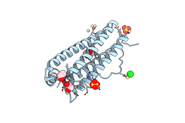
Crystal Structure Of Horse Spleen L-Ferritin Mutant (E53F/E56F/E57F/R59F/E60F/E63F) With Coumarin 153
Organism: Equus caballus
Method: X-RAY DIFFRACTION Resolution:1.64 Å Release Date: 2025-03-05 Classification: METAL BINDING PROTEIN Ligands: CD, SO4, A1L54, EDO, CL |
 |
Crystal Structure Of Horse Spleen L-Ferritin Mutant (E56F/R59F) With Nile Red
Organism: Equus caballus
Method: X-RAY DIFFRACTION Resolution:1.50 Å Release Date: 2025-03-05 Classification: METAL BINDING PROTEIN Ligands: CD, CL, SO4, EDO, A1L52 |