
Search Count: 2,097
 |
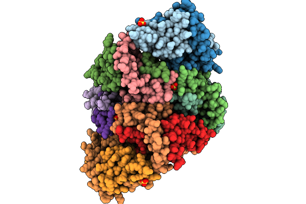
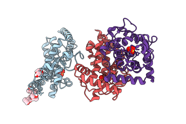
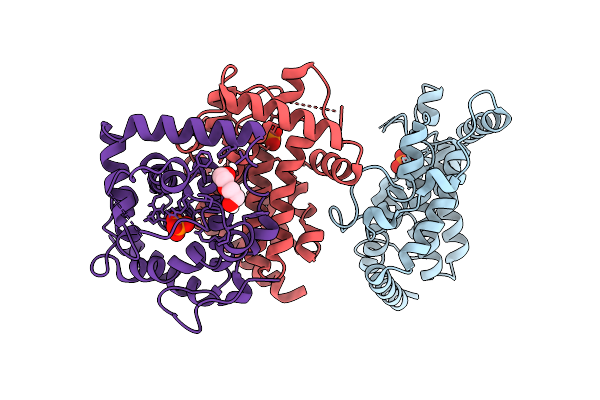
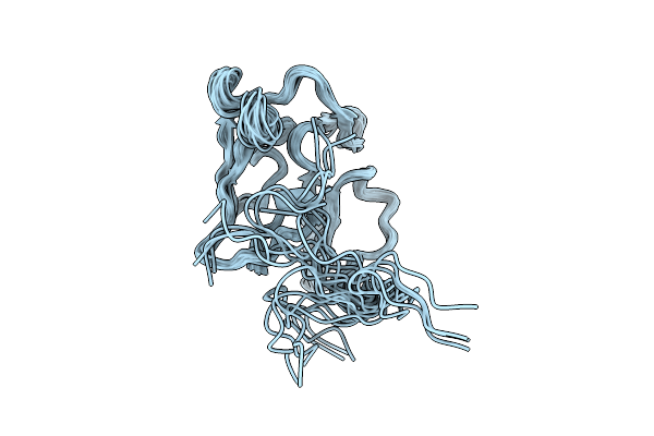
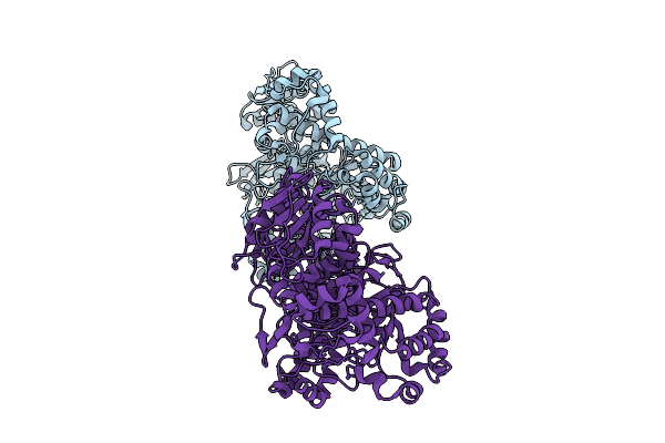
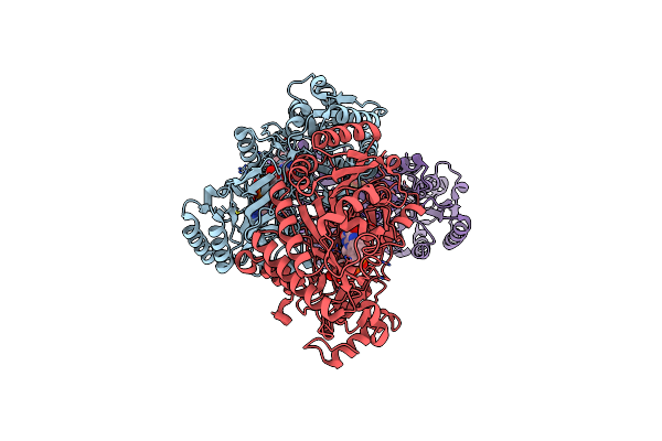
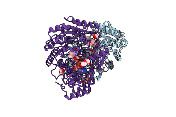
Cryo-Em Structure Of Reduced Form Of Formatedehydrogenase From Rhodobacter Aestuarii (Rafdh) With Nadh
Organism: Rhodobacter aestuarii
Method: ELECTRON MICROSCOPY Resolution:2.90 Å Release Date: 2026-06-03 Classification: OXIDOREDUCTASE Ligands: FES, MGD, 6MO, SF4, NAD, FMN |
 |
The Histone Fold Domain Heterodimer Of Oocyst Rupture Proteins 1 And 2 From Plasmodium Berghei
Organism: Plasmodium berghei
Method: X-RAY DIFFRACTION Resolution:3.10 Å Release Date: 2026-01-21 Classification: UNKNOWN FUNCTION Ligands: SO4, EDO |
 |
Organism: Morganella morganii
Method: X-RAY DIFFRACTION Resolution:2.00 Å Release Date: 2026-01-14 Classification: BIOSYNTHETIC PROTEIN Ligands: MPD, PEG, SO4 |
 |
Organism: Morganella morganii
Method: X-RAY DIFFRACTION Resolution:2.24 Å Release Date: 2026-01-14 Classification: BIOSYNTHETIC PROTEIN Ligands: PO4, PEG |
 |
Three-Dimensional Structure Of The Merozoite Surface Protein 1 C-Terminal Domain
Organism: Plasmodium berghei
Method: SOLUTION NMR Release Date: 2025-12-24 Classification: MEMBRANE PROTEIN |
 |
Organism: Wenyingzhuangia aestuarii
Method: X-RAY DIFFRACTION Resolution:2.00 Å Release Date: 2025-10-01 Classification: HYDROLASE |
 |
Organism: Clostridium phage phicd111
Method: X-RAY DIFFRACTION Resolution:1.40 Å Release Date: 2025-09-24 Classification: ANTIBIOTIC Ligands: ZN |
 |
Organism: Homo sapiens, Recombinant vesicular stomatitis indiana virus rvsv-g/gfp, Bos taurus
Method: ELECTRON MICROSCOPY Release Date: 2025-07-23 Classification: PROTEIN BINDING Ligands: GTP, MG, GDP, TA1 |
 |
Organism: Kalmanozyma brasiliensis ghg001
Method: X-RAY DIFFRACTION Resolution:3.46 Å Release Date: 2025-04-02 Classification: HYDROLASE Ligands: NAG |
 |
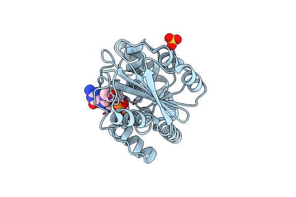
Vanillyl-Alcohol Dehydrogenase From Marinicaulis Flavus: P151L Mutant Bound To Eugenol
Organism: Marinicaulis flavus
Method: X-RAY DIFFRACTION Resolution:1.80 Å Release Date: 2025-01-15 Classification: OXIDOREDUCTASE Ligands: FAD, NO3, PEG, 1PE, PE4, EOL |
 |
Organism: Marinicaulis flavus
Method: X-RAY DIFFRACTION Resolution:2.35 Å Release Date: 2025-01-15 Classification: OXIDOREDUCTASE Ligands: FAD |
 |
Organism: Marinicaulis flavus
Method: X-RAY DIFFRACTION Resolution:2.40 Å Release Date: 2025-01-15 Classification: OXIDOREDUCTASE Ligands: FAD, PEG, GOL |
 |
Organism: Marinicaulis flavus
Method: X-RAY DIFFRACTION Resolution:1.90 Å Release Date: 2025-01-15 Classification: OXIDOREDUCTASE Ligands: FAD, GOL, PEG |
 |
Organism: Marinicaulis flavus
Method: X-RAY DIFFRACTION Resolution:2.50 Å Release Date: 2025-01-15 Classification: OXIDOREDUCTASE Ligands: FAD |
 |
Organism: Marinicaulis flavus
Method: X-RAY DIFFRACTION Resolution:2.10 Å Release Date: 2025-01-15 Classification: OXIDOREDUCTASE Ligands: FAD, PEG, CL |
 |
Organism: Marinicaulis flavus
Method: X-RAY DIFFRACTION Resolution:1.80 Å Release Date: 2025-01-15 Classification: OXIDOREDUCTASE Ligands: FAD, NO3, PEG, SO4 |
 |
Organism: Marinicaulis flavus
Method: X-RAY DIFFRACTION Resolution:2.10 Å Release Date: 2025-01-15 Classification: OXIDOREDUCTASE Ligands: FAD, CL, PEG |
 |
Organism: Nocardioides sp. s-1144
Method: X-RAY DIFFRACTION Resolution:1.55 Å Release Date: 2024-12-04 Classification: HYDROLASE Ligands: SO4 |
 |
Organism: Lacticaseibacillus casei
Method: X-RAY DIFFRACTION Resolution:2.25 Å Release Date: 2024-11-20 Classification: SUGAR BINDING PROTEIN Ligands: SO4 |
 |
Organism: Thermoanaerobacterium xylanolyticum lx-11
Method: X-RAY DIFFRACTION Resolution:1.90 Å Release Date: 2024-10-02 Classification: HYDROLASE Ligands: CA, CL, SO4, XHR |