
Search Count: 493
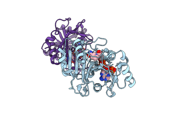 |
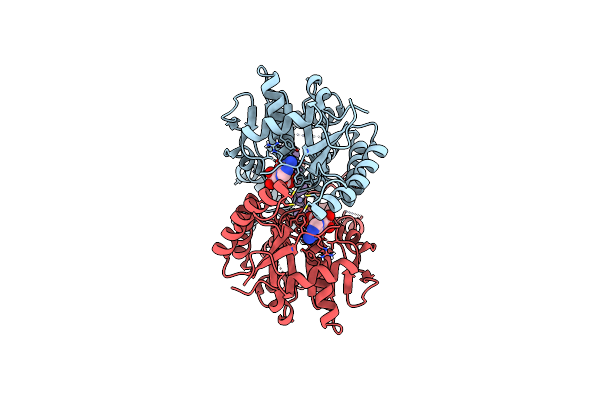
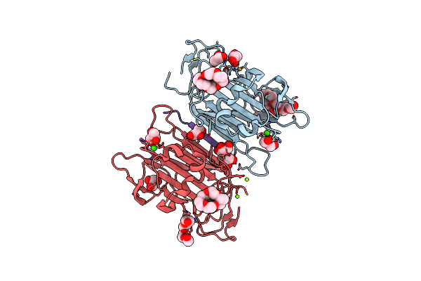
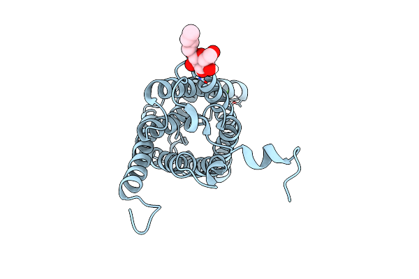
Crystal Structure Of Pprib7 In Complex Nadph (P1 Form)
Organism: Komagataella pastoris
Method: X-RAY DIFFRACTION Resolution:1.60 Å Release Date: 2025-12-17 Classification: BIOSYNTHETIC PROTEIN Ligands: NAP |
Organism: Komagataella pastoris
Method: X-RAY DIFFRACTION
Release Date: 2025-12-17
Ligands: NAP
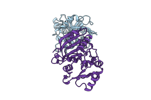 |
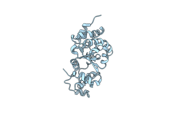
Crystal Structure Of Pprib7
Organism: Komagataella pastoris
Method: X-RAY DIFFRACTION Resolution:2.10 Å Release Date: 2025-12-17 Classification: BIOSYNTHETIC PROTEIN |
Organism: Komagataella pastoris
Method: X-RAY DIFFRACTION
Release Date: 2025-12-17
 |
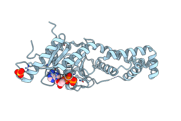
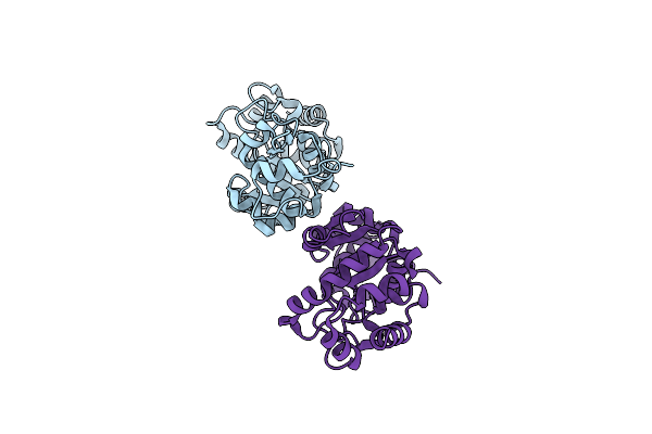
Crystal Structure Of Pprib7 In Complex With Nadph (P212121 Form)
Organism: Komagataella pastoris
Method: X-RAY DIFFRACTION Resolution:2.08 Å Release Date: 2025-12-17 Classification: BIOSYNTHETIC PROTEIN Ligands: NAP |
Organism: Komagataella pastoris
Method: X-RAY DIFFRACTION
Release Date: 2025-12-17
Ligands: NAP
 |
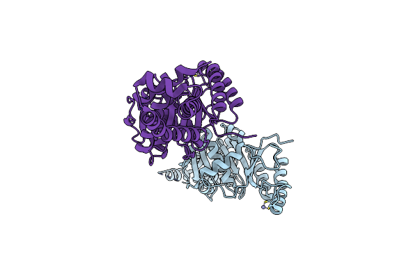
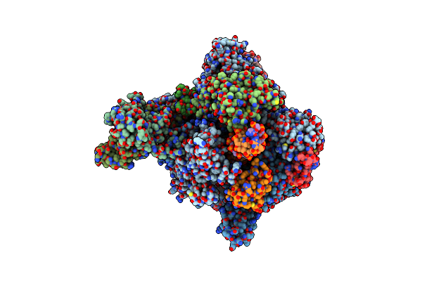
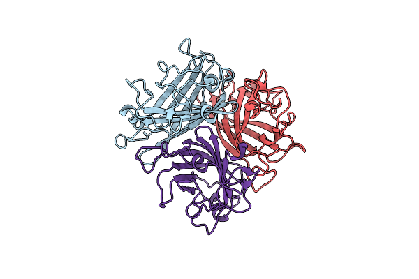
Crystal Structure Of Pichia Pastoris Pex8
Organism: Komagataella pastoris
Method: X-RAY DIFFRACTION Resolution:2.41 Å Release Date: 2025-01-29 Classification: PROTEIN TRANSPORT |
Organism: Komagataella pastoris
Method: X-RAY DIFFRACTION
Release Date: 2025-01-29
 |
Get3 Bound To Atp From G. Intestinalis In The Closed Form
Organism: Giardia intestinalis (strain atcc 50803 / wb clone c6)
Method: X-RAY DIFFRACTION Resolution:2.23 Å Release Date: 2022-07-20 Classification: CHAPERONE Ligands: ATP, MG, ZN, SO4 |
Organism: Giardia intestinalis (strain atcc 50803 / wb clone c6)
Method: X-RAY DIFFRACTION
Release Date: 2022-07-20
Ligands: ATP, MG, ZN, SO4
 |
Nucleotide-Free Get3 In Two Open Forms
Organism: Giardia lamblia atcc 50803
Method: X-RAY DIFFRACTION Resolution:3.00 Å Release Date: 2022-07-20 Classification: CHAPERONE Ligands: ZN |
Organism: Giardia lamblia atcc 50803
Method: X-RAY DIFFRACTION
Release Date: 2022-07-20
Ligands: ZN
 |
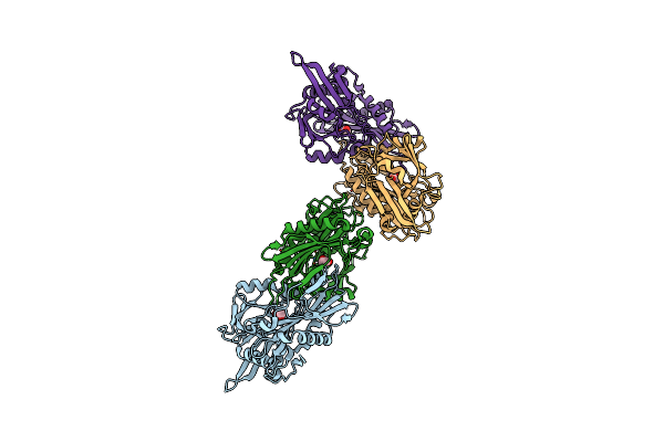
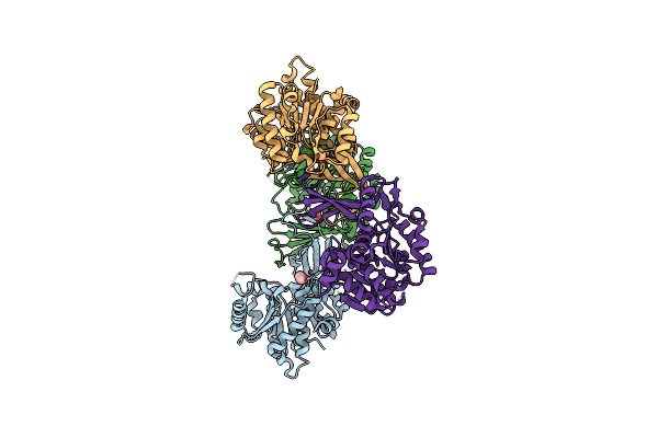
Get3 Bound To Adp And The Transmembrane Domain Of The Tail-Anchored Protein Bos1
Organism: Giardia intestinalis (strain atcc 50803 / wb clone c6), Saccharomyces cerevisiae
Method: ELECTRON MICROSCOPY Release Date: 2022-07-20 Classification: CHAPERONE Ligands: ADP, ZN, MG |
Organism: Giardia intestinalis (strain atcc 50803 / wb clone c6), Saccharomyces cerevisiae
Method: ELECTRON MICROSCOPY
Release Date: 2022-07-20
Ligands: ADP, ZN, MG
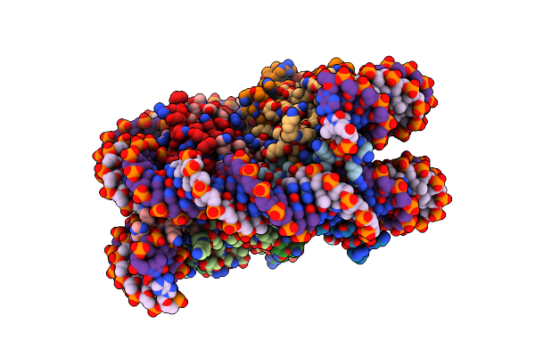 |
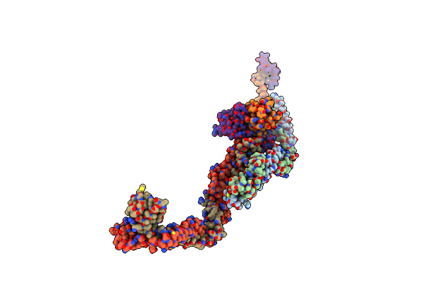
Cryo-Em Structure Of The Nucleosome Containing Komagataella Pastoris Histones
Organism: Komagataella pastoris, Synthetic construct
Method: ELECTRON MICROSCOPY Release Date: 2022-07-13 Classification: GENE REGULATION/DNA |
Organism: Komagataella pastoris, Synthetic construct
Method: ELECTRON MICROSCOPY
Release Date: 2022-07-13
 |
Crystal Structure Of Branched-Chain Amino Acid Aminotransferase From Giardia Lamblia Atcc 50803
Organism: Giardia intestinalis
Method: X-RAY DIFFRACTION Resolution:2.10 Å Release Date: 2021-03-17 Classification: TRANSFERASE Ligands: EDO, MG, CL |
Organism: Giardia intestinalis
Method: X-RAY DIFFRACTION
Release Date: 2021-03-17
Ligands: EDO, MG, CL
 |
Structure Of Ribokinase From Giardia Lamblia
Organism: Giardia lamblia
Method: X-RAY DIFFRACTION Resolution:2.65 Å Release Date: 2020-04-22 Classification: TRANSFERASE Ligands: EDO |
Organism: Giardia lamblia
Method: X-RAY DIFFRACTION
Release Date: 2020-04-22
Ligands: EDO
 |
Crystal Structure Of A Fungal Catalase At 2.3 Angstroms
Organism: Komagataella pastoris
Method: X-RAY DIFFRACTION Resolution:2.30 Å Release Date: 2020-03-04 Classification: OXIDOREDUCTASE Ligands: HEM, NDP, GOL, SO4, CL, K, NA, PEG |
Organism: Komagataella pastoris
Method: X-RAY DIFFRACTION
Release Date: 2020-03-04
Ligands: HEM, NDP, GOL, SO4, CL, K, NA, PEG
 |
Rna Polymerase Ii Elongation Complex Bound With Elf1 And Spt4/5, Stalled At Shl(-5) Of The Nucleosome
Organism: Komagataella phaffii (strain gs115 / atcc 20864), Komagataella pastoris, Homo sapiens, Synthetic construct, Komagataella phaffii (strain atcc 76273 / cbs 7435 / cect 11047 / nrrl y-11430 / wegner 21-1)
Method: ELECTRON MICROSCOPY Release Date: 2019-02-20 Classification: TRANSCRIPTION/RNA/DNA Ligands: ZN, MG |
 |
Rna Polymerase Ii Elongation Complex Bound With Spt4/5 And Foreign Dna, Stalled At Shl(-1) Of The Nucleosome
Organism: Komagataella phaffii (strain gs115 / atcc 20864), Komagataella pastoris, Homo sapiens, Synthetic construct, Komagataella phaffii (strain atcc 76273 / cbs 7435 / cect 11047 / nrrl y-11430 / wegner 21-1)
Method: ELECTRON MICROSCOPY Release Date: 2019-02-20 Classification: TRANSCRIPTION/RNA/DNA Ligands: ZN, MG |
 |
Structure Of The Kpflo2 Adhesin Domain In Complex With Glycerol
Organism: Komagataella pastoris
Method: X-RAY DIFFRACTION Resolution:2.15 Å Release Date: 2018-10-24 Classification: CELL ADHESION Ligands: CA, GOL, P33, MG, PG4 |
Organism: Komagataella pastoris
Method: X-RAY DIFFRACTION
Release Date: 2018-10-24
Ligands: CA, GOL, P33, MG, PG4
 |
The Wt Ung Crystal Structure From Nitratifractor Salsuginis
Organism: Nitratifractor salsuginis
Method: X-RAY DIFFRACTION Resolution:2.02 Å Release Date: 2017-10-18 Classification: DNA BINDING PROTEIN |
Organism: Nitratifractor salsuginis
Method: X-RAY DIFFRACTION
Release Date: 2017-10-18
 |
The Y81G Mutant Of The Ung Crystal Structure From Nitratifractor Salsuginis
Organism: Nitratifractor salsuginis (strain dsm 16511 / jcm 12458 / e9i37-1)
Method: X-RAY DIFFRACTION Resolution:2.50 Å Release Date: 2017-10-18 Classification: DNA BINDING PROTEIN |
 |
Rna Polymerase Ii Elongation Complex Bound With Spt5 Kow5 And Elf1
Organism: Komagataella pastoris, Komagataella phaffii, Synthetic construct, Komagataella phaffii (strain gs115 / atcc 20864), Komagataella phaffii (strain atcc 76273 / cbs 7435 / cect 11047 / nrrl y-11430 / wegner 21-1)
Method: X-RAY DIFFRACTION Resolution:3.00 Å Release Date: 2017-08-16 Classification: TRANSCRIPTION Ligands: ZN, MG, APC |
 |
Room Temperature Structure Of Pichia Pastoris Aquaporin At 1.3 A
Organism: Komagataella pastoris
Method: X-RAY DIFFRACTION Resolution:1.30 Å Release Date: 2016-06-29 Classification: TRANSPORT PROTEIN Ligands: BOG, CL, CA |
Organism: Komagataella pastoris
Method: X-RAY DIFFRACTION
Release Date: 2016-06-29
Ligands: BOG, CL, CA
 |
Structure Of The Adenovirus 14P1 Knob Domain
Organism: Human adenovirus 14p1
Method: X-RAY DIFFRACTION Resolution:3.20 Å Release Date: 2015-09-09 Classification: VIRAL PROTEIN |
Organism: Human adenovirus 14p1
Method: X-RAY DIFFRACTION
Release Date: 2015-09-09
 |
Crystal Structure Of Giardia Lamblia Hop2-Mnd1 Complex
Organism: Giardia lamblia atcc 50803, Giardia lamblia
Method: X-RAY DIFFRACTION Resolution:3.20 Å Release Date: 2015-03-18 Classification: CELL CYCLE |
Organism: Giardia lamblia atcc 50803, Giardia lamblia
Method: X-RAY DIFFRACTION
Release Date: 2015-03-18