

Search Count: 22
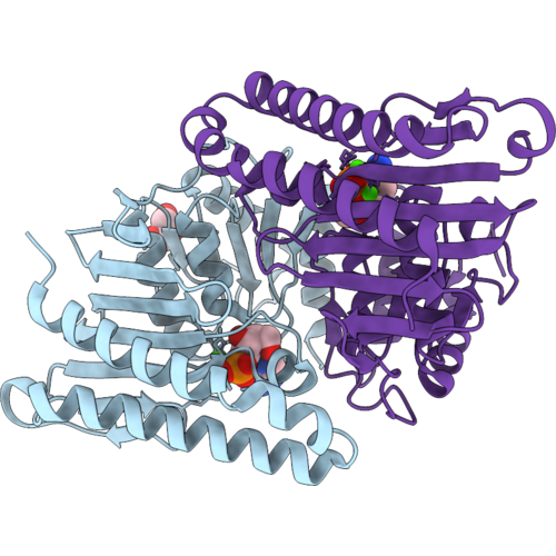 |
Crystal Structure Of Pseudomonas Aeruginosa Suhb In Complex With Adenosine 2'-Monophosphate
Organism: Pseudomonas aeruginosa (strain atcc 15692 / dsm 22644 / cip 104116 / jcm 14847 / lmg 12228 / 1c / prs 101 / pao1)
Method: X-RAY DIFFRACTION Resolution:2.00 Å Release Date: 2026-09-09 Classification: HYDROLASE Ligands: 2AM, GOL, CA |
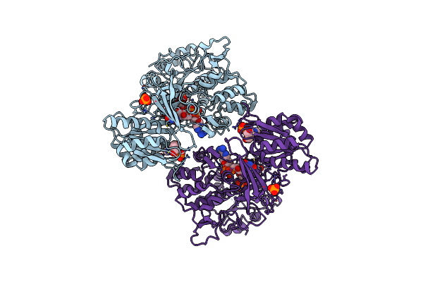 |
Rat Cypor/D632A With 2'-Amp
Organism: Rattus norvegicus
Method: X-RAY DIFFRACTION Resolution:2.70 Å Release Date: 2018-02-14 Classification: OXIDOREDUCTASE Ligands: FMN, FAD, 2AM, PO4 |
Organism: Rattus norvegicus
Method: X-RAY DIFFRACTION
Release Date: 2018-02-14
Ligands: FMN, FAD, 2AM, PO4
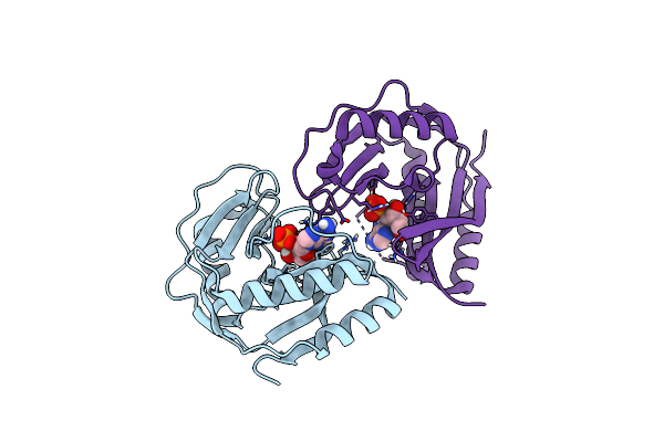 |
Crystal Structure Of E.Coli Ligt Complexed With 2'-Amp
Organism: Escherichia coli (strain b / bl21-de3)
Method: X-RAY DIFFRACTION Resolution:2.46 Å Release Date: 2017-01-18 Classification: HYDROLASE Ligands: 2AM, CL |
Organism: Escherichia coli (strain b / bl21-de3)
Method: X-RAY DIFFRACTION
Release Date: 2017-01-18
Ligands: 2AM, CL
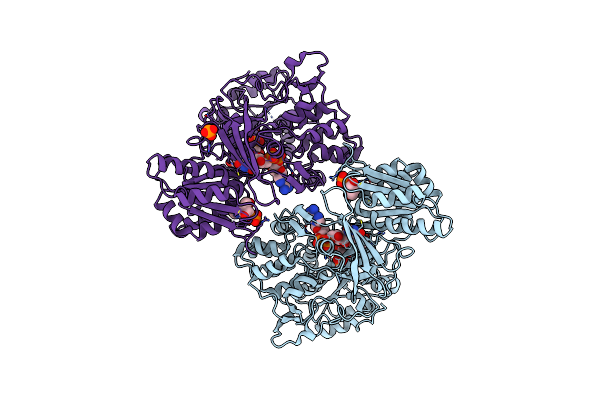 |
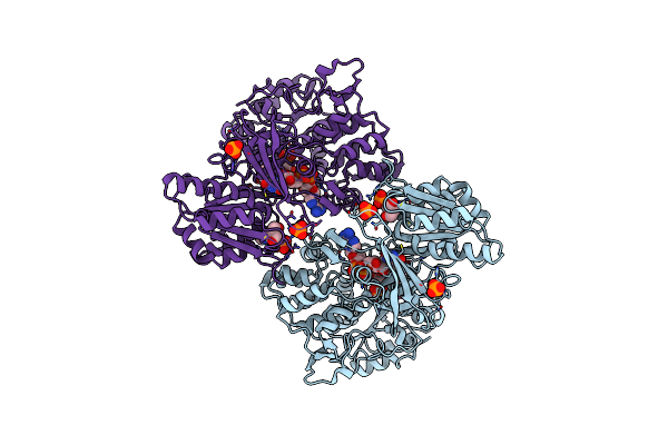
Rat Cypor With 2'-Amp
Organism: Rattus norvegicus
Method: X-RAY DIFFRACTION Resolution:1.91 Å Release Date: 2016-03-09 Classification: OXIDOREDUCTASE Ligands: FMN, FAD, 2AM, PO4 |
Organism: Rattus norvegicus
Method: X-RAY DIFFRACTION
Release Date: 2016-03-09
Ligands: FMN, FAD, 2AM, PO4
 |
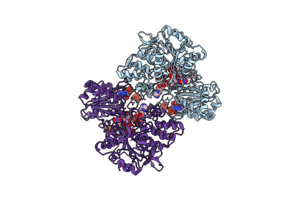
Reduced Cypor With 2'-Amp
Organism: Rattus norvegicus
Method: X-RAY DIFFRACTION Resolution:1.88 Å Release Date: 2016-03-02 Classification: OXIDOREDUCTASE Ligands: FMN, FAD, 2AM, PO4 |
Organism: Rattus norvegicus
Method: X-RAY DIFFRACTION
Release Date: 2016-03-02
Ligands: FMN, FAD, 2AM, PO4
 |
Reduced Cypor Mutant - G143Del
Organism: Rattus norvegicus
Method: X-RAY DIFFRACTION Resolution:2.50 Å Release Date: 2016-03-02 Classification: OXIDOREDUCTASE Ligands: FMN, FAD, 2AM, PO4 |
Organism: Rattus norvegicus
Method: X-RAY DIFFRACTION
Release Date: 2016-03-02
Ligands: FMN, FAD, 2AM, PO4
 |
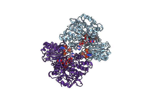
Reduced Cypor Mutant - G141Del/E142N
Organism: Rattus norvegicus
Method: X-RAY DIFFRACTION Resolution:2.20 Å Release Date: 2016-03-02 Classification: OXIDOREDUCTASE Ligands: FMN, FAD, 2AM, PO4 |
Organism: Rattus norvegicus
Method: X-RAY DIFFRACTION
Release Date: 2016-03-02
Ligands: FMN, FAD, 2AM, PO4
 |
Reduced Cypor Mutant - G141Del
Organism: Rattus norvegicus
Method: X-RAY DIFFRACTION Resolution:2.00 Å Release Date: 2016-03-02 Classification: OXIDOREDUCTASE Ligands: FMN, FAD, 2AM, PO4 |
Organism: Rattus norvegicus
Method: X-RAY DIFFRACTION
Release Date: 2016-03-02
Ligands: FMN, FAD, 2AM, PO4
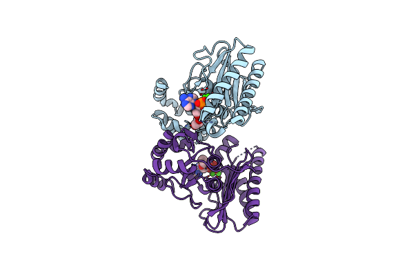 |
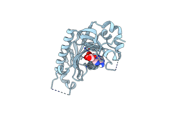
Crystal Structure Of 2'Amp Bound Saimpase-Ii
Organism: Staphylococcus aureus
Method: X-RAY DIFFRACTION Resolution:2.40 Å Release Date: 2015-12-09 Classification: HYDROLASE Ligands: CA, 2AM, GOL |
Organism: Staphylococcus aureus
Method: X-RAY DIFFRACTION
Release Date: 2015-12-09
Ligands: CA, 2AM, GOL
 |
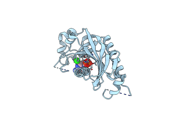
Catalytic Domain Of Mouse 2',3'-Cyclic Nucleotide 3'- Phosphodiesterase, With Mutation T232A, Complexed With 2'-Amp
Organism: Mus musculus
Method: X-RAY DIFFRACTION Resolution:2.00 Å Release Date: 2015-09-30 Classification: HYDROLASE Ligands: 2AM |
Organism: Mus musculus
Method: X-RAY DIFFRACTION
Release Date: 2015-09-30
Ligands: 2AM
 |
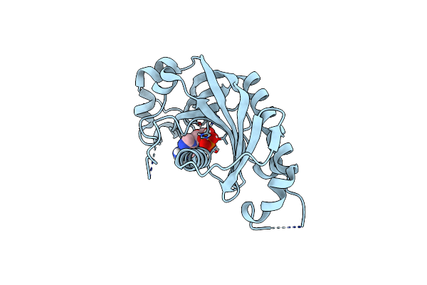
Catalytic Domain Of Mouse 2',3'-Cyclic Nucleotide 3'- Phosphodiesterase, With Mutation P225G
Organism: Mus musculus
Method: X-RAY DIFFRACTION Resolution:2.70 Å Release Date: 2015-09-23 Classification: HYDROLASE Ligands: 2AM, CL |
Organism: Mus musculus
Method: X-RAY DIFFRACTION
Release Date: 2015-09-23
Ligands: 2AM, CL
 |
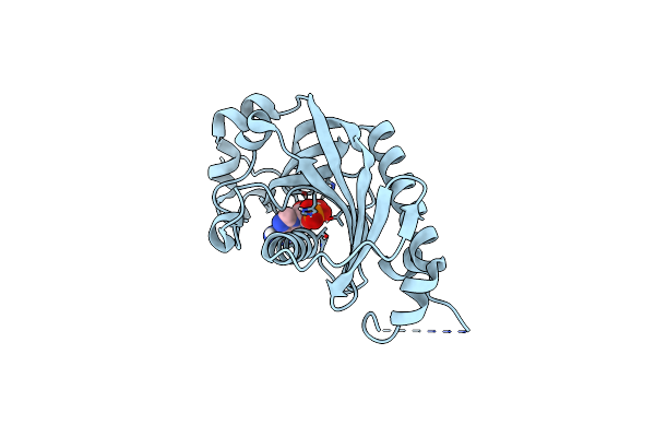
Catalytic Domain Of Mouse 2',3'-Cyclic Nucleotide 3'- Phosphodiesterase, With Mutation P296G, Complexed With 2'-Amp
Organism: Mus musculus
Method: X-RAY DIFFRACTION Resolution:1.85 Å Release Date: 2015-09-23 Classification: HYDROLASE Ligands: 2AM |
Organism: Mus musculus
Method: X-RAY DIFFRACTION
Release Date: 2015-09-23
Ligands: 2AM
 |
Catalytic Domain Of Mouse 2',3'-Cyclic Nucleotide 3'- Phosphodiesterase, With Mutation R307Q, Complexed With 2'-Amp
Organism: Mus musculus
Method: X-RAY DIFFRACTION Resolution:1.60 Å Release Date: 2015-09-23 Classification: HYDROLASE Ligands: 2AM, CL |
Organism: Mus musculus
Method: X-RAY DIFFRACTION
Release Date: 2015-09-23
Ligands: 2AM, CL
 |
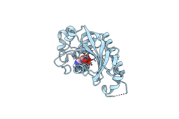
Crystal Structure Of Phosphoesterase
Organism: Escherichia coli
Method: X-RAY DIFFRACTION Resolution:2.03 Å Release Date: 2014-10-22 Classification: HYDROLASE Ligands: GOL, 2AM |
Organism: Escherichia coli
Method: X-RAY DIFFRACTION
Release Date: 2014-10-22
Ligands: GOL, 2AM
 |
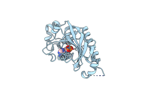
Catalytic Domain Of Mouse 2',3'-Cyclic Nucleotide 3'- Phosphodiesterase, With Mutation V321A, Crystallized With 2'-Amp
Organism: Mus musculus
Method: X-RAY DIFFRACTION Resolution:1.90 Å Release Date: 2013-07-10 Classification: HYDROLASE Ligands: 2AM |
Organism: Mus musculus
Method: X-RAY DIFFRACTION
Release Date: 2013-07-10
Ligands: 2AM
 |
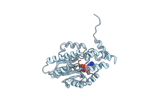
Catalytic Domain Of Mouse 2',3'-Cyclic Nucleotide 3'- Phosphodiesterase, Soaked With 2',3'-Cyclic Amp
Organism: Mus musculus
Method: X-RAY DIFFRACTION Resolution:2.40 Å Release Date: 2012-03-21 Classification: HYDROLASE Ligands: 2AM |
Organism: Mus musculus
Method: X-RAY DIFFRACTION
Release Date: 2012-03-21
Ligands: 2AM
 |
Structure Of Recombinant Haemophilus Influenzae E(P4) Acid Phosphatase Mutant D66N Complexed With 2'-Amp
Organism: Haemophilus influenzae
Method: X-RAY DIFFRACTION Resolution:1.90 Å Release Date: 2010-10-20 Classification: HYDROLASE Ligands: 2AM, MG |
Organism: Haemophilus influenzae
Method: X-RAY DIFFRACTION
Release Date: 2010-10-20
Ligands: 2AM, MG
 |
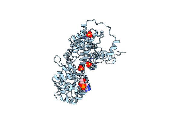
Crystal Structure Of Ustilago Sphaerogena Ribonuclease U2 Complexed With Adenosine 2'-Monophosphate
Organism: Ustilago sphaerogena
Method: X-RAY DIFFRACTION Resolution:1.03 Å Release Date: 2010-07-07 Classification: HYDROLASE Ligands: 2AM, CA |
Organism: Ustilago sphaerogena
Method: X-RAY DIFFRACTION
Release Date: 2010-07-07
Ligands: 2AM, CA
 |
Crystallographic Study Of Coenzyme, Coenzyme Analogue And Substrate Binding In 6-Phosphogluconate Dehydrogenase: Implications For Nadp Specificity And The Enzyme Mechanism
Organism: Ovis aries
Method: X-RAY DIFFRACTION Resolution:3.17 Å Release Date: 1995-02-27 Classification: OXIDOREDUCTASE (CHOH(D)-NADP+(A)) Ligands: SO4, 2AM |
Organism: Ovis aries
Method: X-RAY DIFFRACTION
Release Date: 1995-02-27
Ligands: SO4, 2AM
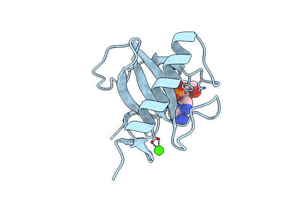 |
Rnase T1 Mutant Glu46Gln Binds The Inhibitors 2'Gmp And 2'Amp At The 3' Subsite
Organism: Aspergillus oryzae
Method: X-RAY DIFFRACTION Resolution:1.87 Å Release Date: 1993-01-15 Classification: HYDROLASE(ENDORIBONUCLEASE) Ligands: CA, 2AM |
Organism: Aspergillus oryzae
Method: X-RAY DIFFRACTION
Release Date: 1993-01-15
Ligands: CA, 2AM