
Search Count: 50,056
 |
Organism: Streptococcus intermedius b196
Method: X-RAY DIFFRACTION Resolution:1.48 Å Release Date: 2026-06-24 Classification: STRUCTURAL PROTEIN |
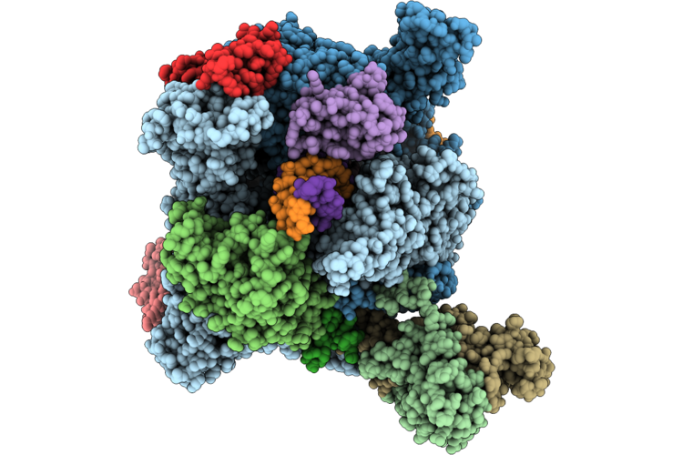 |
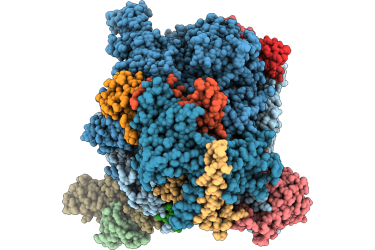
Structure Of Yeast Pol Ii In Complex With An Oxaliplatin-Icl Lesion At +7 Position.
Organism: Saccharomyces cerevisiae s288c, Synthetic construct
Method: ELECTRON MICROSCOPY Release Date: 2026-06-24 Classification: TRANSCRIPTION Ligands: ZN, MG, DNH, PT |
 |
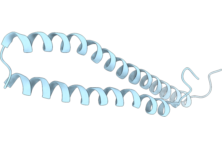
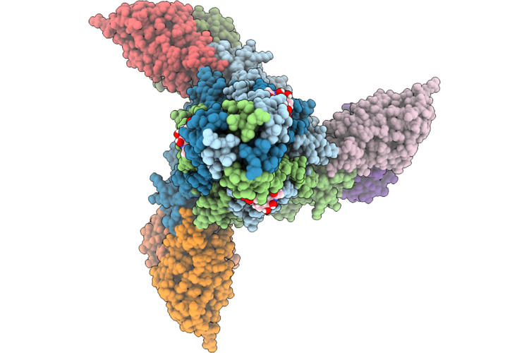
Averaged Protofilament Of Heimdallarchaeales Alpha/Beta Tubulin Microtubule With A Single Seam
Organism: Candidatus heimdallarchaeaceae
Method: ELECTRON MICROSCOPY Release Date: 2026-06-24 Classification: PROTEIN FIBRIL Ligands: GDP, MG |
 |
Crystal Structure Of The Substrate-Binding Domain Of S. Cerevisiae Ssc1 In Complex With Peptide Lslppvklhc
Organism: Saccharomyces cerevisiae s288c
Method: X-RAY DIFFRACTION Resolution:2.23 Å Release Date: 2026-06-24 Classification: CHAPERONE |
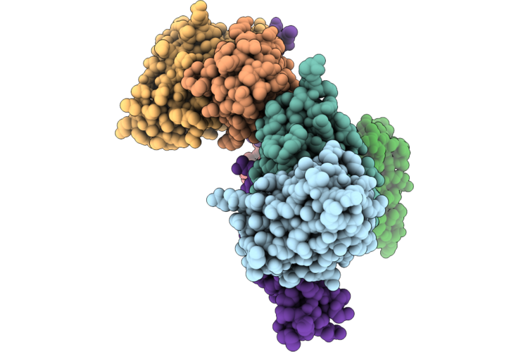 |
Cryo-Electron Microscopy Structure Of Pfripr Bound To Monoclonal Antibodies Rp.047, Rp.057 And Rp.035
Organism: Mus musculus, Plasmodium falciparum 3d7
Method: ELECTRON MICROSCOPY Resolution:3.35 Å Release Date: 2026-06-24 Classification: IMMUNE SYSTEM |
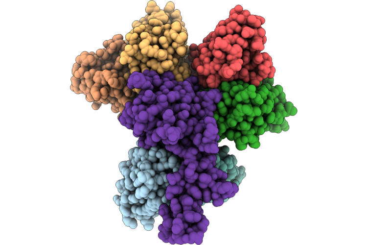 |
Cryo-Electron Microscopy Structure Of Pfripr Bound To Monoclonal Antibodies Rp.093, Rp.073 And Rp.063
Organism: Mus musculus, Plasmodium falciparum 3d7
Method: ELECTRON MICROSCOPY Resolution:2.97 Å Release Date: 2026-06-24 Classification: IMMUNE SYSTEM |
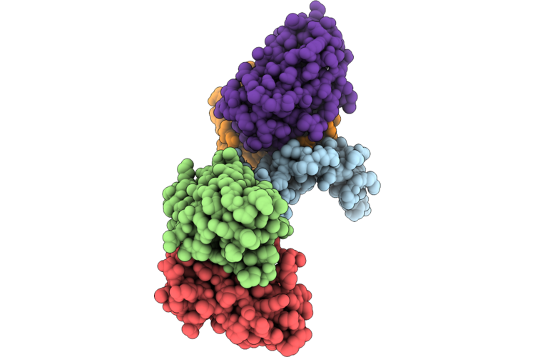 |
Cryo-Electron Microscopy Structure Of Pfripr Bound To Monoclonal Antibodies Rp.092 And Rp.052
Organism: Plasmodium falciparum 3d7, Mus musculus
Method: ELECTRON MICROSCOPY Resolution:4.01 Å Release Date: 2026-06-24 Classification: IMMUNE SYSTEM |
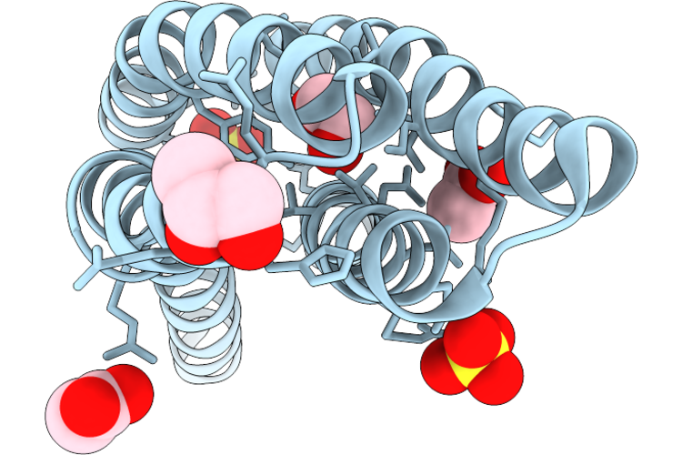 |
Ligand Binding Domain Of The Pacg Chemoreceptor From Pectobacterium Atrosepticum
Organism: Pectobacterium atrosepticum
Method: X-RAY DIFFRACTION Resolution:1.40 Å Release Date: 2026-06-24 Classification: SIGNALING PROTEIN Ligands: GOL, ACT, EDO, SO4 |
 |
Dimeric Crystallization Of The Ligand Binding Domain Of The Pacg Chemoreceptor From Pectobacterium Atrosepticum
Organism: Pectobacterium atrosepticum
Method: X-RAY DIFFRACTION Resolution:1.80 Å Release Date: 2026-06-24 Classification: SIGNALING PROTEIN Ligands: GOL, SO4, NA |
 |
Crystal Structure Of Rhizobium Etli L-Asparaginase Reav K138A Mutant In Complex With L-Asn And L-Asp
Organism: Rhizobium etli
Method: X-RAY DIFFRACTION Resolution:1.30 Å Release Date: 2026-06-24 Classification: HYDROLASE Ligands: EDO, ASN, PEG, CD, ASP |
 |
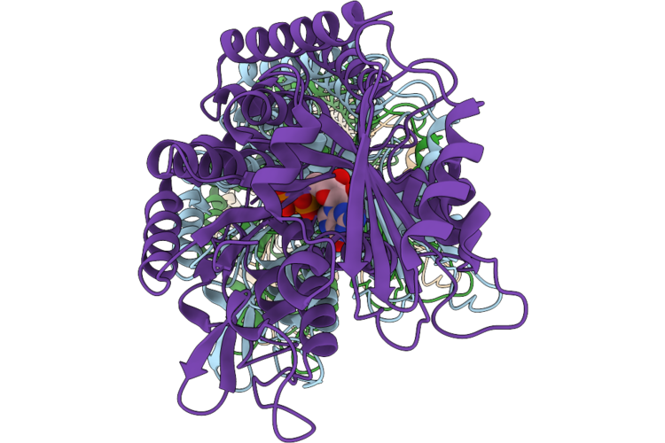
Structure Of A Membrane-Bound Inositol Phosphorylceramide Synthase And Aureobasidin A Complex
Organism: Saccharomyces cerevisiae (strain atcc 204508 / s288c)
Method: ELECTRON MICROSCOPY Release Date: 2026-06-24 Classification: LIPID BINDING PROTEIN Ligands: C14, 46E |
 |
Organism: Human alphaherpesvirus 1 strain 17
Method: ELECTRON MICROSCOPY Resolution:3.70 Å Release Date: 2026-06-24 Classification: VIRAL PROTEIN/DNA |
 |
Organism: Human alphaherpesvirus 1 strain 17
Method: ELECTRON MICROSCOPY Release Date: 2026-06-24 Classification: VIRAL PROTEIN |
 |
Structure Of Yeast Pol Ii-Elf1 Complex Containing A Cisplatin-Icl Lesion At +7 Position.
Organism: Saccharomyces cerevisiae (strain atcc 204508 / s288c), Synthetic construct, Saccharomyces cerevisiae s288c
Method: ELECTRON MICROSCOPY Resolution:2.72 Å Release Date: 2026-06-24 Classification: TRANSCRIPTION Ligands: ZN, MG, CPT |
 |
Organism: Borreliella garinii
Method: X-RAY DIFFRACTION Resolution:2.55 Å Release Date: 2026-06-17 Classification: IMMUNE SYSTEM |
 |
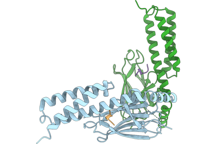
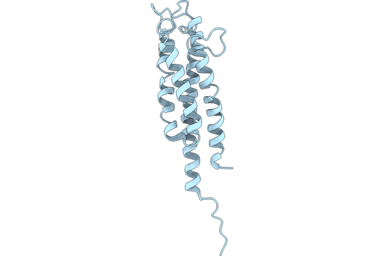
Cryo-Em Structure Of Vaccine Elicited Antibody 22F5 Bound To The Post-Fusion Conformation Of The Layv-F Glycoprotein
Organism: Langya virus, Mus musculus
Method: ELECTRON MICROSCOPY Release Date: 2026-06-17 Classification: IMMUNE SYSTEM/VIRAL PROTEIN Ligands: NAG |
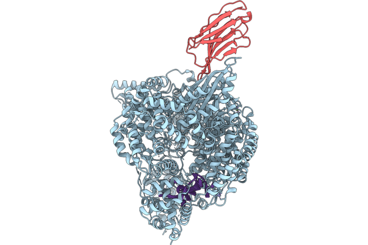 |
Structure Of Crimean Congo Hemorrhagic Fever Virus (Cchfv) L Protein Bound To 5' Vrna And Nanobody 20096.
Organism: Lama glama, Crimean-congo hemorrhagic fever virus strain ibar10200
Method: ELECTRON MICROSCOPY Release Date: 2026-06-17 Classification: VIRAL PROTEIN Ligands: ZN, MG |
 |
Organism: Crimean-congo hemorrhagic fever virus strain ibar10200
Method: ELECTRON MICROSCOPY Release Date: 2026-06-17 Classification: VIRAL PROTEIN Ligands: ZN, MG |
 |
Crystal Structure Of Anthocyanin 4'-O-Methyltransferase From Vitis Vinifera L.
Organism: Vitis vinifera
Method: X-RAY DIFFRACTION Resolution:2.78 Å Release Date: 2026-06-17 Classification: TRANSFERASE Ligands: SAH |
 |
Organism: Candidatus heimdallarchaeaceae
Method: ELECTRON MICROSCOPY Release Date: 2026-06-10 Classification: PROTEIN FIBRIL Ligands: GDP, MG |