

Search Count: 1,182
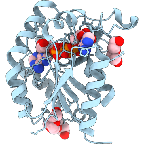 |
Crystal Structure Of An Adenylate Kinase From Leishmania Major (Tetragonal Form, Adp And Amp Bound)
Organism: Leishmania major strain friedlin
Method: X-RAY DIFFRACTION Resolution:1.40 Å Release Date: 2026-09-16 Classification: TRANSFERASE Ligands: 1PE, PEG, ADP, AMP, MG |
Organism: Leishmania major strain friedlin
Method: X-RAY DIFFRACTION
Release Date: 2026-09-16
Ligands: 1PE, PEG, ADP, AMP, MG
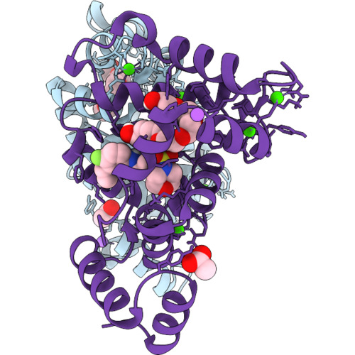 |
Ncs-1 Bound To Xchem Ligand 1
Organism: Homo sapiens
Method: X-RAY DIFFRACTION Resolution:2.15 Å Release Date: 2026-09-16 Classification: METAL BINDING PROTEIN Ligands: CA, ACT, A1I63, P6G, NA, 1PE |
Organism: Homo sapiens
Method: X-RAY DIFFRACTION
Release Date: 2026-09-16
Ligands: CA, ACT, A1I63, P6G, NA, 1PE
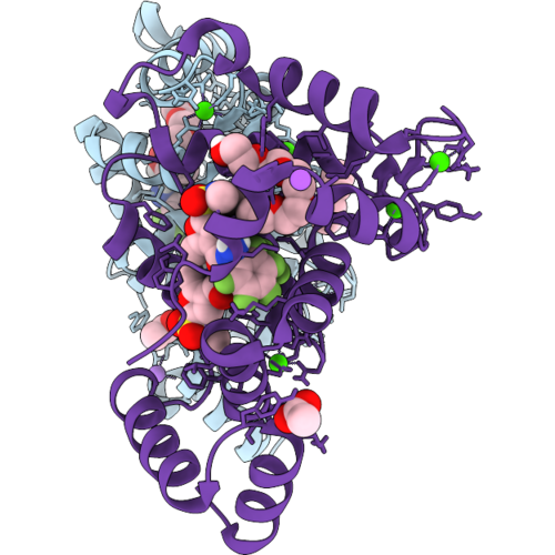 |
Ncs-1 Bound To Xchem Ligand 3
Organism: Homo sapiens
Method: X-RAY DIFFRACTION Resolution:2.33 Å Release Date: 2026-09-16 Classification: METAL BINDING PROTEIN Ligands: CA, ACT, 1PE, A1I7M, NA |
Organism: Homo sapiens
Method: X-RAY DIFFRACTION
Release Date: 2026-09-16
Ligands: CA, ACT, 1PE, A1I7M, NA
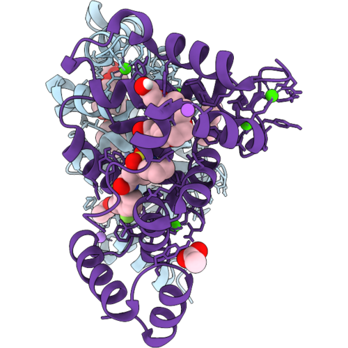 |
Ncs-1 Bound To Xchem Ligand 4
Organism: Homo sapiens
Method: X-RAY DIFFRACTION Resolution:2.82 Å Release Date: 2026-09-16 Classification: METAL BINDING PROTEIN Ligands: CA, ACT, A1I7N, P6G, NA, 1PE |
Organism: Homo sapiens
Method: X-RAY DIFFRACTION
Release Date: 2026-09-16
Ligands: CA, ACT, A1I7N, P6G, NA, 1PE
 |
Ncs-1 Bound To Xchem Ligand 5
Organism: Homo sapiens
Method: X-RAY DIFFRACTION Resolution:2.01 Å Release Date: 2026-09-16 Classification: METAL BINDING PROTEIN Ligands: CA, ACT, 1PE, A1I7A, NA |
Organism: Homo sapiens
Method: X-RAY DIFFRACTION
Release Date: 2026-09-16
Ligands: CA, ACT, 1PE, A1I7A, NA
 |
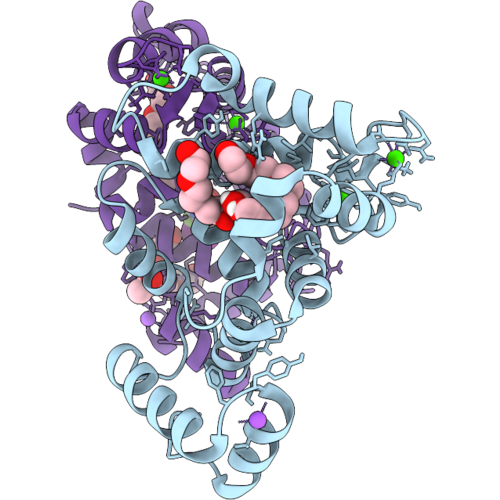
Ncs-1 Bound To Xchem Ligand 6
Organism: Homo sapiens
Method: X-RAY DIFFRACTION Resolution:1.97 Å Release Date: 2026-09-16 Classification: METAL BINDING PROTEIN Ligands: CA, ACT, 1PE, A1I7D, NA |
Organism: Homo sapiens
Method: X-RAY DIFFRACTION
Release Date: 2026-09-16
Ligands: CA, ACT, 1PE, A1I7D, NA
 |
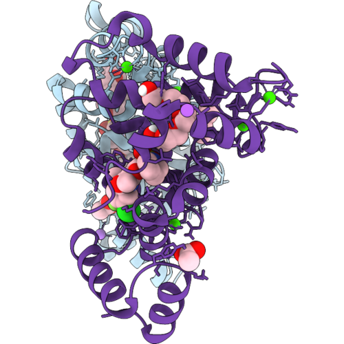
Ncs-1 Bound To Xchem Ligand 7
Organism: Homo sapiens
Method: X-RAY DIFFRACTION Resolution:2.00 Å Release Date: 2026-09-16 Classification: METAL BINDING PROTEIN Ligands: CA, P6G, NA, ACT, 1PE, A1I7C |
Organism: Homo sapiens
Method: X-RAY DIFFRACTION
Release Date: 2026-09-16
Ligands: CA, P6G, NA, ACT, 1PE, A1I7C
 |
Ncs-1 Bound To Xchem Ligand 8
Organism: Homo sapiens
Method: X-RAY DIFFRACTION Resolution:2.93 Å Release Date: 2026-09-16 Classification: METAL BINDING PROTEIN Ligands: CA, ACT, 1PE, A1I7O, NA, P6G |
Organism: Homo sapiens
Method: X-RAY DIFFRACTION
Release Date: 2026-09-16
Ligands: CA, ACT, 1PE, A1I7O, NA, P6G
 |
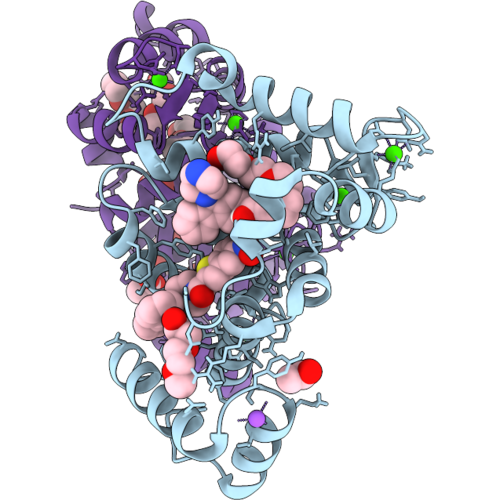
Ncs-1 Bound To Xchem Ligand 9
Organism: Homo sapiens
Method: X-RAY DIFFRACTION Resolution:2.15 Å Release Date: 2026-09-16 Classification: METAL BINDING PROTEIN Ligands: CA, ACT, 1PE, A1I7P, NA |
Organism: Homo sapiens
Method: X-RAY DIFFRACTION
Release Date: 2026-09-16
Ligands: CA, ACT, 1PE, A1I7P, NA
 |
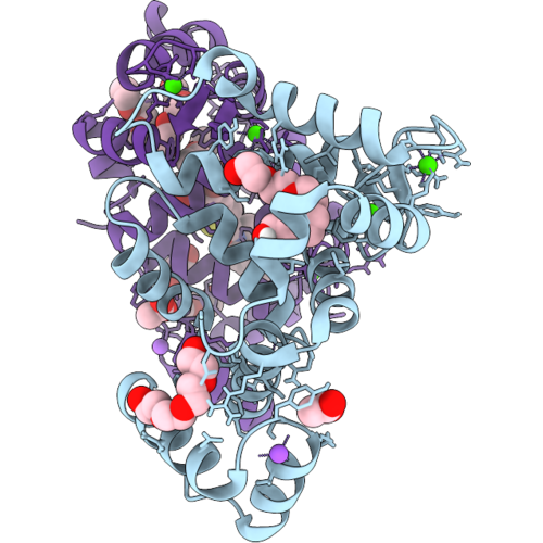
Ncs-1 Bound To Xchem Ligand 10
Organism: Homo sapiens
Method: X-RAY DIFFRACTION Resolution:2.10 Å Release Date: 2026-09-16 Classification: METAL BINDING PROTEIN Ligands: CA, P6G, NA, ACT, 1PE, A1I7V |
Organism: Homo sapiens
Method: X-RAY DIFFRACTION
Release Date: 2026-09-16
Ligands: CA, P6G, NA, ACT, 1PE, A1I7V
 |
Ncs-1 Bound To Xchem Ligand 11
Organism: Homo sapiens
Method: X-RAY DIFFRACTION Resolution:1.73 Å Release Date: 2026-09-16 Classification: METAL BINDING PROTEIN Ligands: CA, P6G, NA, ACT, 1PE, A1I7Q |
Organism: Homo sapiens
Method: X-RAY DIFFRACTION
Release Date: 2026-09-16
Ligands: CA, P6G, NA, ACT, 1PE, A1I7Q
 |
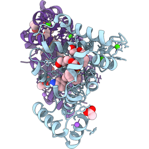
Ncs-1 Bound To Xchem Ligand 12
Organism: Homo sapiens
Method: X-RAY DIFFRACTION Resolution:1.90 Å Release Date: 2026-09-16 Classification: METAL BINDING PROTEIN Ligands: CA, P6G, NA, ACT, 1PE, A1I7R |
Organism: Homo sapiens
Method: X-RAY DIFFRACTION
Release Date: 2026-09-16
Ligands: CA, P6G, NA, ACT, 1PE, A1I7R
 |
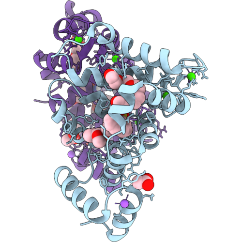
Ncs-1 Bound To Xchem Ligand 13
Organism: Homo sapiens
Method: X-RAY DIFFRACTION Resolution:1.78 Å Release Date: 2026-09-16 Classification: METAL BINDING PROTEIN Ligands: CA, P6G, NA, ACT, 1PE, A1I7B |
Organism: Homo sapiens
Method: X-RAY DIFFRACTION
Release Date: 2026-09-16
Ligands: CA, P6G, NA, ACT, 1PE, A1I7B
 |
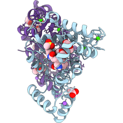
Ncs-1 Bound To Xchem Ligand 14
Organism: Homo sapiens
Method: X-RAY DIFFRACTION Resolution:2.09 Å Release Date: 2026-09-16 Classification: METAL BINDING PROTEIN Ligands: CA, P6G, NA, ACT, 1PE, A1I7S |
Organism: Homo sapiens
Method: X-RAY DIFFRACTION
Release Date: 2026-09-16
Ligands: CA, P6G, NA, ACT, 1PE, A1I7S
 |
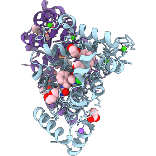
Ncs-1 Bound To Xchem Ligand 15
Organism: Homo sapiens
Method: X-RAY DIFFRACTION Resolution:2.40 Å Release Date: 2026-09-16 Classification: METAL BINDING PROTEIN Ligands: CA, P6G, NA, ACT, 1PE, A1I7T, PG5 |
Organism: Homo sapiens
Method: X-RAY DIFFRACTION
Release Date: 2026-09-16
Ligands: CA, P6G, NA, ACT, 1PE, A1I7T, PG5
 |
Ncs-1 Bound To Xchem Ligand 16
Organism: Homo sapiens
Method: X-RAY DIFFRACTION Resolution:2.26 Å Release Date: 2026-09-16 Classification: METAL BINDING PROTEIN Ligands: CA, P6G, NA, ACT, 1PE, A1I7U, CL |
Organism: Homo sapiens
Method: X-RAY DIFFRACTION
Release Date: 2026-09-16
Ligands: CA, P6G, NA, ACT, 1PE, A1I7U, CL
 |
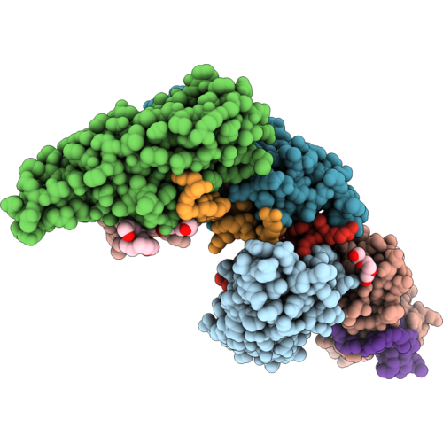
Structure Of Thermus Thermophilus Mfd
Organism: Thermus thermophilus hb8
Method: X-RAY DIFFRACTION Resolution:2.60 Å Release Date: 2026-09-09 Classification: DNA BINDING PROTEIN Ligands: SO4, 1PE |
Organism: Thermus thermophilus hb8
Method: X-RAY DIFFRACTION
Release Date: 2026-09-09
Ligands: SO4, 1PE
 |
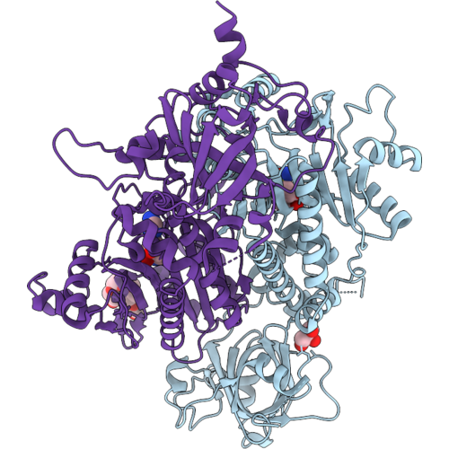
Crystal Structure Hasf1A 156-Cr17
Organism: Homo sapiens
Method: X-RAY DIFFRACTION Resolution:1.70 Å Release Date: 2026-09-02 Classification: CHAPERONE Ligands: GOL, PGE, 1PE, P6G, CL, PEG |
Organism: Homo sapiens
Method: X-RAY DIFFRACTION
Release Date: 2026-09-02
Ligands: GOL, PGE, 1PE, P6G, CL, PEG
 |
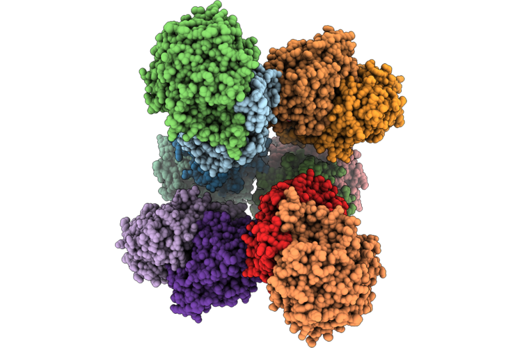
Plasmodium Vivax Aspartyl-Trna Synthetase In Complex With Ams, Mg Ion, Iodides And Pentaethylene Glycol
Organism: Plasmodium vivax
Method: X-RAY DIFFRACTION Resolution:2.23 Å Release Date: 2026-09-02 Classification: LIGASE Ligands: LMS, IOD, GOL, MG, 1PE |
Organism: Plasmodium vivax
Method: X-RAY DIFFRACTION
Release Date: 2026-09-02
Ligands: LMS, IOD, GOL, MG, 1PE
 |
Anammox-Specific Heterodimeric Ketoacyl-Acp Synthase (Amxfabfhet) From Brocadia Fulgida
Organism: Candidatus brocadia fulgida
Method: X-RAY DIFFRACTION Resolution:2.60 Å Release Date: 2026-08-12 Classification: BIOSYNTHETIC PROTEIN Ligands: MG, EPE, 1PE, PO4, CL, GOL, TAM |
Organism: Candidatus brocadia fulgida
Method: X-RAY DIFFRACTION
Release Date: 2026-08-12
Ligands: MG, EPE, 1PE, PO4, CL, GOL, TAM