

Search Count: 61
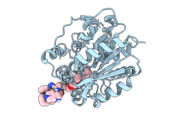 |
Halotag Bound To Compound Mrc71
Organism: Rhodococcus sp. (in: high g+c gram-positive bacteria)
Method: X-RAY DIFFRACTION Resolution:2.04 Å Release Date: 2025-10-08 Classification: LIGASE Ligands: CL, A1JC1 |
Organism: Rhodococcus sp. (in: high g+c gram-positive bacteria)
Method: X-RAY DIFFRACTION
Release Date: 2025-10-08
Ligands: CL, A1JC1
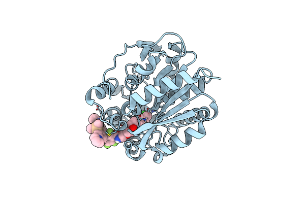 |
Crystal Structure Of Halotag7 Bound To Jf669-Halotag Ligand
Organism: Rhodococcus sp. (in: high g+c gram-positive bacteria)
Method: X-RAY DIFFRACTION Resolution:1.90 Å Release Date: 2024-06-05 Classification: HYDROLASE Ligands: XSR, CL |
Organism: Rhodococcus sp. (in: high g+c gram-positive bacteria)
Method: X-RAY DIFFRACTION
Release Date: 2024-06-05
Ligands: XSR, CL
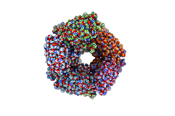 |
Cryo-Em Structure Of A Bacterial Nitrilase Filament With A Covalent Adduct Derived From Benzonitrile Hydrolysis
Organism: Rhodococcus sp. (in: high g+c gram-positive bacteria)
Method: ELECTRON MICROSCOPY Release Date: 2024-05-01 Classification: HYDROLASE Ligands: HBX |
Organism: Rhodococcus sp. (in: high g+c gram-positive bacteria)
Method: ELECTRON MICROSCOPY
Release Date: 2024-05-01
Ligands: HBX
 |
Crystal Structure Of Halotag Complexed With Btta
Organism: Rhodococcus sp. (in: high g+c gram-positive bacteria)
Method: X-RAY DIFFRACTION Resolution:1.99 Å Release Date: 2024-04-17 Classification: HYDROLASE Ligands: TN9, CL, SO4 |
Organism: Rhodococcus sp. (in: high g+c gram-positive bacteria)
Method: X-RAY DIFFRACTION
Release Date: 2024-04-17
Ligands: TN9, CL, SO4
 |
X-Ray Structure Of The Haloalkane Dehalogenase Halotag7 Circular Permutated At Positions 141-156 (Cphalotagdelta)
Organism: Rhodococcus sp.
Method: X-RAY DIFFRACTION Resolution:2.30 Å Release Date: 2023-10-11 Classification: HYDROLASE Ligands: NHE |
Organism: Rhodococcus sp.
Method: X-RAY DIFFRACTION
Release Date: 2023-10-11
Ligands: NHE
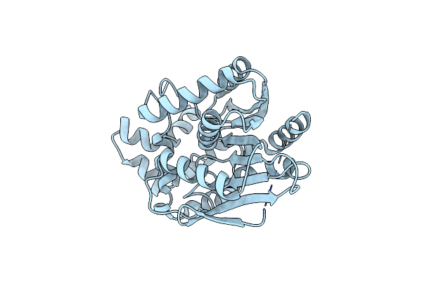 |
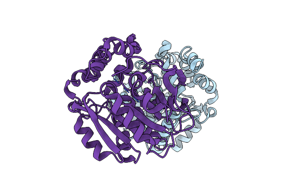
X-Ray Structure Of The Haloalkane Dehalogenase Halotag7 Circular Permutated At Positions 141-156 (Cphalotagdelta) Fused To M13
Organism: Rhodococcus sp.
Method: X-RAY DIFFRACTION Resolution:2.00 Å Release Date: 2023-10-11 Classification: HYDROLASE Ligands: CL |
Organism: Rhodococcus sp.
Method: X-RAY DIFFRACTION
Release Date: 2023-10-11
Ligands: CL
 |
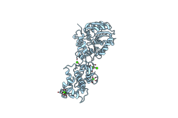
X-Ray Structure Of The Haloalkane Dehalogenase Halotag7 Circular Permutated At Positions 154-156 (Cphalotag7_154-156)
Organism: Rhodococcus sp.
Method: X-RAY DIFFRACTION Resolution:1.10 Å Release Date: 2023-10-11 Classification: HYDROLASE Ligands: CL |
Organism: Rhodococcus sp.
Method: X-RAY DIFFRACTION
Release Date: 2023-10-11
Ligands: CL
 |
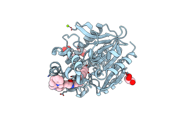
X-Ray Structure Of The Haloalkane Dehalogenase Halotag7 With An Insertion Of Calmodulin-M13 Fusion At Position 154-156 That Mimic The Structure Of Caprola, An Calcium Gated Protein Labeling Technology
Organism: Rhodococcus sp. (in: high g+c gram-positive bacteria), Homo sapiens
Method: X-RAY DIFFRACTION Resolution:2.60 Å Release Date: 2023-10-11 Classification: HYDROLASE Ligands: CL, CA |
Organism: Rhodococcus sp. (in: high g+c gram-positive bacteria), Homo sapiens
Method: X-RAY DIFFRACTION
Release Date: 2023-10-11
Ligands: CL, CA
 |
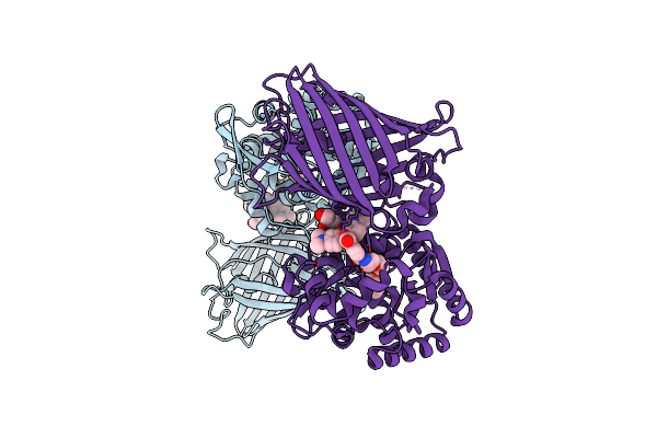
X-Ray Structure Of The Haloalkane Dehalogenase Halotag7 Labeled With A Chloroalkane Cyanine3 Fluorophore Substrate
Organism: Rhodococcus sp.
Method: X-RAY DIFFRACTION Resolution:1.50 Å Release Date: 2023-07-26 Classification: HYDROLASE Ligands: PJI, CL, GOL, MG |
Organism: Rhodococcus sp.
Method: X-RAY DIFFRACTION
Release Date: 2023-07-26
Ligands: PJI, CL, GOL, MG
 |
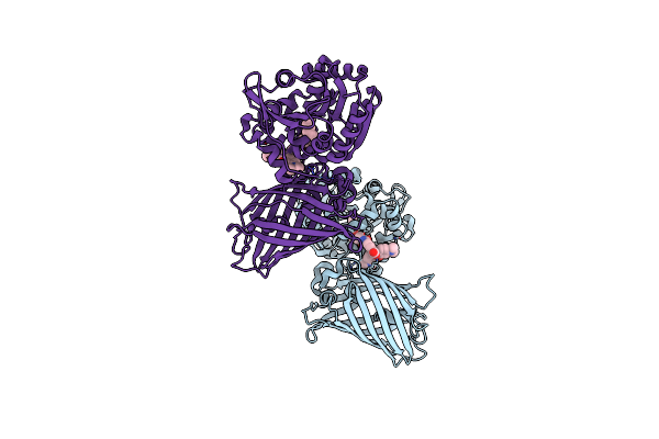
X-Ray Structure Of The Haloalkane Dehalogenase Halotag7 Fusion To The Green Fluorescent Protein Gfp (Chemog1) Labeled With A Chloroalkane Tetramethylrhodamine Fluorophore Substrate
Organism: Rhodococcus sp.
Method: X-RAY DIFFRACTION Resolution:1.80 Å Release Date: 2023-07-26 Classification: HYDROLASE Ligands: OEH, CL, GOL |
Organism: Rhodococcus sp.
Method: X-RAY DIFFRACTION
Release Date: 2023-07-26
Ligands: OEH, CL, GOL
 |
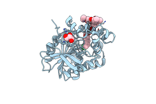
X-Ray Structure Of The Interface Optimized Haloalkane Dehalogenase Halotag7 Fusion To The Green Fluorescent Protein Gfp (Chemog5-Tmr) Labeled With A Chloroalkane Tetramethylrhodamine Fluorophore Substrate
Organism: Rhodococcus sp.
Method: X-RAY DIFFRACTION Resolution:2.00 Å Release Date: 2023-07-26 Classification: HYDROLASE Ligands: OEH, CL |
Organism: Rhodococcus sp.
Method: X-RAY DIFFRACTION
Release Date: 2023-07-26
Ligands: OEH, CL
 |
X-Ray Structure Of The Haloalkane Dehalogenase Dead Variant Halotag7-D106A Bound To A Chloroalkane Tetramethylrhodamine Fluorophore Ligand (Ca-Tmr)
Organism: Rhodococcus sp.
Method: X-RAY DIFFRACTION Resolution:1.40 Å Release Date: 2023-04-19 Classification: HYDROLASE Ligands: OEH, CL, GOL |
Organism: Rhodococcus sp.
Method: X-RAY DIFFRACTION
Release Date: 2023-04-19
Ligands: OEH, CL, GOL
 |
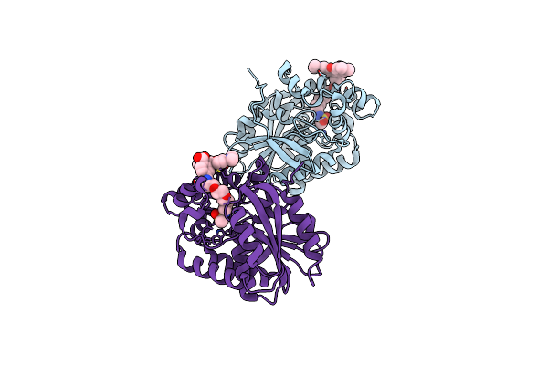
X-Ray Structure Of The Haloalkane Dehalogenase Halotag7 Bound To A Butyltrifluoromethanesulfonamide Tetramethylrhodamine Ligand (Tmr-T4)
Organism: Rhodococcus sp.
Method: X-RAY DIFFRACTION Resolution:1.99 Å Release Date: 2023-04-19 Classification: HYDROLASE Ligands: IYE |
Organism: Rhodococcus sp.
Method: X-RAY DIFFRACTION
Release Date: 2023-04-19
Ligands: IYE
 |
X-Ray Structure Of The Haloalkane Dehalogenase Halotag7 Bound To A Butylmethanesulfonamide Tetramethylrhodamine Ligand (Tmr-S4)
Organism: Rhodococcus sp.
Method: X-RAY DIFFRACTION Resolution:2.39 Å Release Date: 2023-04-19 Classification: HYDROLASE Ligands: IYU |
Organism: Rhodococcus sp.
Method: X-RAY DIFFRACTION
Release Date: 2023-04-19
Ligands: IYU
 |
X-Ray Structure Of The Haloalkane Dehalogenase Halotag7 Bound To A Pentyltrifluoromethanesulfonamide Tetramethylrhodamine Ligand (Tmr-T5)
Organism: Rhodococcus sp.
Method: X-RAY DIFFRACTION Resolution:1.70 Å Release Date: 2023-02-22 Classification: HYDROLASE Ligands: IYI, CA |
Organism: Rhodococcus sp.
Method: X-RAY DIFFRACTION
Release Date: 2023-02-22
Ligands: IYI, CA
 |
X-Ray Structure Of The Dead Variant Haloalkane Dehalogenase Halotag7-D106A Bound To A Pentanol Tetramethylrhodamine Ligand (Tmr-Hy5)
Organism: Rhodococcus sp.
Method: X-RAY DIFFRACTION Resolution:1.50 Å Release Date: 2023-02-22 Classification: HYDROLASE Ligands: IYL, CL, GOL |
Organism: Rhodococcus sp.
Method: X-RAY DIFFRACTION
Release Date: 2023-02-22
Ligands: IYL, CL, GOL
 |
X-Ray Structure Of The Haloalkane Dehalogenase Halotag7 Bound To A Pentylmethanesulfonamide Tetramethylrhodamine Ligand (Tmr-S5)
Organism: Rhodococcus sp.
Method: X-RAY DIFFRACTION Resolution:1.50 Å Release Date: 2023-02-22 Classification: HYDROLASE Ligands: IYO, GOL |
Organism: Rhodococcus sp.
Method: X-RAY DIFFRACTION
Release Date: 2023-02-22
Ligands: IYO, GOL
 |
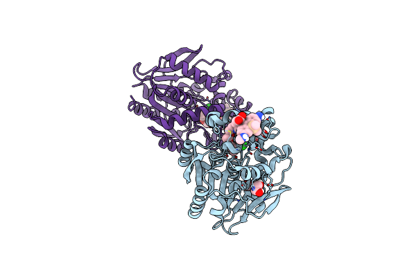
Halotag With Me-Traq-G Ligand
Organism: Rhodococcus sp.
Method: X-RAY DIFFRACTION Resolution:1.23 Å Release Date: 2023-02-01 Classification: HYDROLASE Ligands: GOL, IL7, CL |
Organism: Rhodococcus sp.
Method: X-RAY DIFFRACTION
Release Date: 2023-02-01
Ligands: GOL, IL7, CL
 |
Halotag With Traq-G-Ctrl Ligand
Organism: Rhodococcus sp.
Method: X-RAY DIFFRACTION Resolution:1.95 Å Release Date: 2023-02-01 Classification: HYDROLASE Ligands: ILJ, GOL, CL |
Organism: Rhodococcus sp.
Method: X-RAY DIFFRACTION
Release Date: 2023-02-01
Ligands: ILJ, GOL, CL
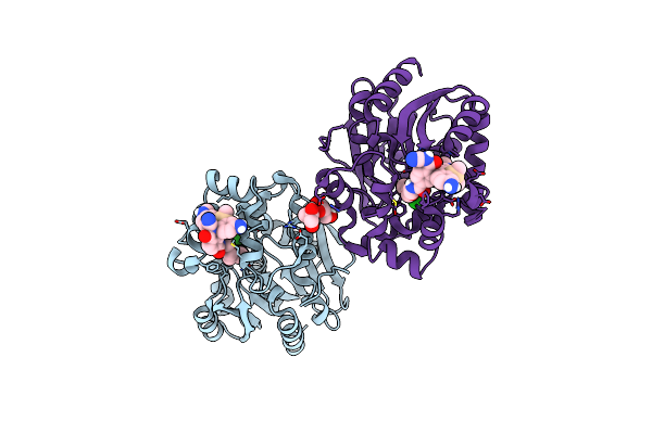 |
Halotag With Traq-G Ligand
Organism: Rhodococcus sp.
Method: X-RAY DIFFRACTION Resolution:1.68 Å Release Date: 2023-02-01 Classification: HYDROLASE Ligands: GOL, ILQ, CL, ILU |
Organism: Rhodococcus sp.
Method: X-RAY DIFFRACTION
Release Date: 2023-02-01
Ligands: GOL, ILQ, CL, ILU