
Search Count: 636
 |
Organism: Streptococcus pneumoniae r6
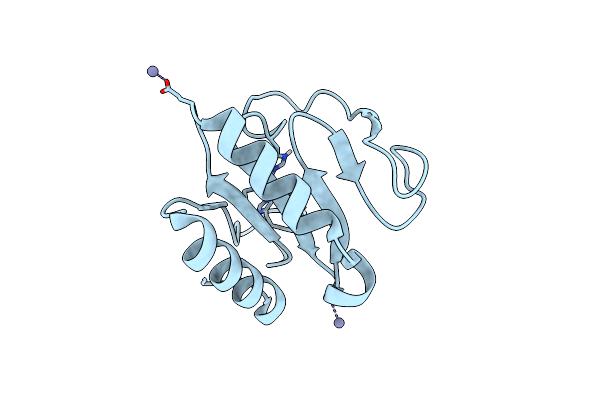
Method: X-RAY DIFFRACTION Resolution:2.80 Å Release Date: 2026-06-10 Classification: CELL CYCLE |
 |
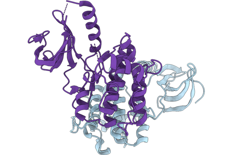
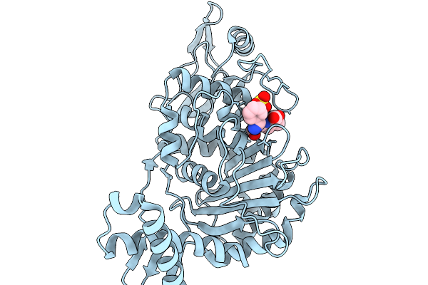
Streptococcus Pneumoniae Stkp Catalytic Domain T167E/T169E Double Mutant In Complex With Amp-Pnp And Mn2+
Organism: Streptococcus pneumoniae r6
Method: X-RAY DIFFRACTION Resolution:1.60 Å Release Date: 2026-06-10 Classification: CELL CYCLE Ligands: ANP, MN |
 |
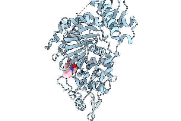
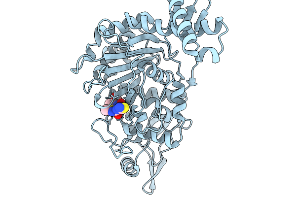
Streptococcus Pneumoniae Stkp Catalytic Domain T167A/T169A Double Mutant In Complex With Amp-Pnp And Mn2+
Organism: Streptococcus pneumoniae r6
Method: X-RAY DIFFRACTION Resolution:1.88 Å Release Date: 2026-06-10 Classification: CELL CYCLE Ligands: ANP, MN |
 |
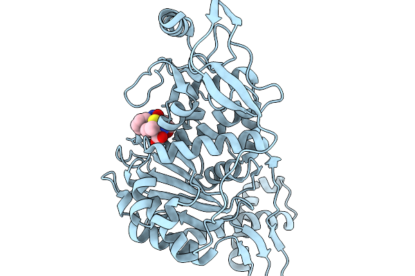
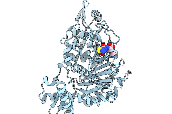
Streptococcus Pneumoniae Stkp Catalytic Domain T167A/T169A Double Mutant In Complex With Amp-Pnp And Mg2+
Organism: Streptococcus pneumoniae r6
Method: X-RAY DIFFRACTION Resolution:1.80 Å Release Date: 2026-06-10 Classification: CELL CYCLE Ligands: ANP, MG, MPD, GOL |
 |
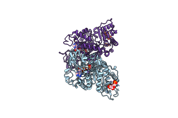
Organism: Escherichia coli, Streptococcus pneumoniae
Method: ELECTRON MICROSCOPY Resolution:3.63 Å Release Date: 2026-04-29 Classification: MEMBRANE PROTEIN |
 |
Penicillin-Binding Protein 1B (Pbp-1B) In Complex With Penicillin G - Streptococcus Pneumoniae R6
Organism: Streptococcus pneumoniae r6
Method: X-RAY DIFFRACTION Resolution:1.51 Å Release Date: 2026-02-18 Classification: TRANSFERASE Ligands: PNM, CL |
 |
Penicillin-Binding Protein 1B (Pbp-1B) In Complex With Ampicillin - Streptococcus Pneumoniae R6
Organism: Streptococcus pneumoniae r6
Method: X-RAY DIFFRACTION Resolution:1.79 Å Release Date: 2026-02-18 Classification: TRANSFERASE Ligands: AIX, CL |
 |
Penicillin-Binding Protein 1B (Pbp-1B) In Complex With Methicillin - Streptococcus Pneumoniae R6
Organism: Streptococcus pneumoniae r6
Method: X-RAY DIFFRACTION Resolution:1.93 Å Release Date: 2026-02-18 Classification: TRANSFERASE Ligands: 7EP, CL |
 |
Penicillin-Binding Protein 1B (Pbp-1B) In Complex With Cephalexin - Streptococcus Pneumoniae R6
Organism: Streptococcus pneumoniae r6
Method: X-RAY DIFFRACTION Resolution:1.64 Å Release Date: 2026-02-18 Classification: TRANSFERASE Ligands: A1JXD, CL, SO3 |
 |
Penicillin-Binding Protein 1B (Pbp-1B) In Complex With Ceftriaxone - Streptococcus Pneumoniae R6
Organism: Streptococcus pneumoniae r6
Method: X-RAY DIFFRACTION Resolution:1.79 Å Release Date: 2026-02-18 Classification: TRANSFERASE Ligands: CEF, CL |
 |
Penicillin-Binding Protein 1B (Pbp-1B) In Complex With Cefditoren - Streptococcus Pneumoniae R6
Organism: Streptococcus pneumoniae r6
Method: X-RAY DIFFRACTION Resolution:1.64 Å Release Date: 2026-02-18 Classification: TRANSFERASE Ligands: CDS, CL |
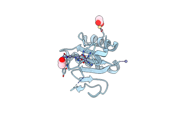 |
Crystal Structure Of The C-Terminal Domain Of Vlde From Streptococcus Pneumoniae Containing Four Zinc Atoms At The Binding Site
Organism: Streptococcus pneumoniae r6
Method: X-RAY DIFFRACTION Resolution:1.50 Å Release Date: 2025-01-22 Classification: METAL BINDING PROTEIN Ligands: ZN, CD, ACT |
 |
Crystal Structure Of The C-Terminal Domain Of Vlde From Streptococcus Pneumoniae Containing Three Zinc Atoms At The Binding Site
Organism: Streptococcus pneumoniae r6
Method: X-RAY DIFFRACTION Resolution:1.60 Å Release Date: 2025-01-22 Classification: METAL BINDING PROTEIN Ligands: ACT, ZN, CD |
 |
Crystal Structure Of The C-Terminal Domain Of Vlde From Streptococcus Pneumoniae Containing Two Zinc Atoms At The Binding Site
Organism: Streptococcus pneumoniae r6
Method: X-RAY DIFFRACTION Resolution:1.85 Å Release Date: 2025-01-22 Classification: METAL BINDING PROTEIN Ligands: ZN, CD |
 |
Crystal Structure Of The C-Terminal Domain Of Vlde From Streptococcus Pneumoniae Containing A Zinc Atom At The Binding Site
Organism: Streptococcus pneumoniae r6
Method: X-RAY DIFFRACTION Resolution:2.80 Å Release Date: 2025-01-22 Classification: METAL BINDING PROTEIN Ligands: ACT, CD, ZN |
 |
Crystal Structure Of The C-Terminal Domain Of Vlde From Streptococcus Pneumoniae In A Catalytically Competent Conformation
Organism: Streptococcus pneumoniae r6
Method: X-RAY DIFFRACTION Resolution:1.50 Å Release Date: 2025-01-22 Classification: METAL BINDING PROTEIN Ligands: ZN |
 |
Crystal Structure Of The C-Terminal Domain Of Vlde H373A From Streptococcus Pneumoniae
Organism: Streptococcus pneumoniae r6
Method: X-RAY DIFFRACTION Resolution:1.14 Å Release Date: 2025-01-22 Classification: METAL BINDING PROTEIN Ligands: ZN |
 |
Crystal Structure Of Streptococcus Pneumoniae Pyruvate Kinase In Complex With Oxalate And Fructose 1,6-Bisphosphate And Atp
Organism: Streptococcus pneumoniae r6
Method: X-RAY DIFFRACTION Resolution:1.99 Å Release Date: 2024-07-31 Classification: TRANSFERASE Ligands: K, MG, OXL, ATP, FBP |
 |
Crystal Structure Of Streptococcus Pneumoniae Pyruvate Kinase In Complex With Oxalate And Fructose 1,6-Bisphosphate And Adp
Organism: Streptococcus pneumoniae r6
Method: X-RAY DIFFRACTION Resolution:2.10 Å Release Date: 2024-07-31 Classification: TRANSFERASE Ligands: MG, K, FBP, OXL, ADP, GOL |
 |
Crystal Structure Of Streptococcus Pneumoniae Pyruvate Kinase In Complex With Oxalate And Fructose 1,6-Bisphosphate And Gdp
Organism: Streptococcus pneumoniae r6
Method: X-RAY DIFFRACTION Resolution:2.00 Å Release Date: 2024-07-31 Classification: TRANSFERASE Ligands: MG, K, FBP, GDP, OXL, GOL |