
Search Count: 4,571
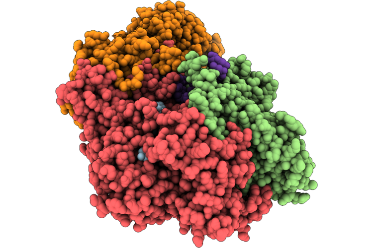 |
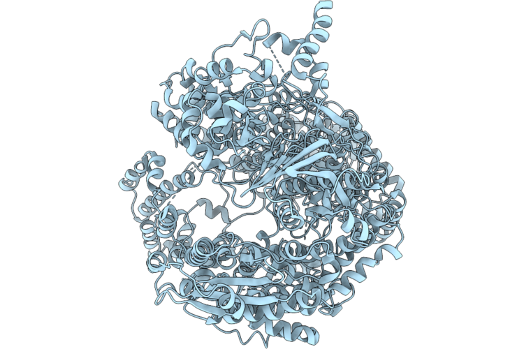
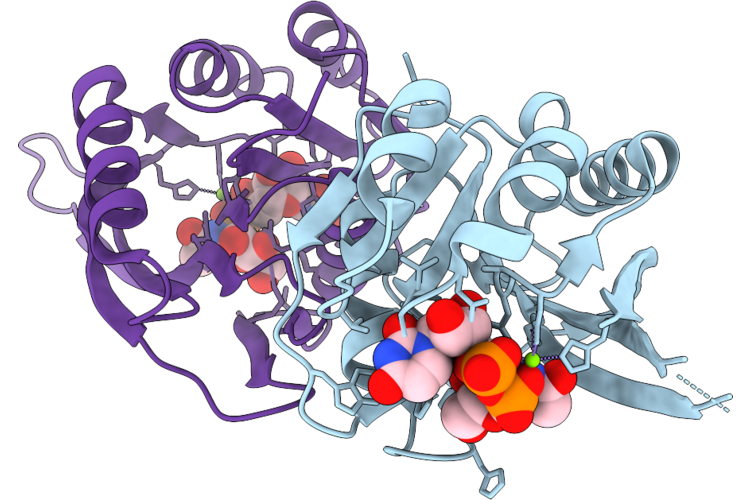
Cryo-Em Structure Of The Type Iii-D2 Crispr-Cas Effector Complex Bound To A Cognate Target Rna In The Pre-Cleavage State
Organism: Gammaproteobacteria bacterium
Method: ELECTRON MICROSCOPY Release Date: 2026-06-24 Classification: IMMUNE SYSTEM Ligands: MG, ANP, SAM, ZN |
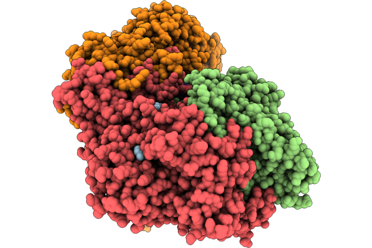 |
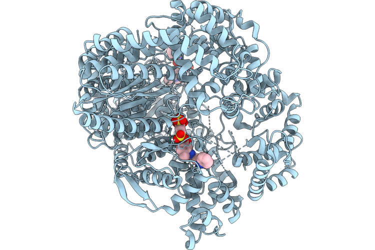
Cryo-Em Structure Of The Type Iii-D2 Crispr-Cas Effector Complex Bound To A Cognate Target Rna In The Post-Cleavage State
Organism: Gammaproteobacteria bacterium
Method: ELECTRON MICROSCOPY Release Date: 2026-06-24 Classification: IMMUNE SYSTEM Ligands: MG, ANP, SAM, ZN |
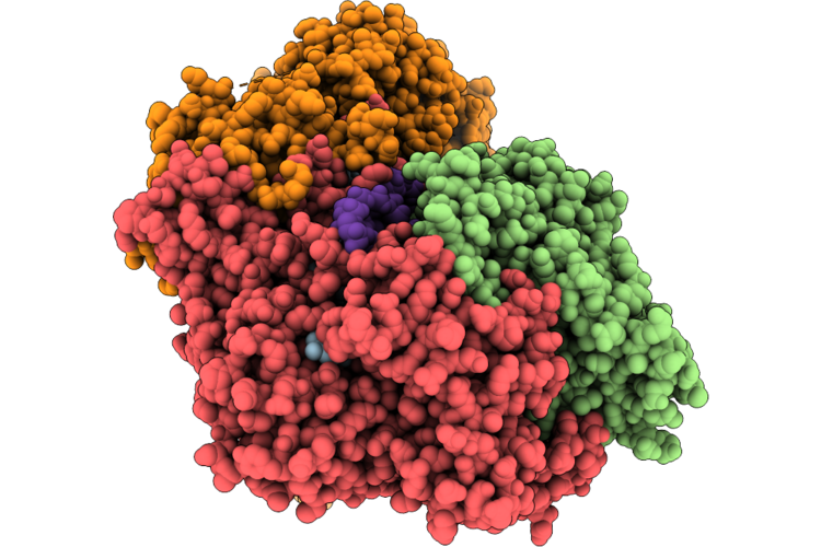 |
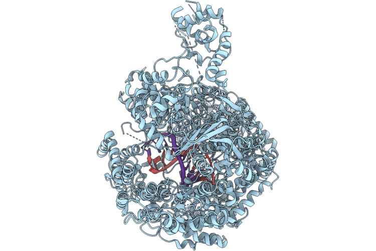
Cryo-Em Structure Of The Type Iii-D2 Crispr-Cas Effector Complex Bound To A Non-Cognate Target Rna In The Pre-Cleavage State
Organism: Gammaproteobacteria bacterium
Method: ELECTRON MICROSCOPY Release Date: 2026-06-24 Classification: IMMUNE SYSTEM Ligands: MG, ANP, SAM, ZN |
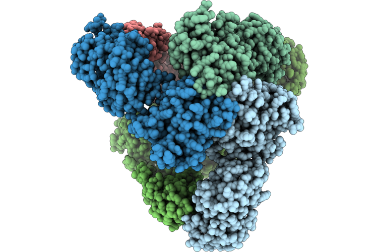 |
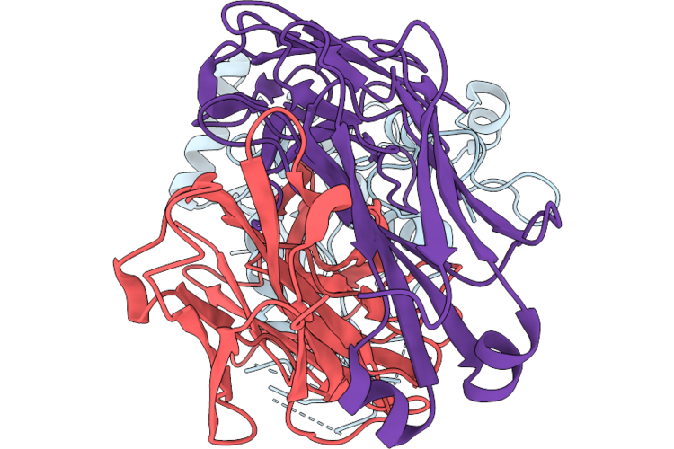
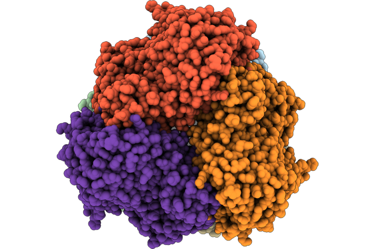
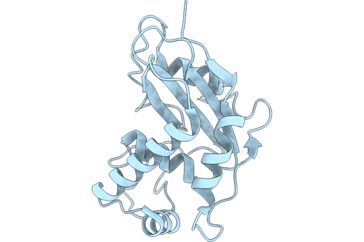
Cryoem Structure Of Hexametric Htra From Borrelia Burgdorferi With Bound Peptides In The Active Sites
Organism: Borreliella burgdorferi, Unidentified
Method: ELECTRON MICROSCOPY Release Date: 2026-06-17 Classification: HYDROLASE |
 |
Organism: Homo sapiens, Niallia circulans
Method: X-RAY DIFFRACTION Resolution:3.00 Å Release Date: 2026-06-03 Classification: MEMBRANE PROTEIN Ligands: A1EUH, A1EUC |
 |
Organism: Homo sapiens, Niallia circulans
Method: X-RAY DIFFRACTION Resolution:3.60 Å Release Date: 2026-06-03 Classification: MEMBRANE PROTEIN Ligands: A1EUC, A1EUD |
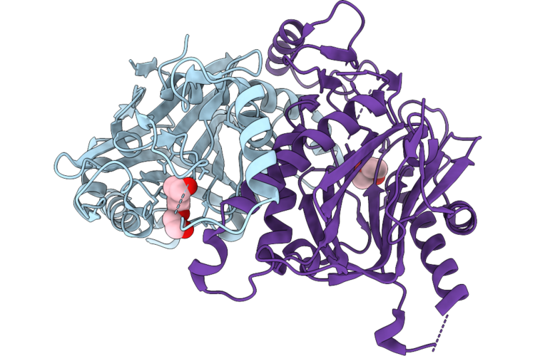 |
Crystal Structure Of The Polycaprolactam (Nylon6) And Poly(Hexamethylene Adipamide) (Nylon66) Hydrolase Nyl12 At Room Temperature
Organism: Gammaproteobacteria bacterium
Method: X-RAY DIFFRACTION Resolution:1.90 Å Release Date: 2026-06-03 Classification: HYDROLASE Ligands: PGE, PEG |
 |
Crystal Structure Of The Polycaprolactam (Nylon6) And Poly(Hexamethylene Adipamide) (Nylon66) Hydrolase Nyl12 At Cryo Temperature
Organism: Gammaproteobacteria bacterium
Method: X-RAY DIFFRACTION Resolution:1.75 Å Release Date: 2026-06-03 Classification: HYDROLASE Ligands: CHT, ACT, EPE, GOL, EDO, PEG |
 |
Crystal Structure Of The Poly(Hexamethylene Adipamide) (Nylon66) Hydrolase Nyl50 Acylenzyme Complex At Room Temperature
Organism: Alphaproteobacteria bacterium
Method: X-RAY DIFFRACTION Resolution:3.10 Å Release Date: 2026-06-03 Classification: HYDROLASE Ligands: A1CZU, NA |
 |
Crystal Structure Of The Poly(Hexamethylene Adipamide) (Nylon66) Hydrolase Nyl50 In Complex With Butyrate At Room Temperature
Organism: Alphaproteobacteria bacterium
Method: X-RAY DIFFRACTION Resolution:2.00 Å Release Date: 2026-06-03 Classification: HYDROLASE Ligands: BUA |
 |
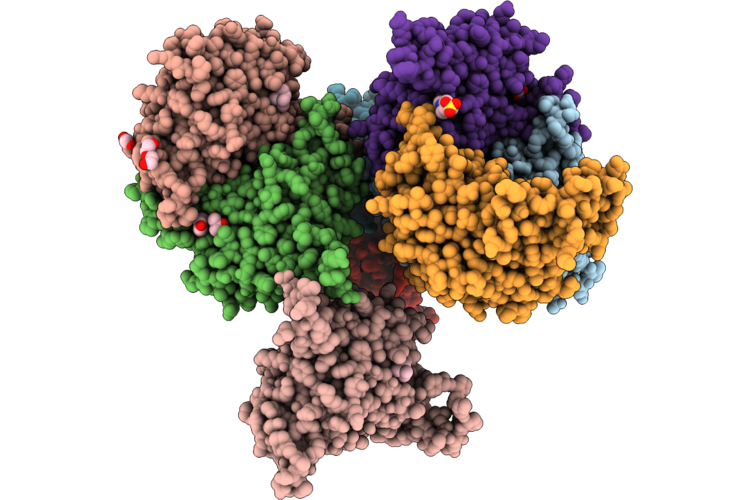
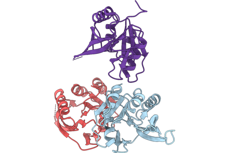
Structure Of The Anthrax Protective Antigen In Complex With A Potent Neutralizing Antibody
Organism: Bacillus anthracis, Homo sapiens
Method: ELECTRON MICROSCOPY Resolution:3.03 Å Release Date: 2026-05-27 Classification: ANTITOXIN |
 |
Organism: Ebinur lake virus
Method: ELECTRON MICROSCOPY Resolution:3.55 Å Release Date: 2026-05-13 Classification: VIRAL PROTEIN |
 |
Organism: Ebinur lake virus
Method: ELECTRON MICROSCOPY Resolution:3.00 Å Release Date: 2026-05-13 Classification: VIRAL PROTEIN Ligands: SVR |
 |
Organism: Ebinur lake virus, Synthetic construct
Method: ELECTRON MICROSCOPY Resolution:3.31 Å Release Date: 2026-05-13 Classification: VIRAL PROTEIN/RNA |
 |
Organism: Heyndrickxia coagulans
Method: ELECTRON MICROSCOPY Release Date: 2026-04-29 Classification: HYDROLASE Ligands: ZN, CL |
 |
Organism: Bacillus cereus
Method: X-RAY DIFFRACTION Resolution:2.80 Å Release Date: 2026-04-22 Classification: TRANSFERASE |
 |
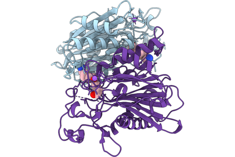
Crystal Structure Of Bacillus Cereus Gmar In Complex With Udp-Glcnac And Mg2+
Organism: Bacillus cereus
Method: X-RAY DIFFRACTION Resolution:2.42 Å Release Date: 2026-04-22 Classification: TRANSFERASE Ligands: UD1, MG |
 |
Organism: Eimeria tenella strain houghton
Method: X-RAY DIFFRACTION Resolution:1.75 Å Release Date: 2026-04-22 Classification: UNKNOWN FUNCTION |
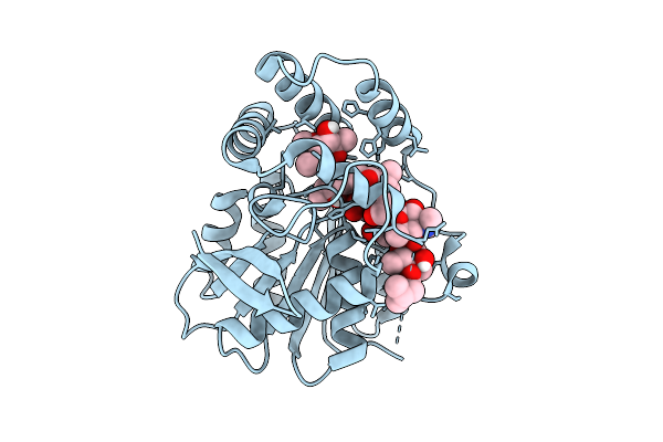 |
Crystal Structure Of Alpha/Beta-Hydrolase Macrolide Esterase Estt From Bacillus Cereus (S102A Mutant) In Complex With Linearized Tylvalosin
Organism: Bacillus cereus
Method: X-RAY DIFFRACTION Resolution:1.70 Å Release Date: 2026-04-08 Classification: HYDROLASE Ligands: A1DAR |
 |
Organism: Anopheles culicifacies
Method: X-RAY DIFFRACTION Resolution:1.34 Å Release Date: 2026-01-28 Classification: LIPID BINDING PROTEIN Ligands: CD, CL |