

Search Count: 27
 |
Streptococcus Gordonii C123 Domain(S)-Structural And Functional Analysis
Organism: Streptococcus gordonii
Method: X-RAY DIFFRACTION Resolution:2.70 Å Release Date: 2021-09-08 Classification: CELL ADHESION Ligands: CA |
Organism: Streptococcus gordonii
Method: X-RAY DIFFRACTION
Release Date: 2021-09-08
Ligands: CA
 |
The Structure Of The Streptococcus Gordonii Surface Protein Sspb In Complex With Tev Peptide Provides Clues To The Adherence Of Oral Streptococcal Adherence To Salivary Agglutinin
Organism: Streptococcus gordonii
Method: X-RAY DIFFRACTION Resolution:2.00 Å Release Date: 2020-08-12 Classification: CELL ADHESION Ligands: CA |
Organism: Streptococcus gordonii
Method: X-RAY DIFFRACTION
Release Date: 2020-08-12
Ligands: CA
 |
The Structure Of The Streptococcus Gordonii Surface Protein Sspb In Complex With Tev Peptide Provides Clues To The Adherence Of Oral Streptococcal Adherence To Salivary Agglutinin
Organism: Streptococcus gordonii
Method: X-RAY DIFFRACTION Resolution:1.80 Å Release Date: 2020-08-12 Classification: CELL ADHESION Ligands: MG, GOL |
Organism: Streptococcus gordonii
Method: X-RAY DIFFRACTION
Release Date: 2020-08-12
Ligands: MG, GOL
 |
Gspb Siglec Domain
Organism: Streptococcus gordonii
Method: X-RAY DIFFRACTION Resolution:1.03 Å Release Date: 2020-02-19 Classification: SUGAR BINDING PROTEIN Ligands: CA |
Organism: Streptococcus gordonii
Method: X-RAY DIFFRACTION
Release Date: 2020-02-19
Ligands: CA
 |
Gspb Siglec Domain
Organism: Streptococcus gordonii
Method: X-RAY DIFFRACTION Resolution:1.30 Å Release Date: 2020-02-19 Classification: SUGAR BINDING PROTEIN |
Organism: Streptococcus gordonii
Method: X-RAY DIFFRACTION
Release Date: 2020-02-19
 |
Gspb Siglec + Unique Domains
Organism: Streptococcus gordonii
Method: X-RAY DIFFRACTION Resolution:1.60 Å Release Date: 2020-02-19 Classification: SUGAR BINDING PROTEIN Ligands: CA |
Organism: Streptococcus gordonii
Method: X-RAY DIFFRACTION
Release Date: 2020-02-19
Ligands: CA
 |
Gspb Siglec Domain
Organism: Streptococcus gordonii
Method: X-RAY DIFFRACTION Resolution:1.90 Å Release Date: 2020-02-19 Classification: SUGAR BINDING PROTEIN Ligands: GOL, CA, SO4 |
Organism: Streptococcus gordonii
Method: X-RAY DIFFRACTION
Release Date: 2020-02-19
Ligands: GOL, CA, SO4
 |
Crystal Structure Of Accessory Secretion Protein 1 And 3
Organism: Streptococcus gordonii
Method: X-RAY DIFFRACTION Resolution:3.11 Å Release Date: 2018-02-21 Classification: CELL ADHESION Modified Residue(s): MSE |
Organism: Streptococcus gordonii
Method: X-RAY DIFFRACTION
Release Date: 2018-02-21
 |
Crystal Structure Of Accessory Secretion Protein 1
Organism: Streptococcus gordonii
Method: X-RAY DIFFRACTION Resolution:2.77 Å Release Date: 2018-02-21 Classification: CELL ADHESION |
Organism: Streptococcus gordonii
Method: X-RAY DIFFRACTION
Release Date: 2018-02-21
 |
Crystal Structure Of The Gspb Siglec Domain With Sialyl T Antigen Bound
Organism: Streptococcus gordonii
Method: X-RAY DIFFRACTION Resolution:1.25 Å Release Date: 2016-04-13 Classification: SUGAR BINDING PROTEIN Ligands: MG |
Organism: Streptococcus gordonii
Method: X-RAY DIFFRACTION
Release Date: 2016-04-13
Ligands: MG
 |
Crystal Structure Of Gtfa/B Complex
Organism: Streptococcus gordonii
Method: X-RAY DIFFRACTION Resolution:2.92 Å Release Date: 2016-03-02 Classification: TRANSFERASE/CHAPERONE Ligands: MG Modified Residue(s): MSE |
Organism: Streptococcus gordonii
Method: X-RAY DIFFRACTION
Release Date: 2016-03-02
Ligands: MG
 |
Crystal Structure Of Gtfa/B Complex Bound To Udp And Glcnac
Organism: Streptococcus gordonii
Method: X-RAY DIFFRACTION Resolution:3.84 Å Release Date: 2016-03-02 Classification: TRANSFERASE/CHAPERONE Ligands: UDP, NAG, MG Modified Residue(s): MSE |
Organism: Streptococcus gordonii
Method: X-RAY DIFFRACTION
Release Date: 2016-03-02
Ligands: UDP, NAG, MG
 |
Gspb
Organism: Streptococcus gordonii
Method: X-RAY DIFFRACTION Resolution:1.40 Å Release Date: 2011-08-10 Classification: SUGAR BINDING PROTEIN Ligands: K, GOL, HDZ, NA |
Organism: Streptococcus gordonii
Method: X-RAY DIFFRACTION
Release Date: 2011-08-10
Ligands: K, GOL, HDZ, NA
 |
Gspb
Organism: Streptococcus gordonii
Method: X-RAY DIFFRACTION Resolution:1.90 Å Release Date: 2011-08-10 Classification: SUGAR BINDING PROTEIN Ligands: CA, GOL, HDZ |
Organism: Streptococcus gordonii
Method: X-RAY DIFFRACTION
Release Date: 2011-08-10
Ligands: CA, GOL, HDZ
 |
Crystal Structure Of The C-Terminal Domain Of Streptococcus Gordonii Surface Protein Sspb
Organism: Streptococcus gordonii
Method: X-RAY DIFFRACTION Resolution:1.50 Å Release Date: 2010-02-16 Classification: CELL ADHESION Ligands: CA Modified Residue(s): MSE |
Organism: Streptococcus gordonii
Method: X-RAY DIFFRACTION
Release Date: 2010-02-16
Ligands: CA
 |
Crystal Structure Of The C-Terminal Domain Of Streptococcus Gordonii Surface Protein Sspb
Organism: Streptococcus gordonii
Method: X-RAY DIFFRACTION Resolution:1.70 Å Release Date: 2010-02-16 Classification: CELL ADHESION Ligands: CA, MG |
Organism: Streptococcus gordonii
Method: X-RAY DIFFRACTION
Release Date: 2010-02-16
Ligands: CA, MG
 |
Two Intramolecular Isopeptide Bonds Are Identified In The Crystal Structure Of The Streptococcus Gordonii Sspb C-Terminal Domain
Organism: Streptococcus gordonii
Method: X-RAY DIFFRACTION Resolution:2.08 Å Release Date: 2010-02-16 Classification: CELL ADHESION Ligands: CA |
Organism: Streptococcus gordonii
Method: X-RAY DIFFRACTION
Release Date: 2010-02-16
Ligands: CA
 |
Crystal Structure Of The Variable Domain Of The Streptococcus Gordonii Surface Protein Sspb
Organism: Streptococcus gordonii
Method: X-RAY DIFFRACTION Resolution:2.30 Å Release Date: 2009-07-28 Classification: CELL ADHESION Ligands: CA, GOL, K |
Organism: Streptococcus gordonii
Method: X-RAY DIFFRACTION
Release Date: 2009-07-28
Ligands: CA, GOL, K
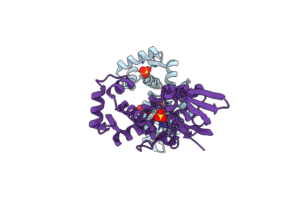 |
Crystal Structure Of The Manganese-Activated Repressor Scar: Apo Form
Organism: Streptococcus gordonii
Method: X-RAY DIFFRACTION Resolution:2.70 Å Release Date: 2009-06-23 Classification: TRANSCRIPTION Ligands: SO4 |
Organism: Streptococcus gordonii
Method: X-RAY DIFFRACTION
Release Date: 2009-06-23
Ligands: SO4
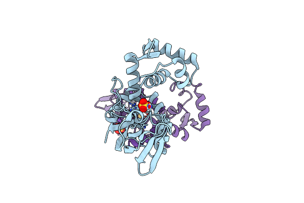 |
Crystal Structure Of Scar With Bound Cd2+
Organism: Streptococcus gordonii
Method: X-RAY DIFFRACTION Resolution:2.80 Å Release Date: 2009-06-23 Classification: TRANSCRIPTION Ligands: SO4, CD |
Organism: Streptococcus gordonii
Method: X-RAY DIFFRACTION
Release Date: 2009-06-23
Ligands: SO4, CD