

Search Count: 15
 |
Hsa Bound To Neu5Gc Alpha2,3 Gal Beta Ome
Organism: Streptococcus
Method: X-RAY DIFFRACTION Resolution:1.45 Å Release Date: 2026-08-26 Classification: SUGAR BINDING PROTEIN |
Organism: Streptococcus
Method: X-RAY DIFFRACTION
Release Date: 2026-08-26
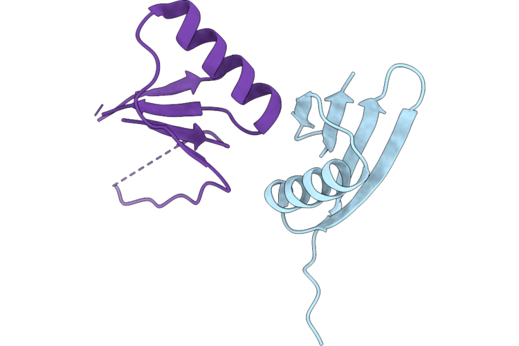 |
Crystal Structure Of The Ustilago Maydis Virulence Factor Pit2
Organism: Streptococcus, Mycosarcoma maydis
Method: X-RAY DIFFRACTION Resolution:2.35 Å Release Date: 2026-07-22 Classification: PEPTIDE BINDING PROTEIN |
Organism: Streptococcus, Mycosarcoma maydis
Method: X-RAY DIFFRACTION
Release Date: 2026-07-22
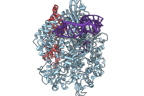 |
Structure Of Spycas9 In Complex With The Anti-Crispr Protein Acriia26
Organism: Streptococcus pyogenes m1 gas, Streptococcus
Method: ELECTRON MICROSCOPY Release Date: 2026-02-04 Classification: IMMUNE SYSTEM |
Organism: Streptococcus pyogenes m1 gas, Streptococcus
Method: ELECTRON MICROSCOPY
Release Date: 2026-02-04
 |
Hsa Bound To Neu5Ac Alpha2,3 Gal Beta Ome
Organism: Streptococcus
Method: X-RAY DIFFRACTION Resolution:1.29 Å Release Date: 2025-11-26 Classification: SUGAR BINDING PROTEIN Ligands: CA, NA |
Organism: Streptococcus
Method: X-RAY DIFFRACTION
Release Date: 2025-11-26
Ligands: CA, NA
 |
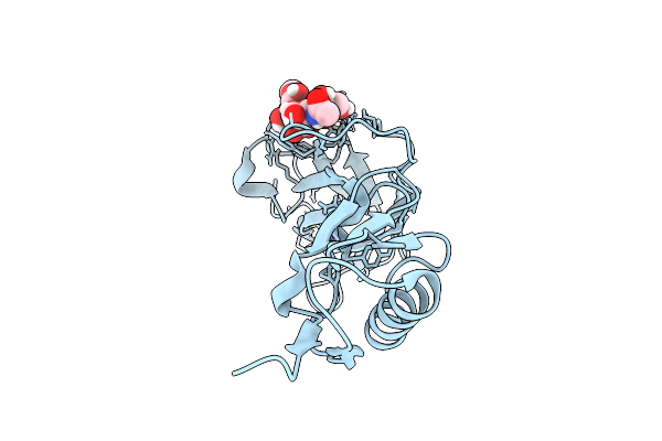
Gspb Siglec Domain Bound To Sialyl T Antigen Linked To Serine-Fmoc
Organism: Streptococcus
Method: X-RAY DIFFRACTION Resolution:1.89 Å Release Date: 2025-09-10 Classification: SUGAR BINDING PROTEIN Ligands: GOL, VP1, SER, CA |
Organism: Streptococcus
Method: X-RAY DIFFRACTION
Release Date: 2025-09-10
Ligands: GOL, VP1, SER, CA
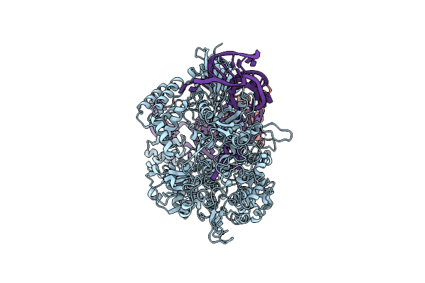 |
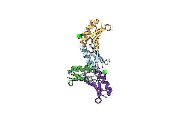
Cryo-Em Structure Of Cas9-Sgrna-A32 Complex
Organism: Streptococcus, Streptococcus pyogenes serotype m1, Streptococcus pyogenes
Method: ELECTRON MICROSCOPY Release Date: 2024-09-11 Classification: IMMUNE SYSTEM/RNA |
Organism: Streptococcus, Streptococcus pyogenes serotype m1, Streptococcus pyogenes
Method: ELECTRON MICROSCOPY
Release Date: 2024-09-11
 |
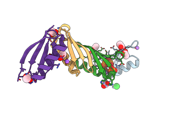
Crystal Structure Of Mutant B1 Immunoglobulin-Binding Domain Of Streptococcal Protein G (T16F, T18A, V21E, T25L, K28Y, V29I, K31R, Q32H, Y33L, N35K, D36H, N37Q)
Organism: Streptococcus
Method: X-RAY DIFFRACTION Resolution:1.40 Å Release Date: 2019-01-23 Classification: DE NOVO PROTEIN Ligands: ZN, CL |
Organism: Streptococcus
Method: X-RAY DIFFRACTION
Release Date: 2019-01-23
Ligands: ZN, CL
 |
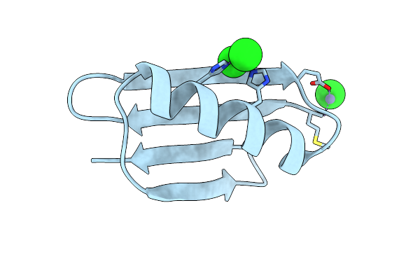
Crystal Structure Of B1 Immunoglobulin-Binding Domain Of Streptococcal Protein G (T16F, T18A, V21H, T25H, K28Y, V29I, K31R, Q32A, Y33L, N35K, D36A, N37Q)
Organism: Streptococcus
Method: X-RAY DIFFRACTION Resolution:1.40 Å Release Date: 2019-01-23 Classification: METAL BINDING PROTEIN Ligands: ZN, ACT, NA, CL, PO4, DPO |
Organism: Streptococcus
Method: X-RAY DIFFRACTION
Release Date: 2019-01-23
Ligands: ZN, ACT, NA, CL, PO4, DPO
 |
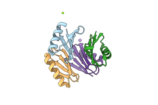
Crystal Structure Of De Novo Designed Metal-Controlled Dimer Of Mutant B1 Immunoglobulin-Binding Domain Of Streptococcal Protein G (L12H, T16L, V29H, Y33H, N37L)-Zinc
Organism: Streptococcus
Method: X-RAY DIFFRACTION Resolution:1.50 Å Release Date: 2019-01-23 Classification: METAL BINDING PROTEIN Ligands: ZN, CL |
Organism: Streptococcus
Method: X-RAY DIFFRACTION
Release Date: 2019-01-23
Ligands: ZN, CL
 |
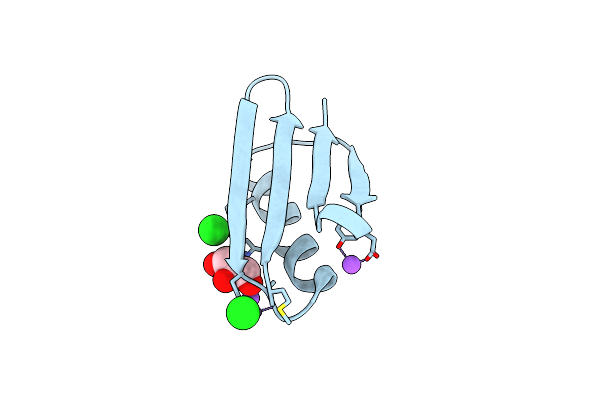
Crystal Structure Of De Novo Designed Metal-Controlled Dimer Of Mutant B1 Immunoglobulin-Binding Domain Of Streptococcal Protein G (L12H, T16L, V29H, Y33H, N37L)-Apo
Organism: Streptococcus
Method: X-RAY DIFFRACTION Resolution:1.70 Å Release Date: 2019-01-23 Classification: DE NOVO PROTEIN Ligands: MG, NA |
Organism: Streptococcus
Method: X-RAY DIFFRACTION
Release Date: 2019-01-23
Ligands: MG, NA
 |
Crystal Structure Of De Novo Designed Metal-Controlled Dimer Of B1 Immunoglobulin-Binding Domain Of Streptococcal Protein G (L12H, E15V, T16L, T18I, V29H, Y33H, N37L)-Zinc
Organism: Streptococcus
Method: X-RAY DIFFRACTION Resolution:1.34 Å Release Date: 2019-01-23 Classification: DE NOVO PROTEIN Ligands: ZN, CL, GOL, NA |
Organism: Streptococcus
Method: X-RAY DIFFRACTION
Release Date: 2019-01-23
Ligands: ZN, CL, GOL, NA
 |
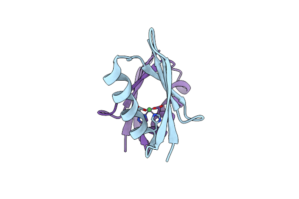
Crystal Structure Of De Novo Designed Metal-Controlled Dimer Of Mutant B1 Immunoglobulin-Binding Domain Of Streptococcal Protein G (L12H, E15V, T16L, T18I, V29H, Y33H, N37L)-Apo
Organism: Streptococcus
Method: X-RAY DIFFRACTION Resolution:2.30 Å Release Date: 2019-01-23 Classification: METAL BINDING PROTEIN Ligands: MG |
Organism: Streptococcus
Method: X-RAY DIFFRACTION
Release Date: 2019-01-23
Ligands: MG
 |
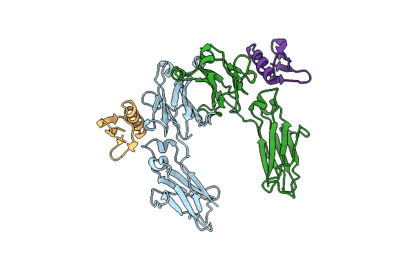
Nickel-Bound Crystal Structure Of A Gb1 Variant
Organism: Streptococcus
Method: X-RAY DIFFRACTION Resolution:2.20 Å Release Date: 2018-12-12 Classification: METAL BINDING PROTEIN Ligands: NI |
Organism: Streptococcus
Method: X-RAY DIFFRACTION
Release Date: 2018-12-12
Ligands: NI
 |
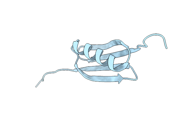
Crystal Structure Of The C2 Fragment Of Streptococcal Protein G In Complex With The Fc Domain Of Human Igg
Organism: Homo sapiens, Streptococcus
Method: X-RAY DIFFRACTION Resolution:3.20 Å Release Date: 1995-04-20 Classification: COMPLEX (ANTIBODY/ANTIGEN) |
Organism: Homo sapiens, Streptococcus
Method: X-RAY DIFFRACTION
Release Date: 1995-04-20
 |
The 1.66 Angstroms X-Ray Structure Of The B2 Immunoglobulin-Binding Domain Of Streptococcal Protein G And Comparison To The Nmr Structure Of The B1 Domain
Organism: Streptococcus
Method: X-RAY DIFFRACTION Resolution:1.66 Å Release Date: 1992-07-15 Classification: IMMUNOGLOBULIN BINDING PROTEIN |
Organism: Streptococcus
Method: X-RAY DIFFRACTION
Release Date: 1992-07-15